DNA methylation are key to understanding regulation of gene expression - Chromatin has a 3D arrangement controlling interaction between gene promoters and regulatory elements - Active vs. repressive compartments - Topologically associating domains - Chromatin loops - Methylation is a dynamic chemical modification occurring at cytosines, affecting expression of genes - Manuscript aim: profile chromatin conformation and DNAm at single-cell resolution to produce an epigenetic atlas of the human brain
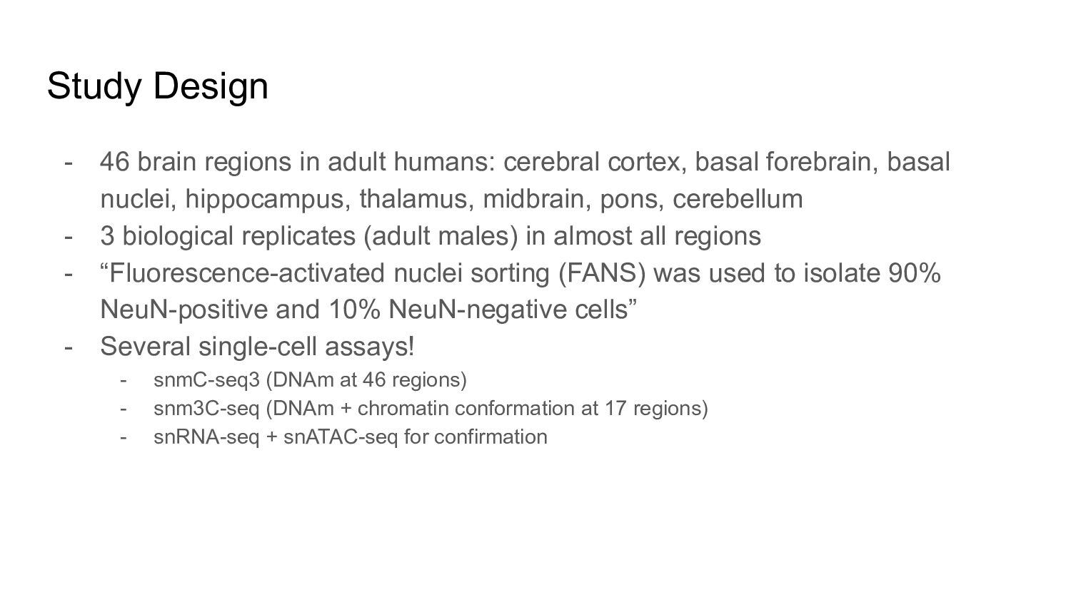
cortex, basal forebrain, basal nuclei, hippocampus, thalamus, midbrain, pons, cerebellum - 3 biological replicates (adult males) in almost all regions - “Fluorescence-activated nuclei sorting (FANS) was used to isolate 90% NeuN-positive and 10% NeuN-negative cells” - Several single-cell assays! - snmC-seq3 (DNAm at 46 regions) - snm3C-seq (DNAm + chromatin conformation at 17 regions) - snRNA-seq + snATAC-seq for confirmation
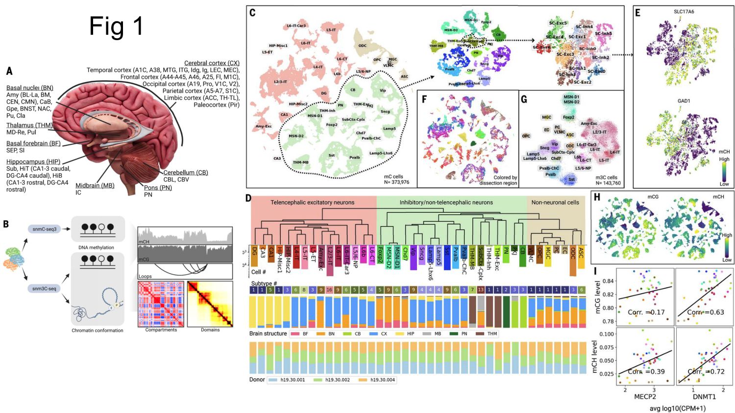
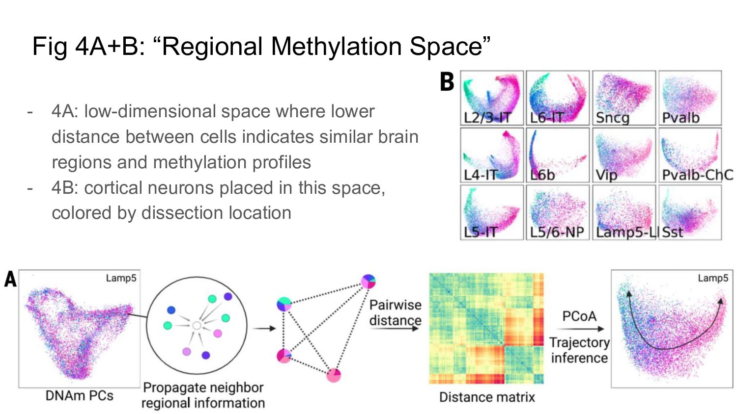
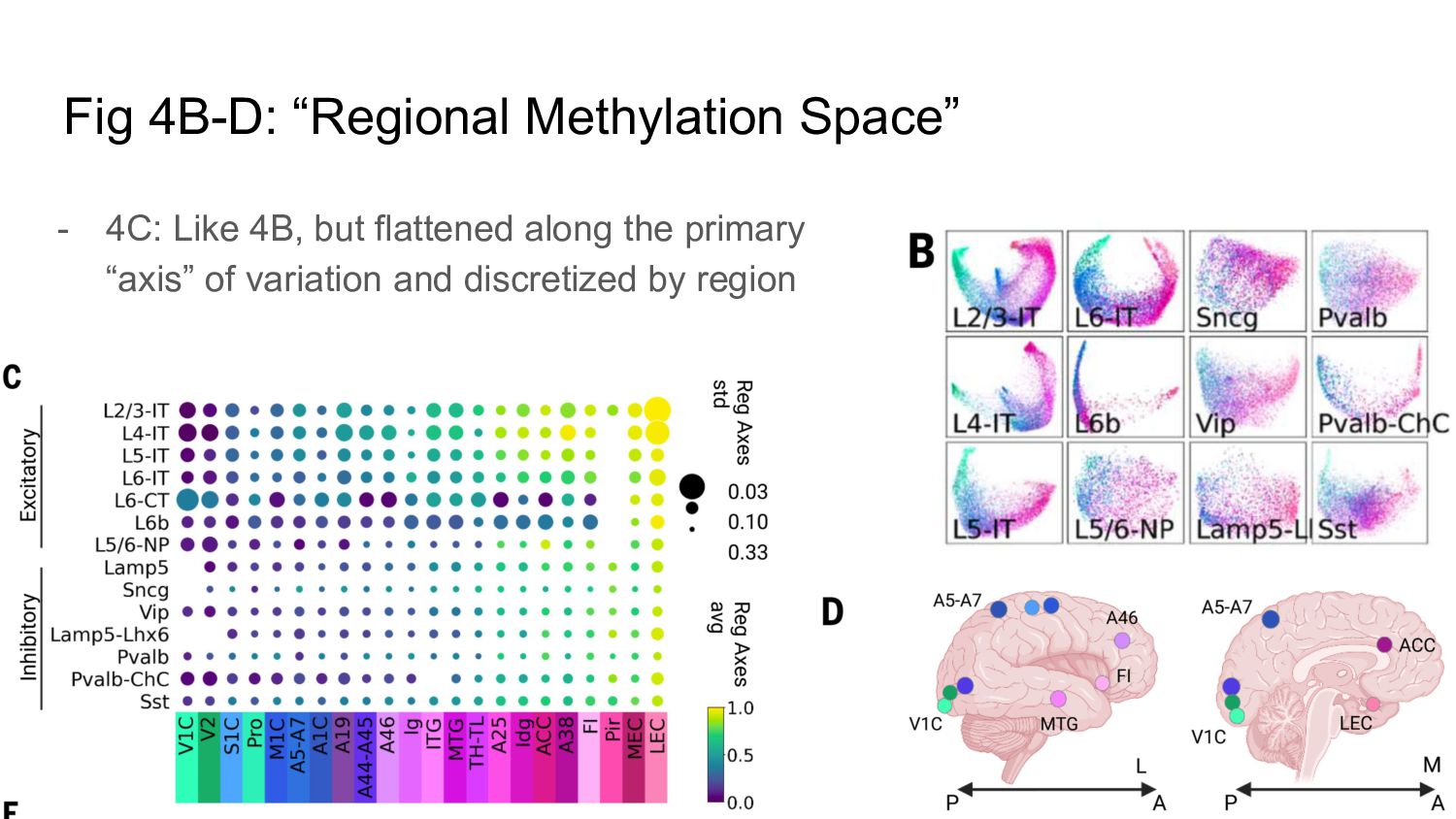
lower distance between cells indicates similar brain regions and methylation profiles - 4B: cortical neurons placed in this space, colored by dissection location
and DMRs across species (in this case human vs mouse) - Chromatin contact distance comparison by cell type (neurons tended to have shorter interaction distances!) - Chromatin features by cell type (compartments, domains, loops) - Chromatin features compared against methlyation
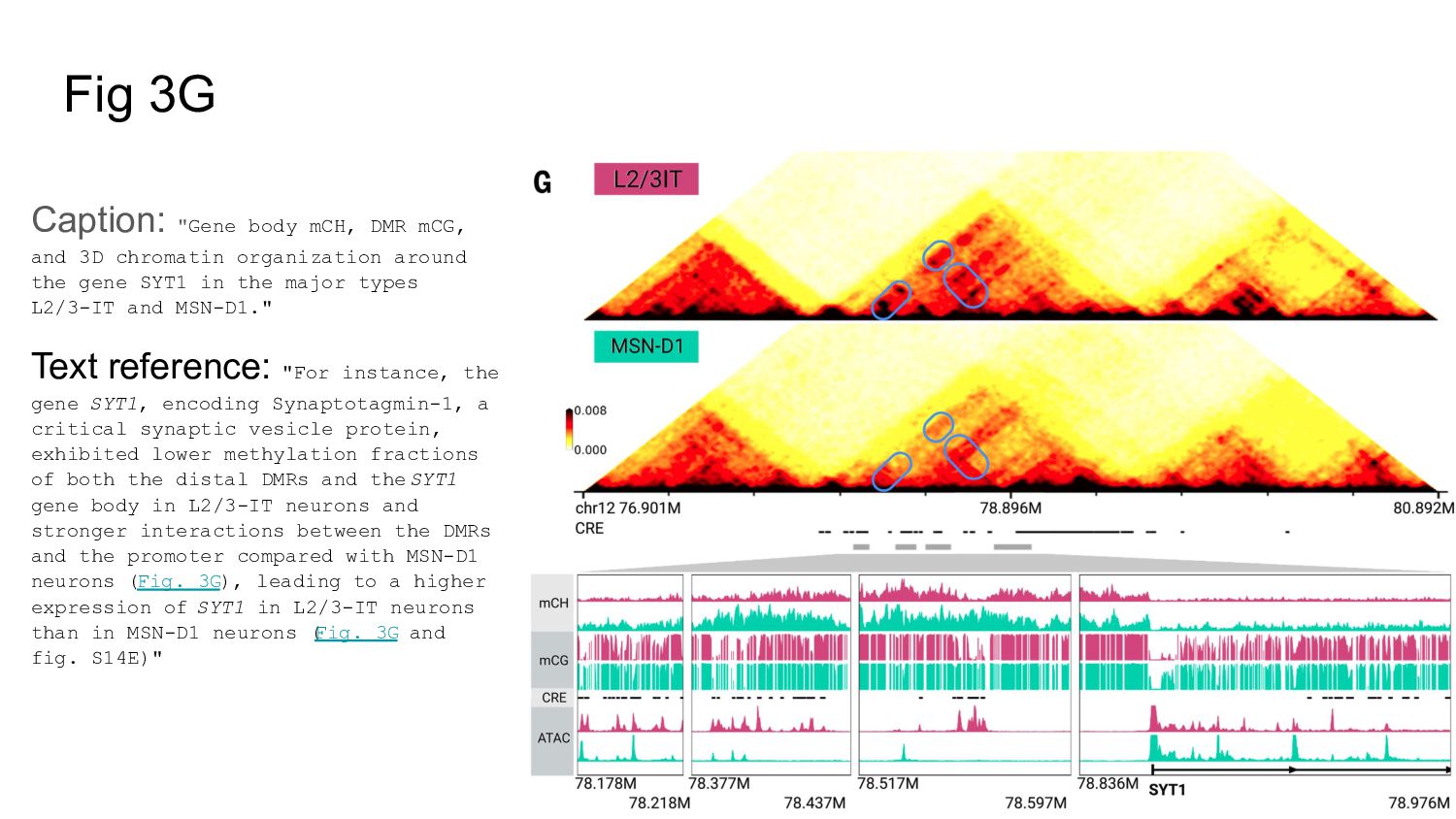
chromatin organization around the gene SYT1 in the major types L2/3-IT and MSN-D1." Text reference: "For instance, the gene SYT1, encoding Synaptotagmin-1, a critical synaptic vesicle protein, exhibited lower methylation fractions of both the distal DMRs and the SYT1 gene body in L2/3-IT neurons and stronger interactions between the DMRs and the promoter compared with MSN-D1 neurons (Fig. 3G), leading to a higher expression of SYT1 in L2/3-IT neurons than in MSN-D1 neurons ( Fig. 3G and fig. S14E)"