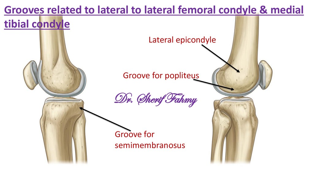
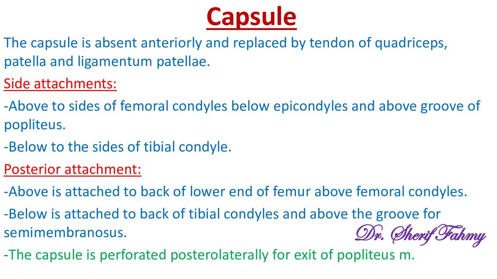
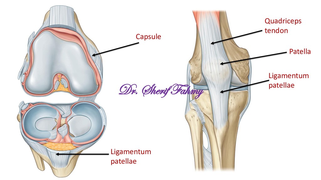
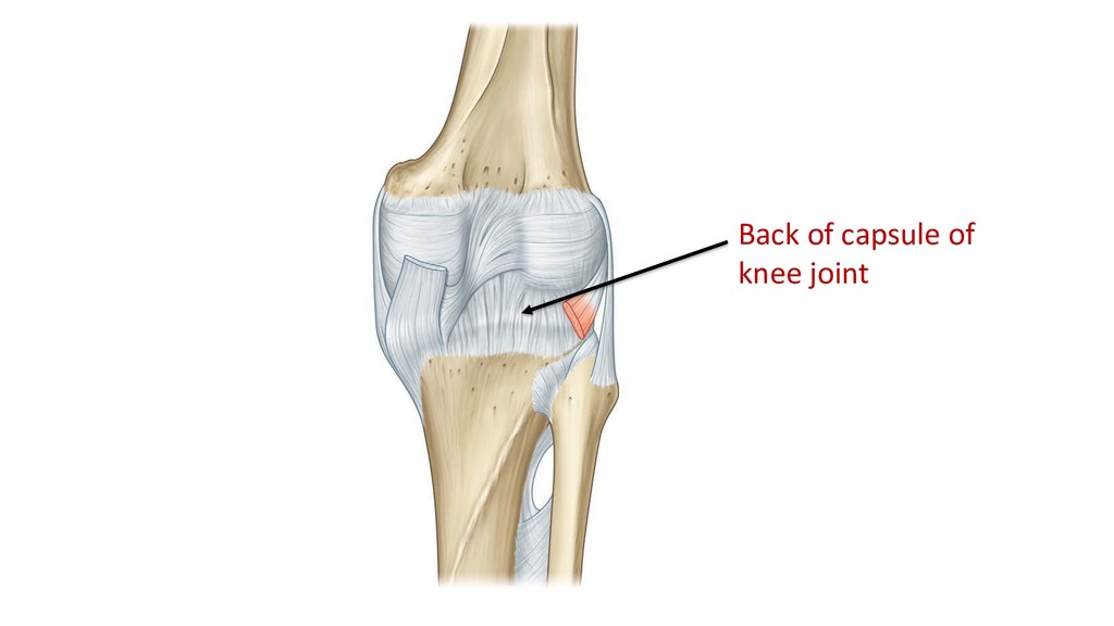
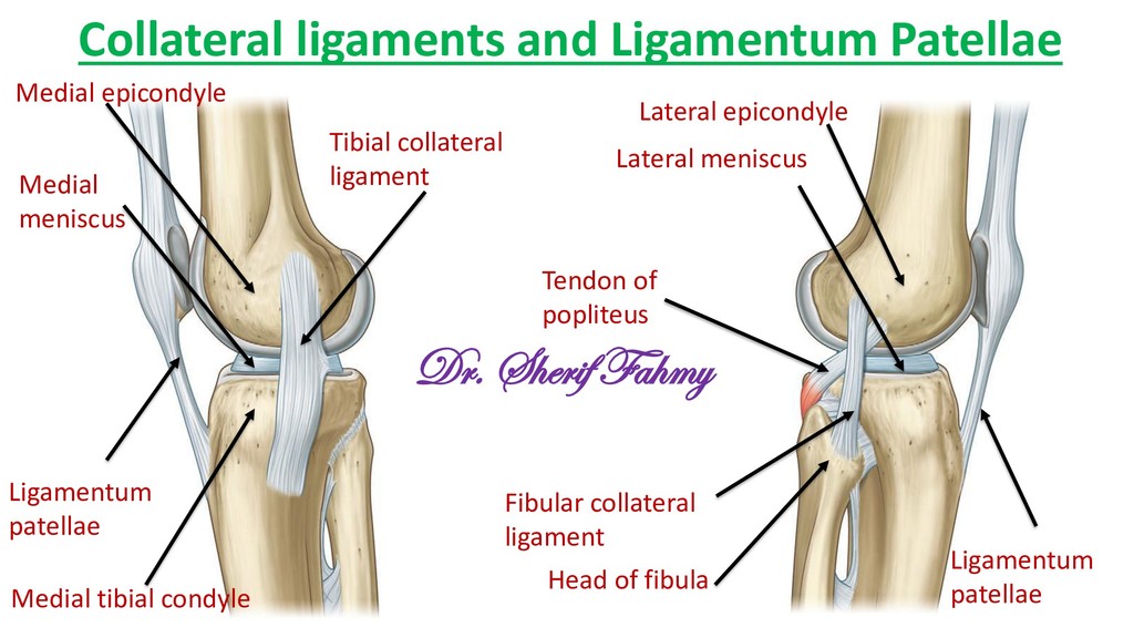
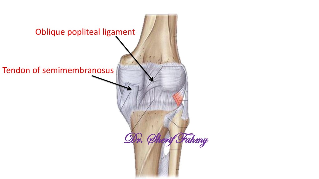
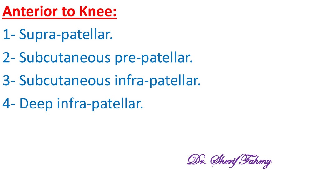
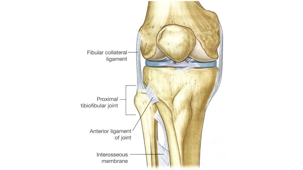
of quadriceps, patella and ligamentum patellae. Side attachments: -Above to sides of femoral condyles below epicondyles and above groove of popliteus. -Below to the sides of tibial condyle. Posterior attachment: -Above is attached to back of lower end of femur above femoral condyles. -Below is attached to back of tibial condyles and above the groove for semimembranosus. -The capsule is perforated posterolaterally for exit of popliteus m. Dr. Sherif Fahmy
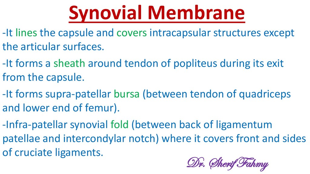
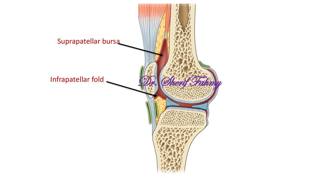
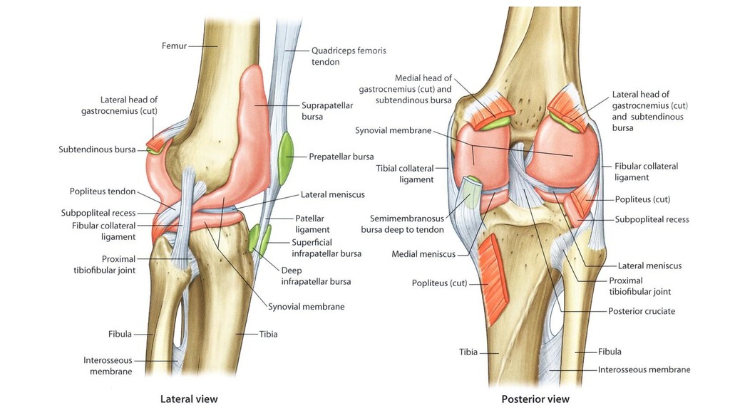
except the articular surfaces. -It forms a sheath around tendon of popliteus during its exit from the capsule. -It forms supra-patellar bursa (between tendon of quadriceps and lower end of femur). -Infra-patellar synovial fold (between back of ligamentum patellae and intercondylar notch) where it covers front and sides of cruciate ligaments. Dr. Sherif Fahmy
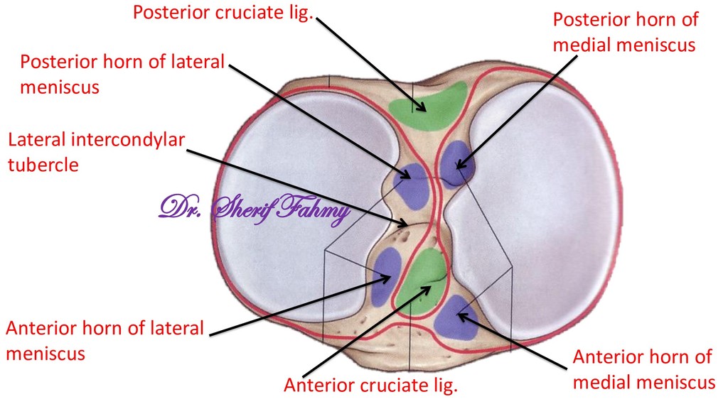
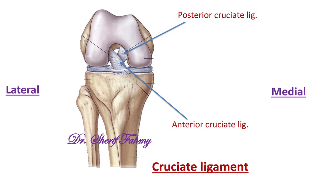
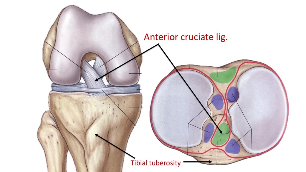
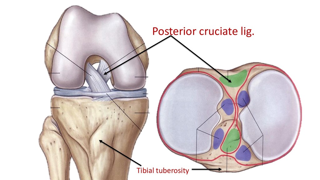
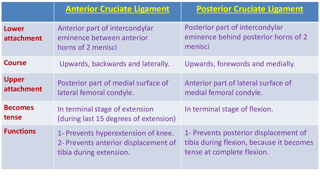
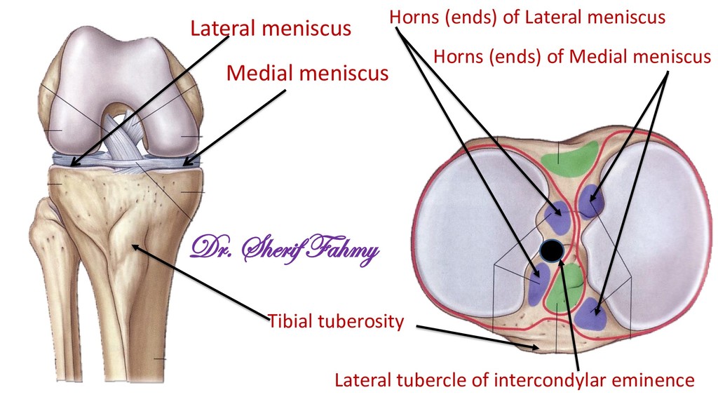
attachment Becomes tense Functions Anterior part of intercondylar eminence between anterior horns of 2 menisci Posterior part of intercondylar eminence behind posterior horns of 2 menisci Upwards, backwards and laterally. Upwards, forewords and medially. Posterior part of medial surface of lateral femoral condyle. Anterior part of lateral surface of medial femoral condyle. In terminal stage of extension (during last 15 degrees of extension) In terminal stage of flexion. 1- Prevents hyperextension of knee. 2- Prevents anterior displacement of tibia during extension. 1- Prevents posterior displacement of tibia during flexion, because it becomes tense at complete flexion.
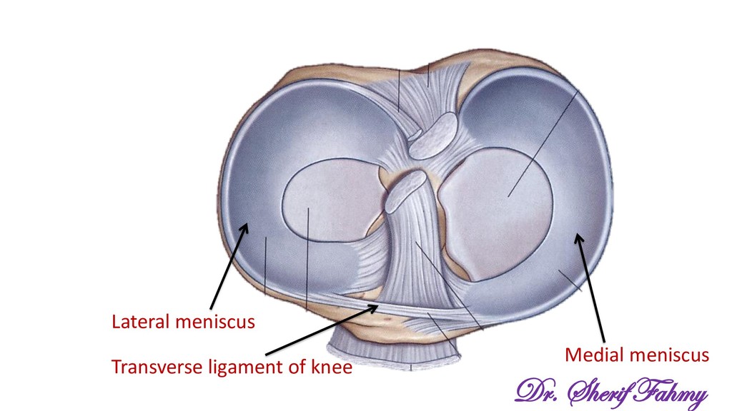
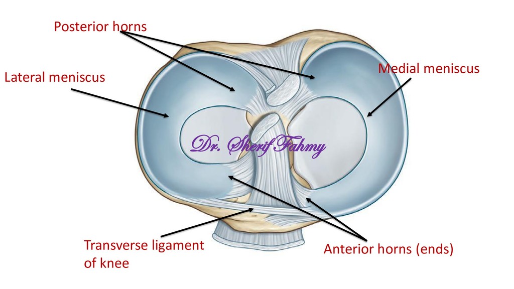
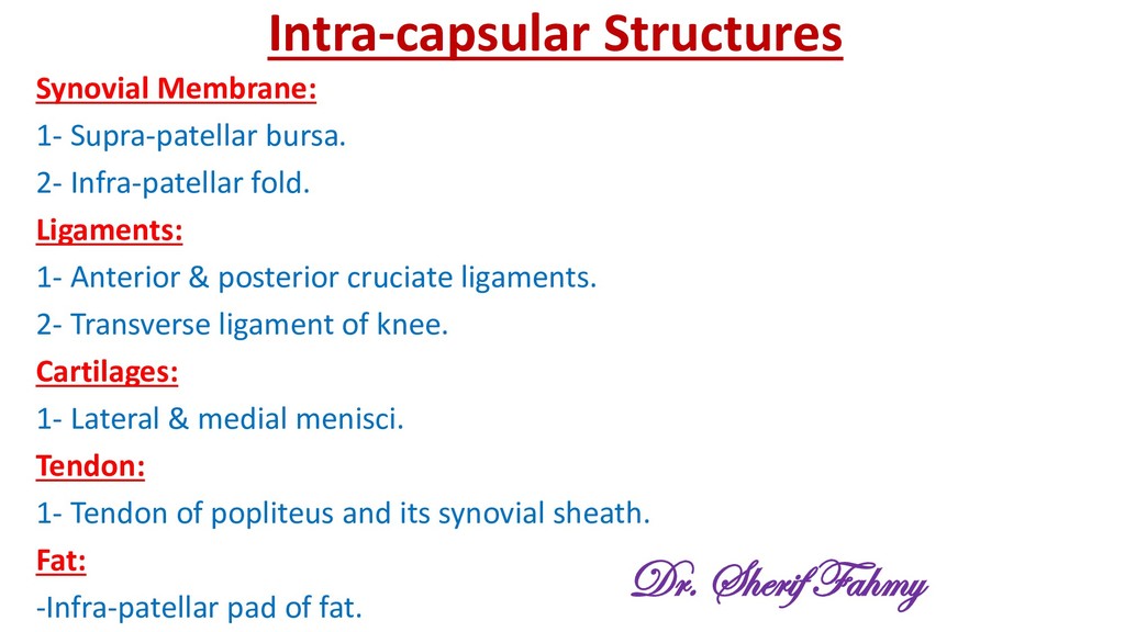
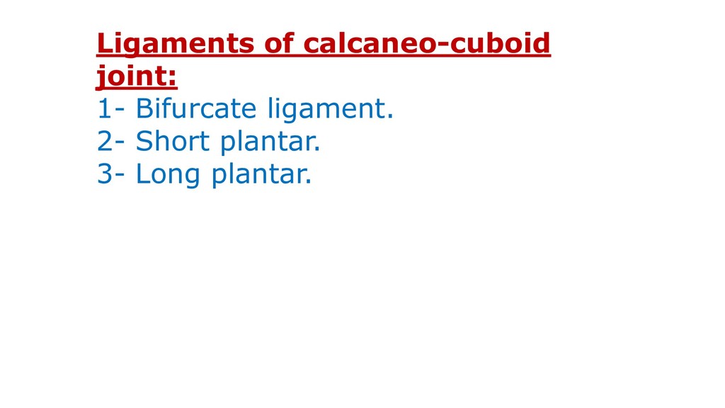
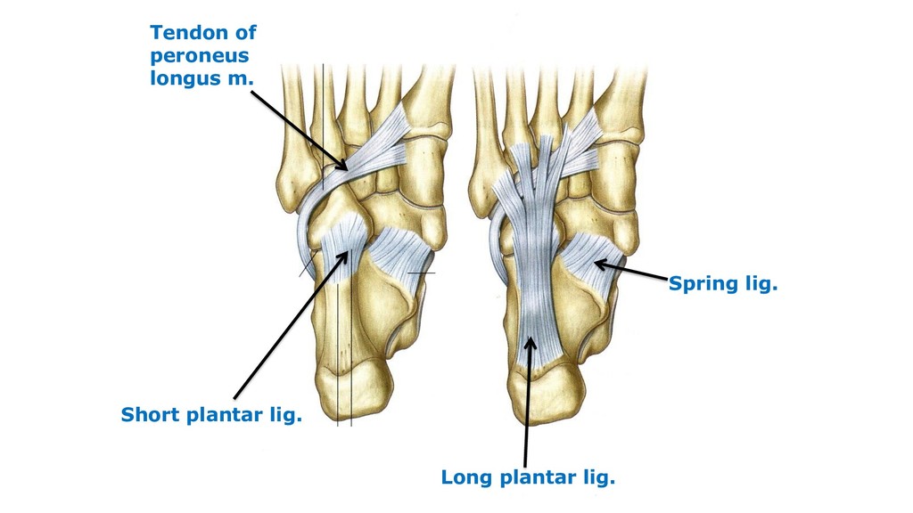
Ligaments: 1- Anterior & posterior cruciate ligaments. 2- Transverse ligament of knee. Cartilages: 1- Lateral & medial menisci. Tendon: 1- Tendon of popliteus and its synovial sheath. Fat: -Infra-patellar pad of fat. Dr. Sherif Fahmy
femoral condyles glide equally over the tibial condyles. -During the last 15 degrees, the anterior cruciate ligament becomes tense. -This will lead to stop of lateral condyle gliding while the medial condyle continues gliding leading to medial rotation of femur over tibia. -This lead to stretch of collateral and oblique popliteal ligament in addition to anterior cruciate lig. and locking of knee. Dr. Sherif Fahmy
tibia, if tibia is free from ground. -Lateral rotation of femur, if tibia is on the ground. -Both movements produced by the action of popliteus helped by hamstrings Dr. Sherif Fahmy
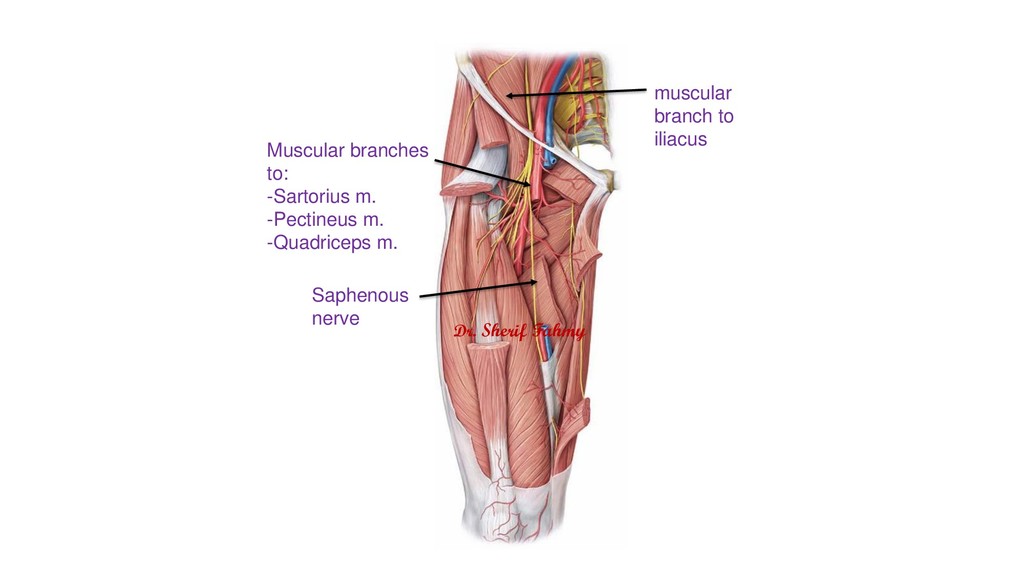
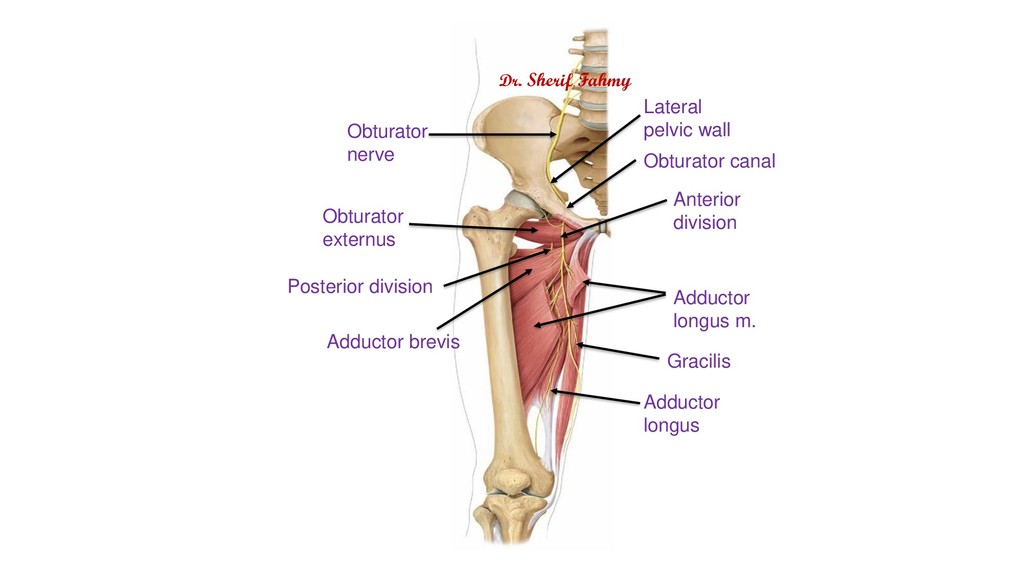
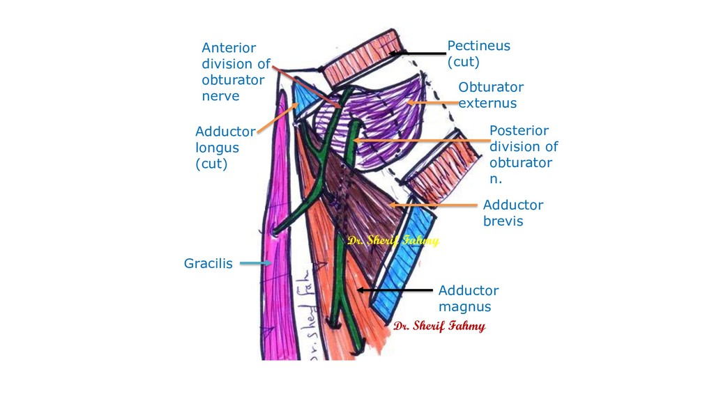
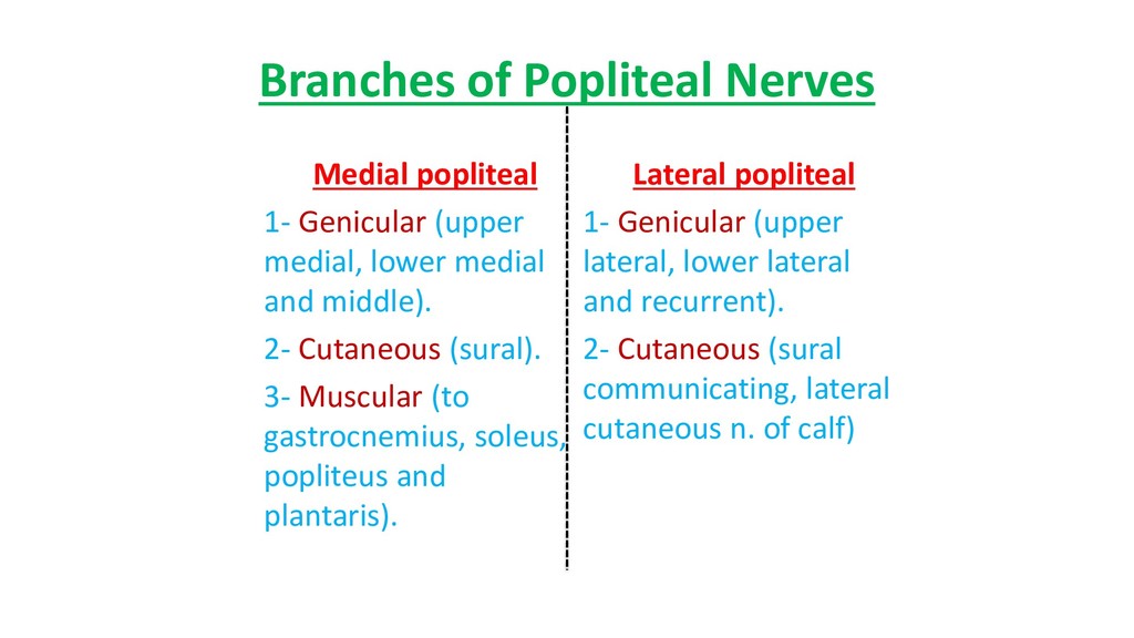
nerve. 2- Genicular branches from common peroneal nerve. 3- Femoral n. through branch to vasti of quadriceps femoris m. 4- Obturator n. from its posterior division. Dr. Sherif Fahmy
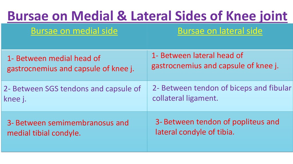
on medial side Bursae on lateral side 1- Between medial head of gastrocnemius and capsule of knee j. 1- Between lateral head of gastrocnemius and capsule of knee j. 2- Between SGS tendons and capsule of knee j. 2- Between tendon of biceps and fibular collateral ligament. 3- Between semimembranosus and medial tibial condyle. 3- Between tendon of popliteus and lateral condyle of tibia.
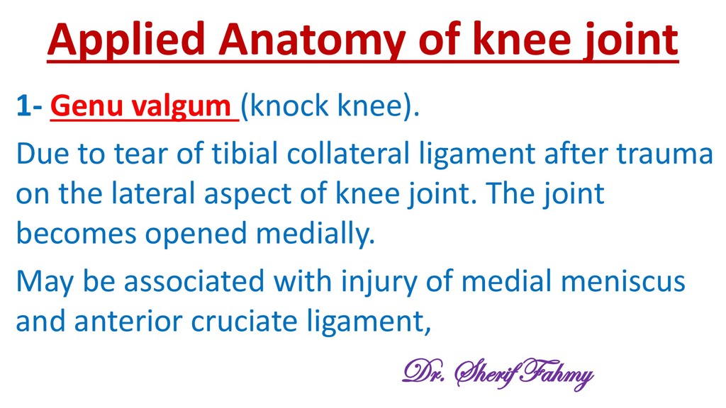
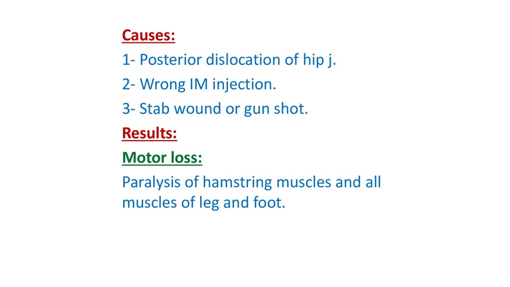
Due to tear of tibial collateral ligament after trauma on the lateral aspect of knee joint. The joint becomes opened medially. May be associated with injury of medial meniscus and anterior cruciate ligament, Dr. Sherif Fahmy
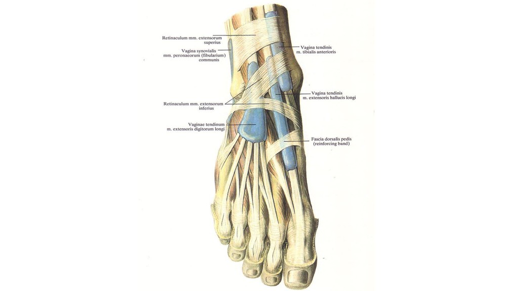
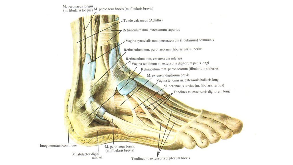
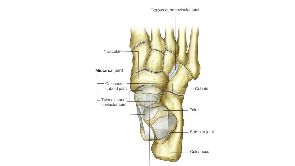
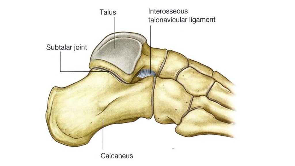
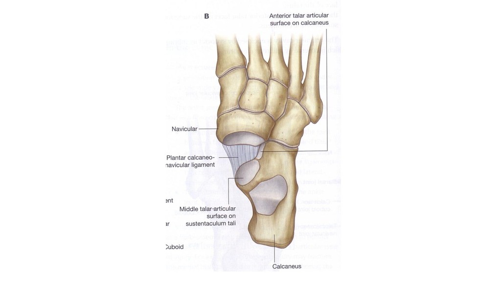
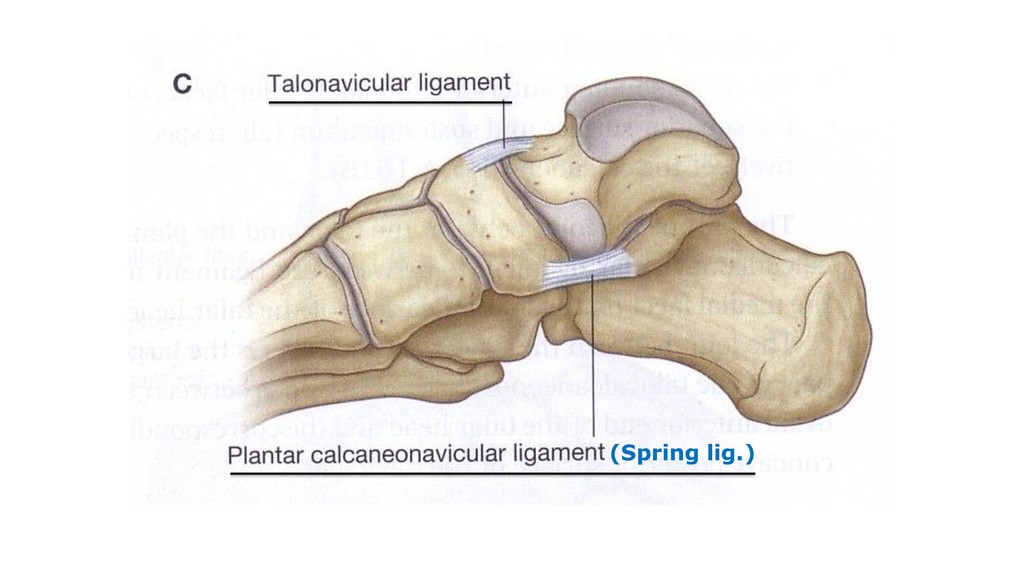
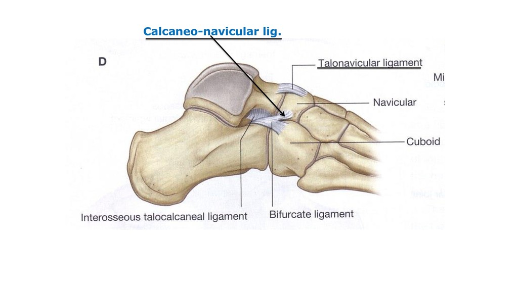
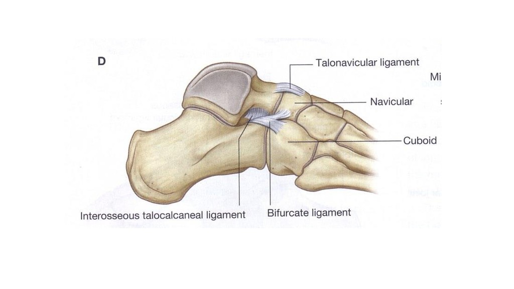
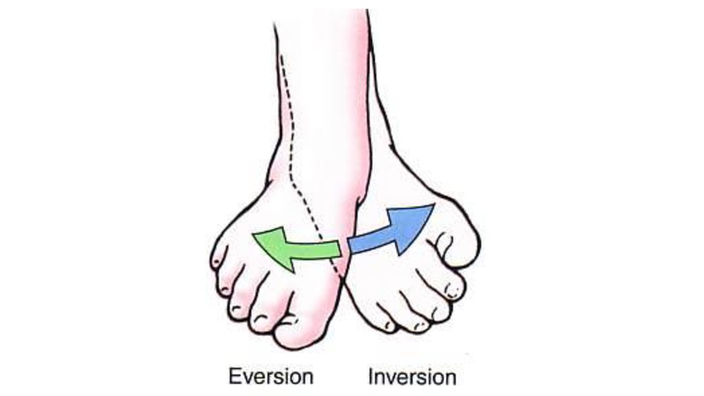
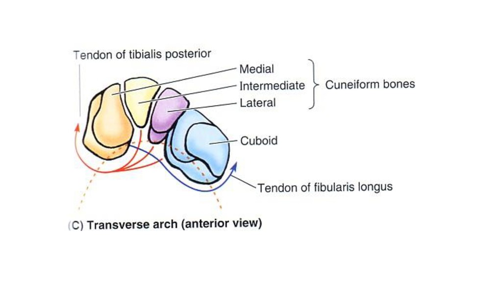
medially. Eversion: Elevation of outer border of foot with sole is directed laterally. Joints: 1- Subtalar joint. 2- Talo-calcaneo-navicular joint. Mechanisms: -Calcaneus and navicular swing around relatively fixed talus. -Muscles: Innvertors & Evertors
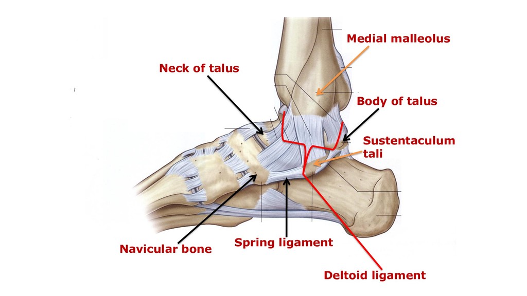
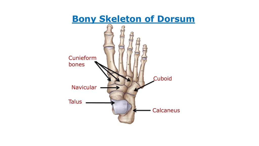
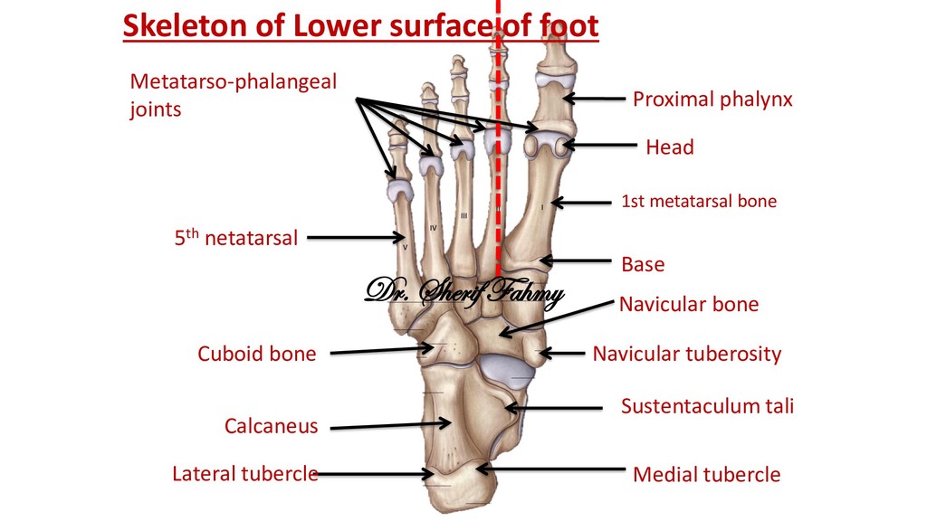
tubercle Sustentaculum tali Cuboid bone Navicular bone Navicular tuberosity 1st metatarsal bone 5th netatarsal Base Head Proximal phalynx Metatarso-phalangeal joints Dr. Sherif Fahmy
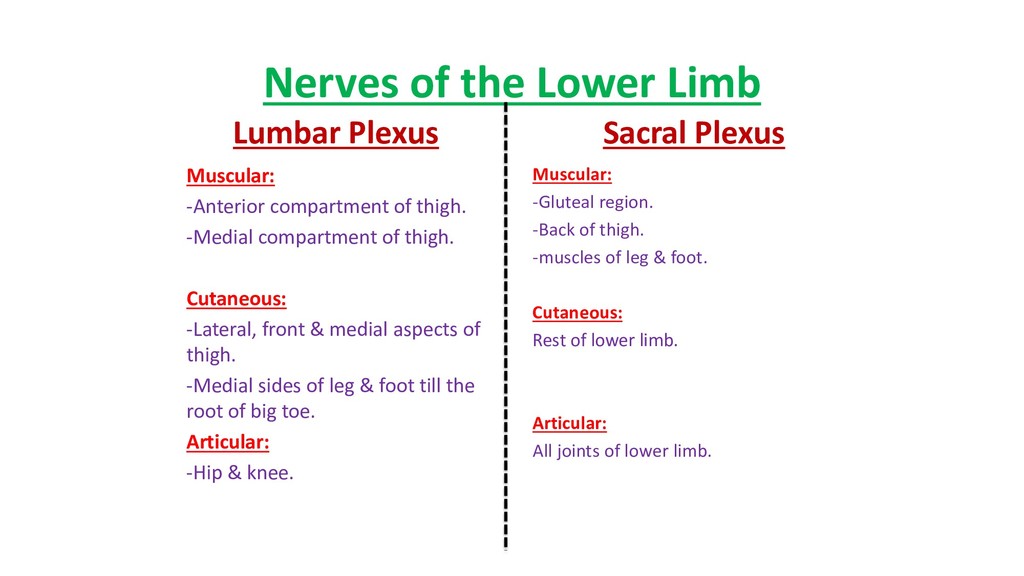
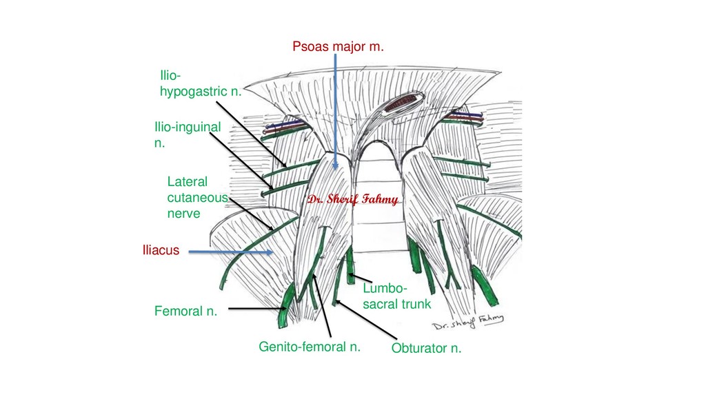
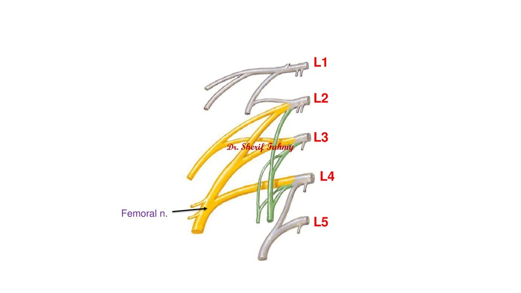
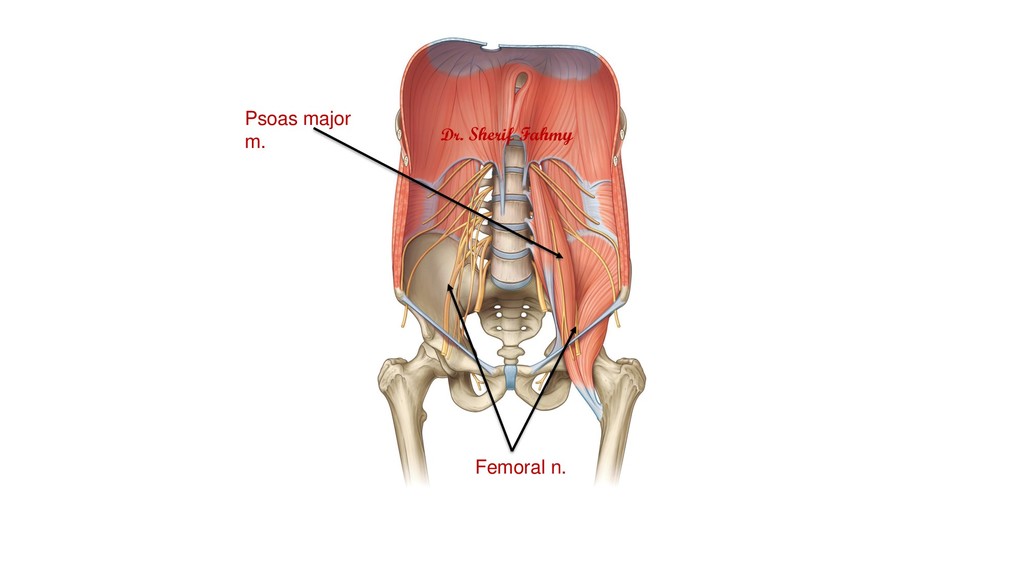
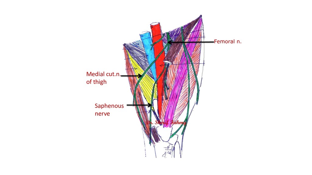
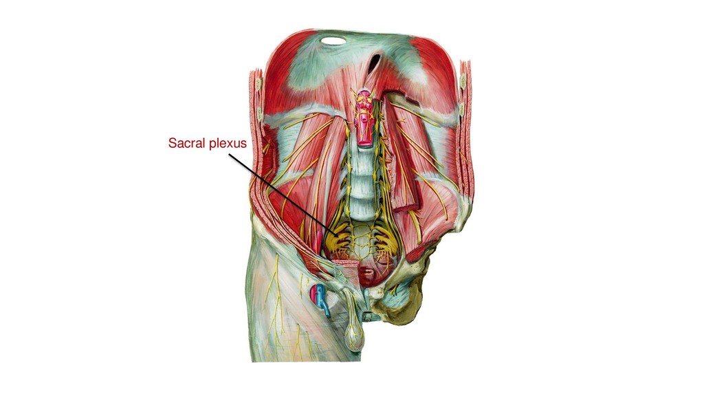
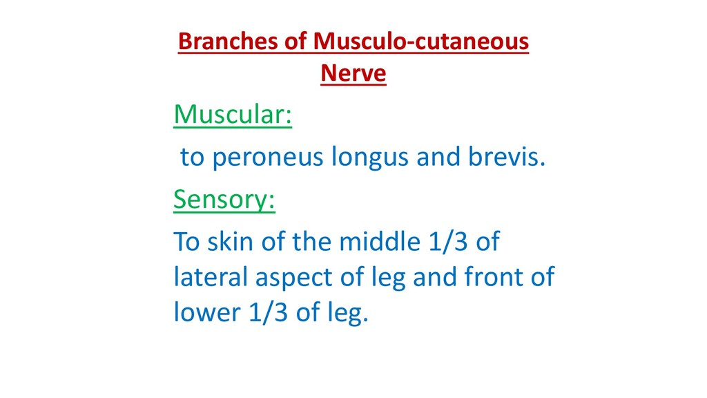
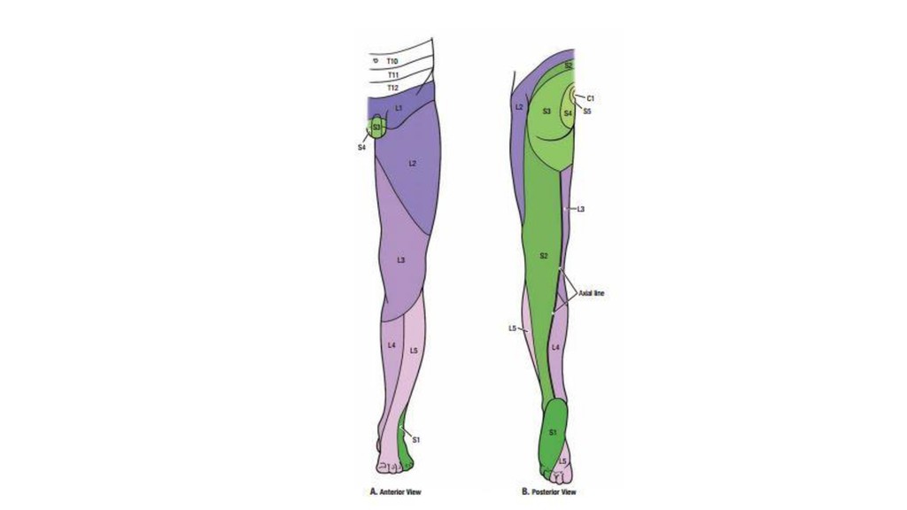
of thigh. -Medial compartment of thigh. Cutaneous: -Lateral, front & medial aspects of thigh. -Medial sides of leg & foot till the root of big toe. Articular: -Hip & knee. Sacral Plexus Muscular: -Gluteal region. -Back of thigh. -muscles of leg & foot. Cutaneous: Rest of lower limb. Articular: All joints of lower limb.
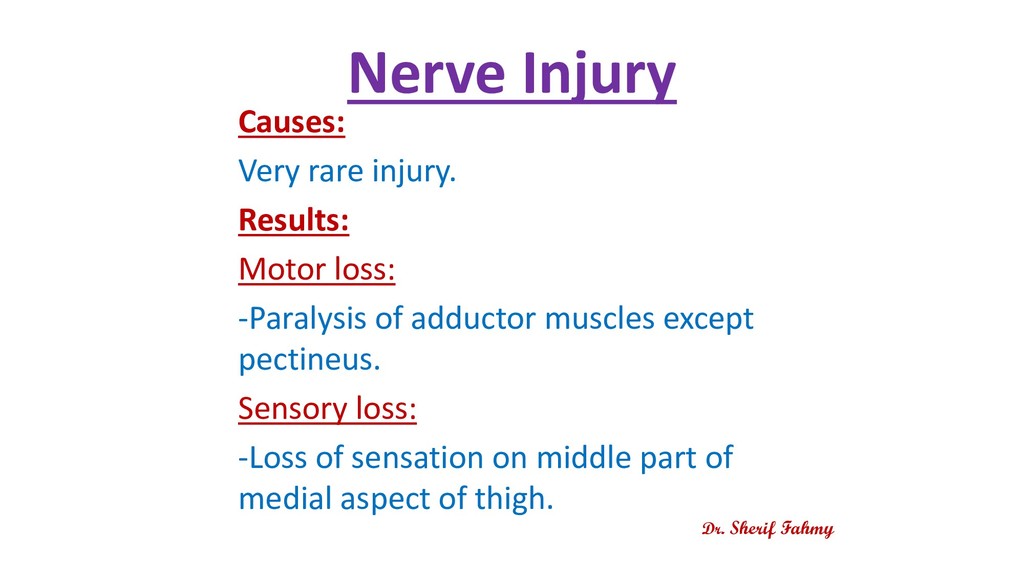
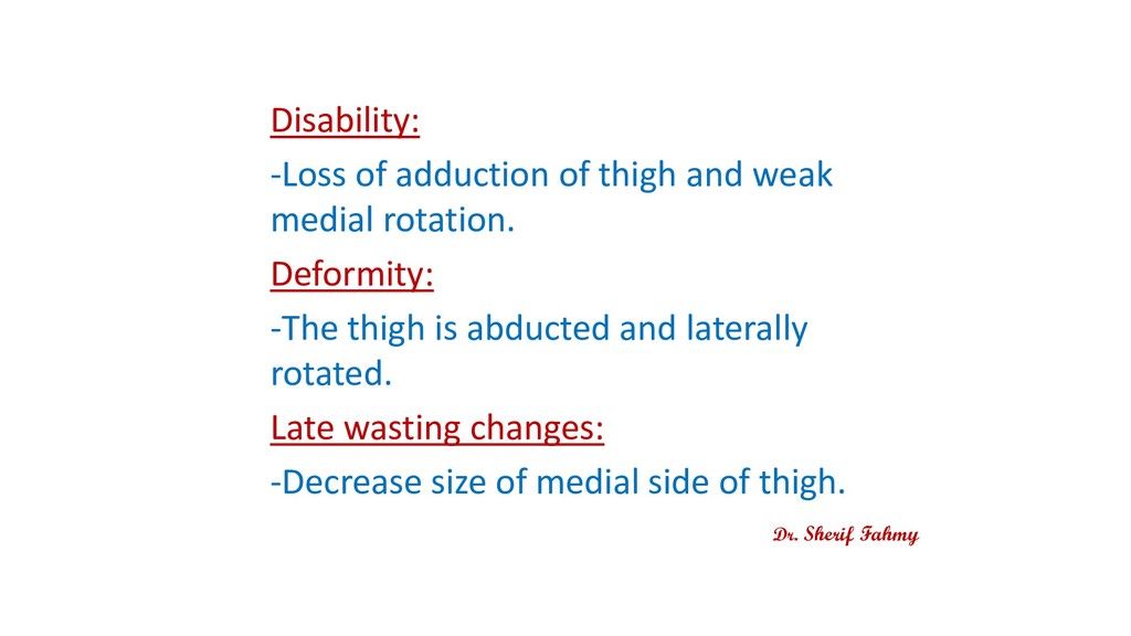
paralyzed muscles. Sensory loss: describe the area of loss of sensation. Disability: describe the lost actions. Deformity: Fixed deformed position of the paralyzed part. Late wasting changes: Decrease the size of paralyzed part.
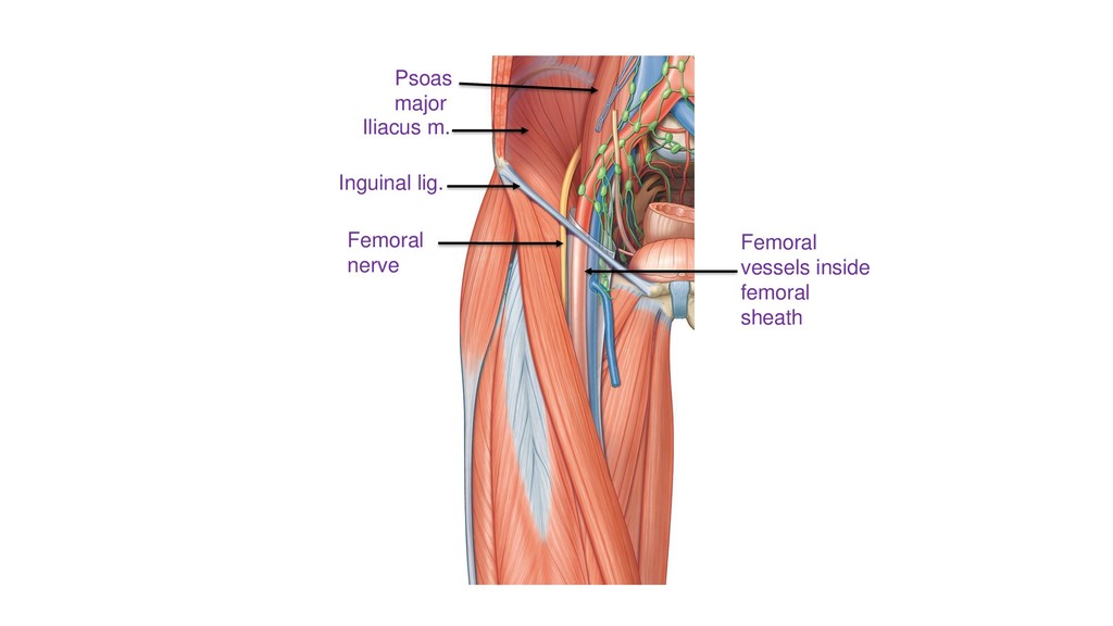
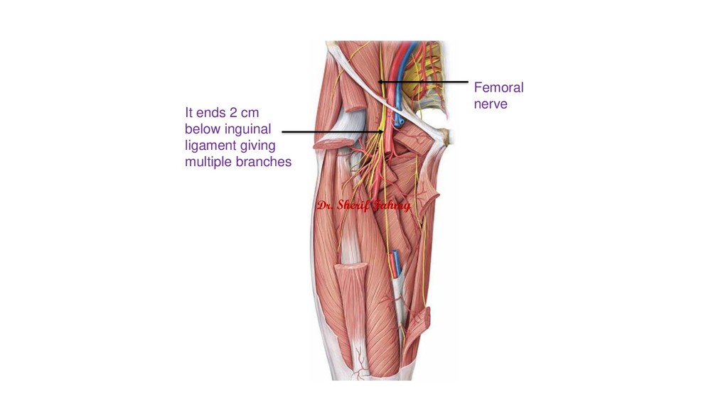
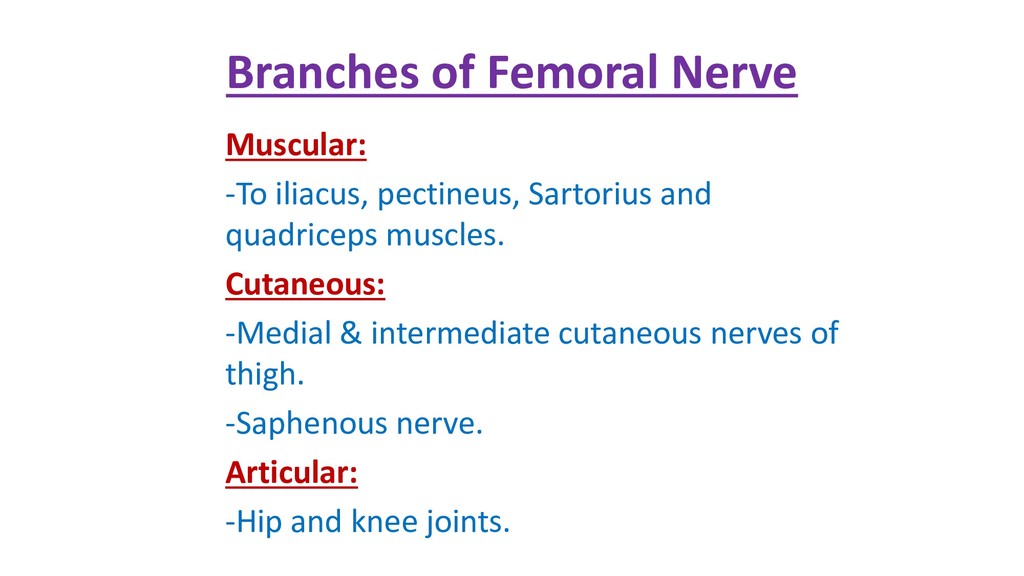
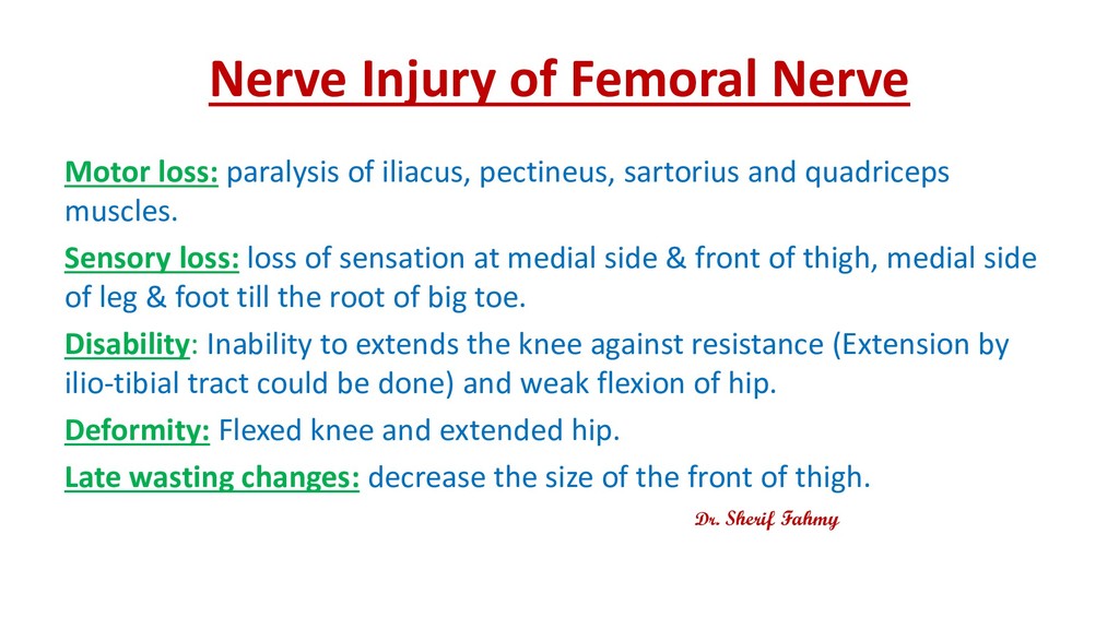
pectineus, sartorius and quadriceps muscles. Sensory loss: loss of sensation at medial side & front of thigh, medial side of leg & foot till the root of big toe. Disability: Inability to extends the knee against resistance (Extension by ilio-tibial tract could be done) and weak flexion of hip. Deformity: Flexed knee and extended hip. Late wasting changes: decrease the size of the front of thigh. Dr. Sherif Fahmy
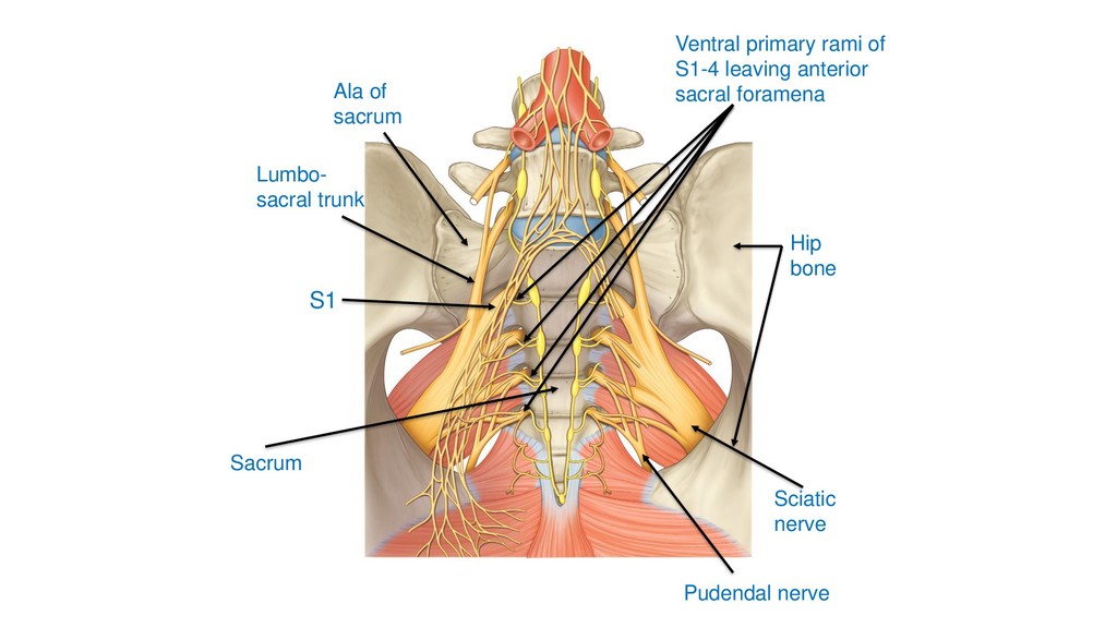
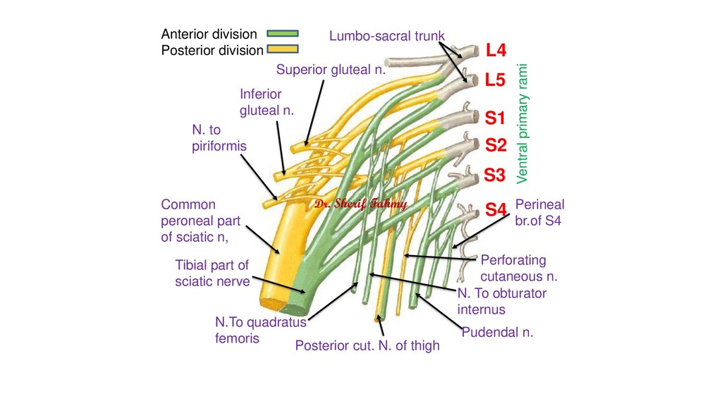
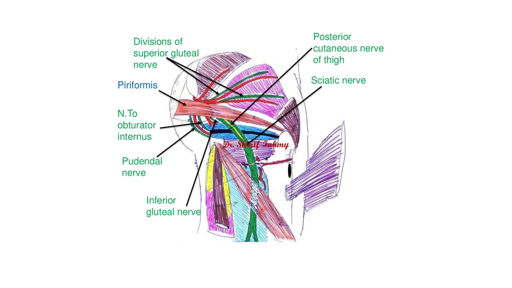
gluteal n. N. to piriformis Common peroneal part of sciatic n, Tibial part of sciatic nerve N. To obturator internus N.To quadratus femoris Perforating cutaneous n. Pudendal n. Posterior cut. N. of thigh Perineal br.of S4 Anterior division Posterior division Lumbo-sacral trunk Ventral primary rami Dr. Sherif Fahmy
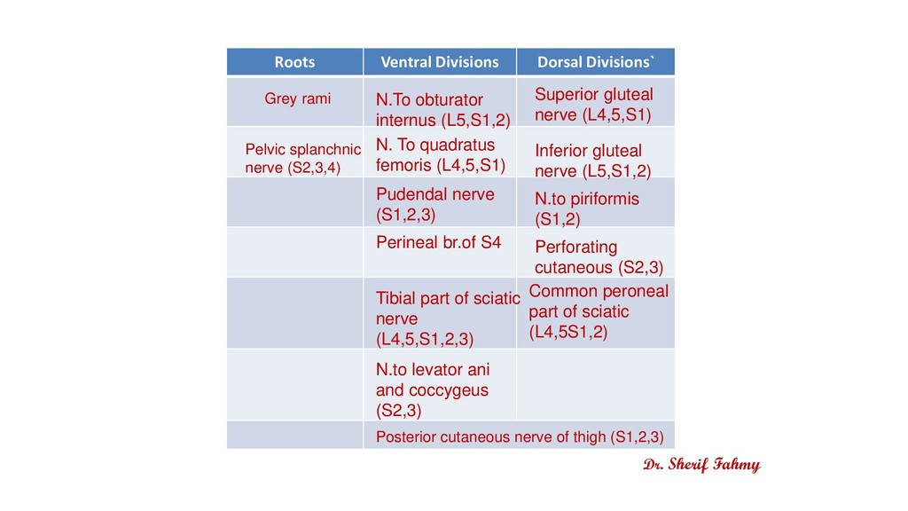
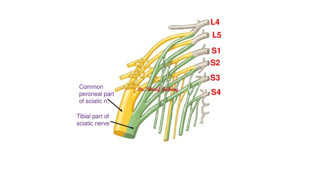
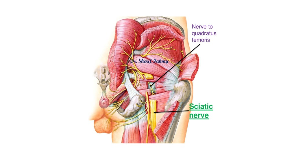
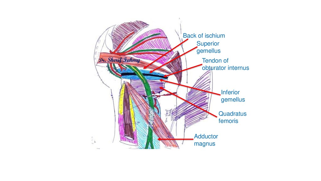
(S2,3,4) N.To obturator internus (L5,S1,2) N. To quadratus femoris (L4,5,S1) Pudendal nerve (S1,2,3) Perineal br.of S4 Tibial part of sciatic nerve (L4,5,S1,2,3) N.to levator ani and coccygeus (S2,3) Superior gluteal nerve (L4,5,S1) Inferior gluteal nerve (L5,S1,2) N.to piriformis (S1,2) Perforating cutaneous (S2,3) Common peroneal part of sciatic (L4,5S1,2) Posterior cutaneous nerve of thigh (S1,2,3) Dr. Sherif Fahmy
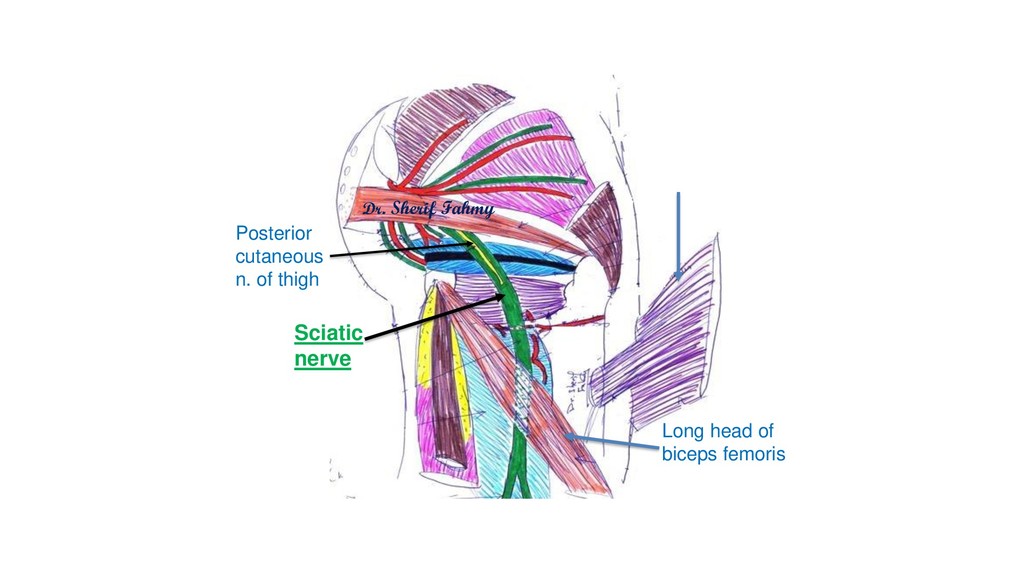
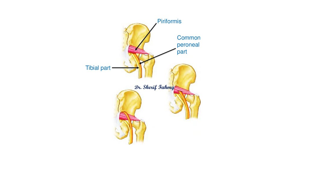
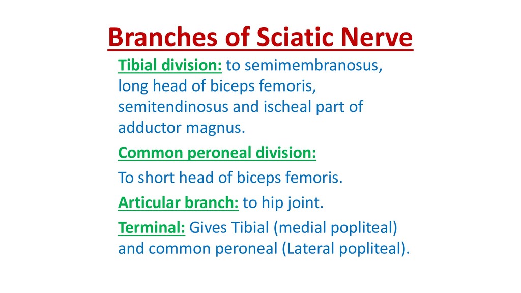
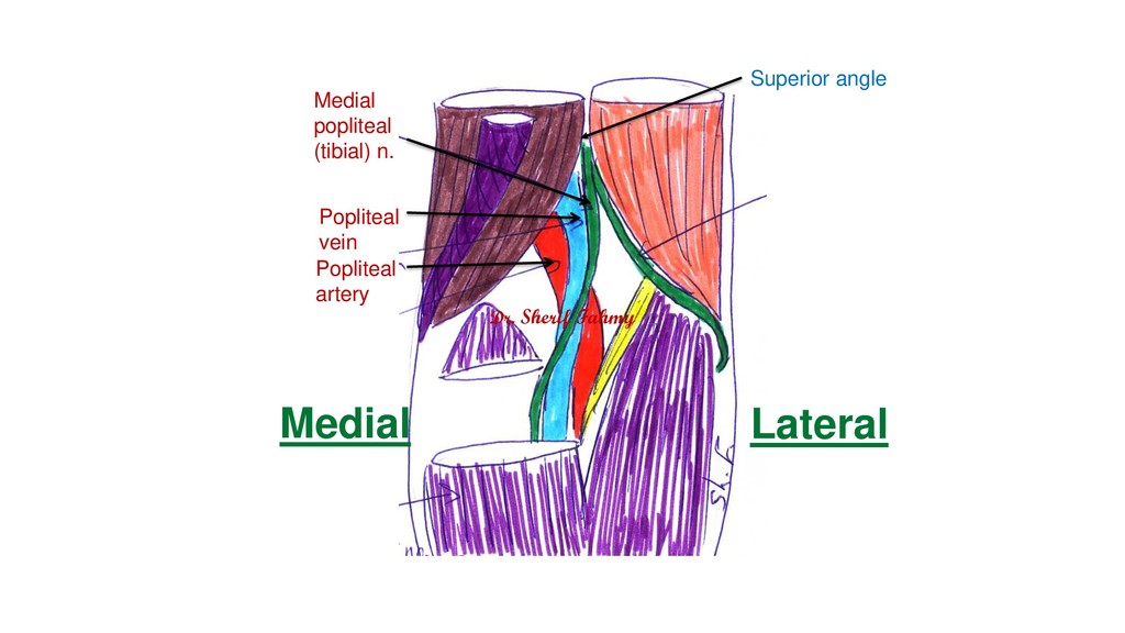
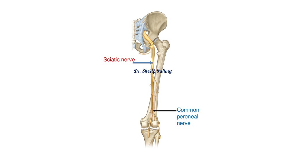
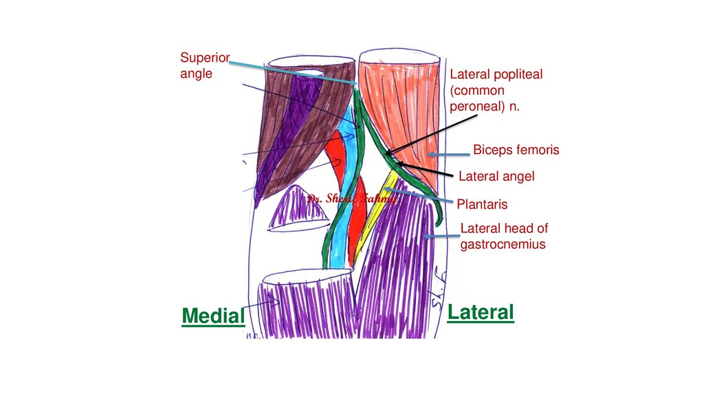
of biceps femoris, semitendinosus and ischeal part of adductor magnus. Common peroneal division: To short head of biceps femoris. Articular branch: to hip joint. Terminal: Gives Tibial (medial popliteal) and common peroneal (Lateral popliteal).
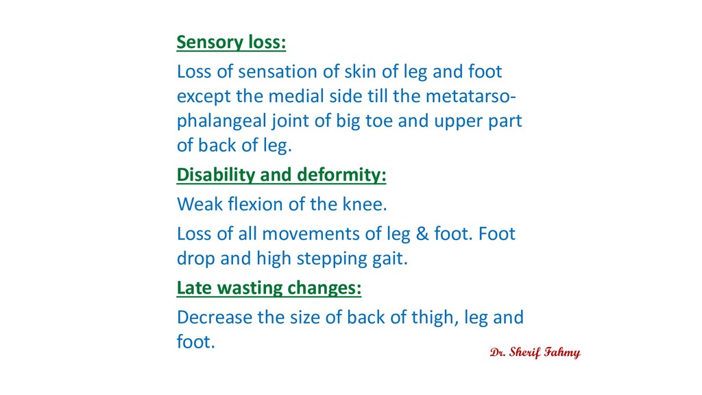
foot except the medial side till the metatarso- phalangeal joint of big toe and upper part of back of leg. Disability and deformity: Weak flexion of the knee. Loss of all movements of leg & foot. Foot drop and high stepping gait. Late wasting changes: Decrease the size of back of thigh, leg and foot. Dr. Sherif Fahmy
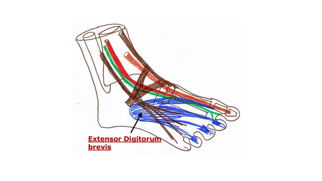
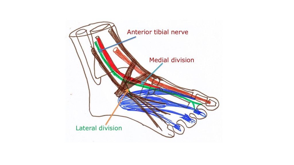
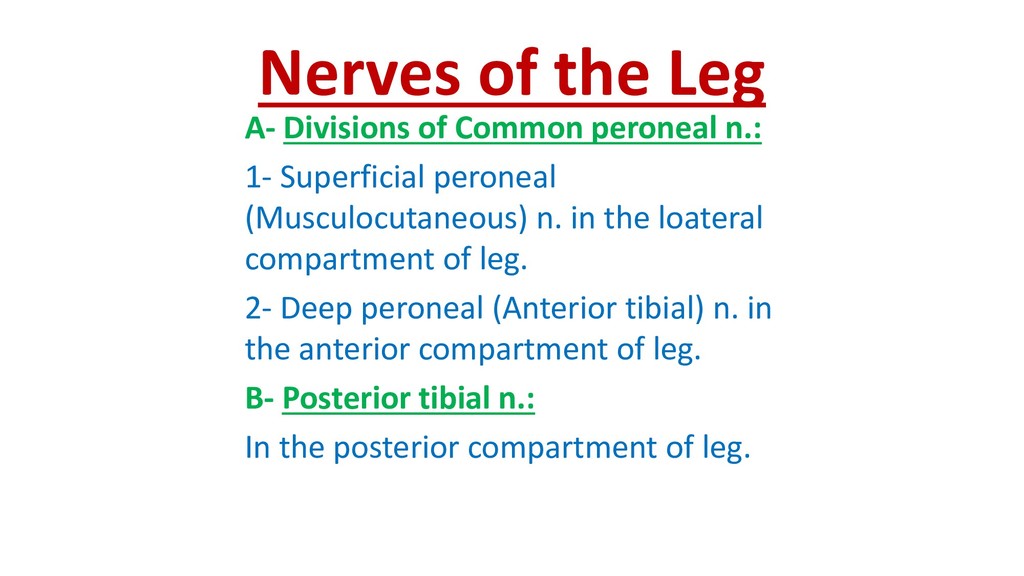
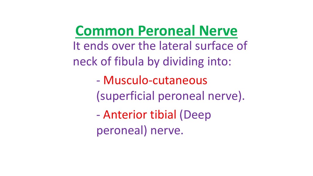
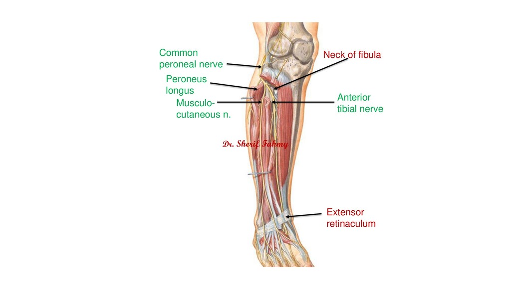
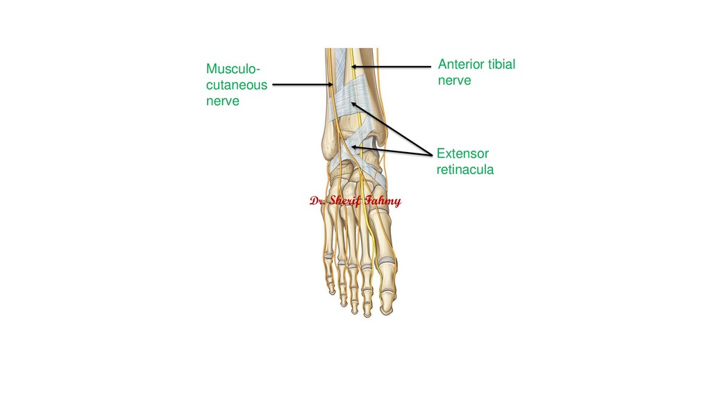
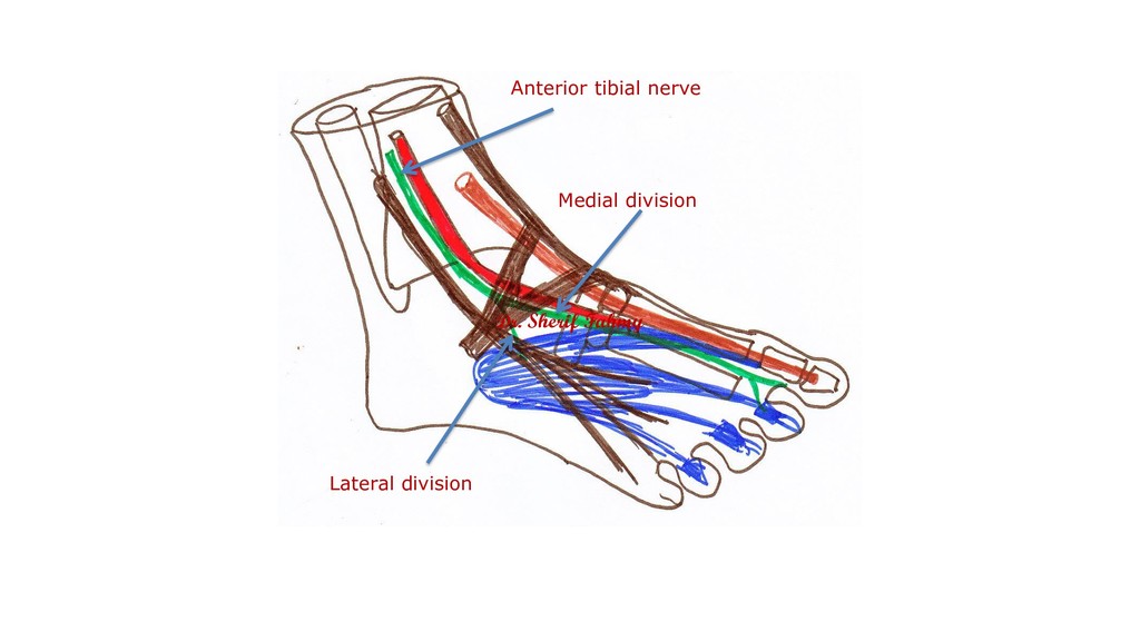
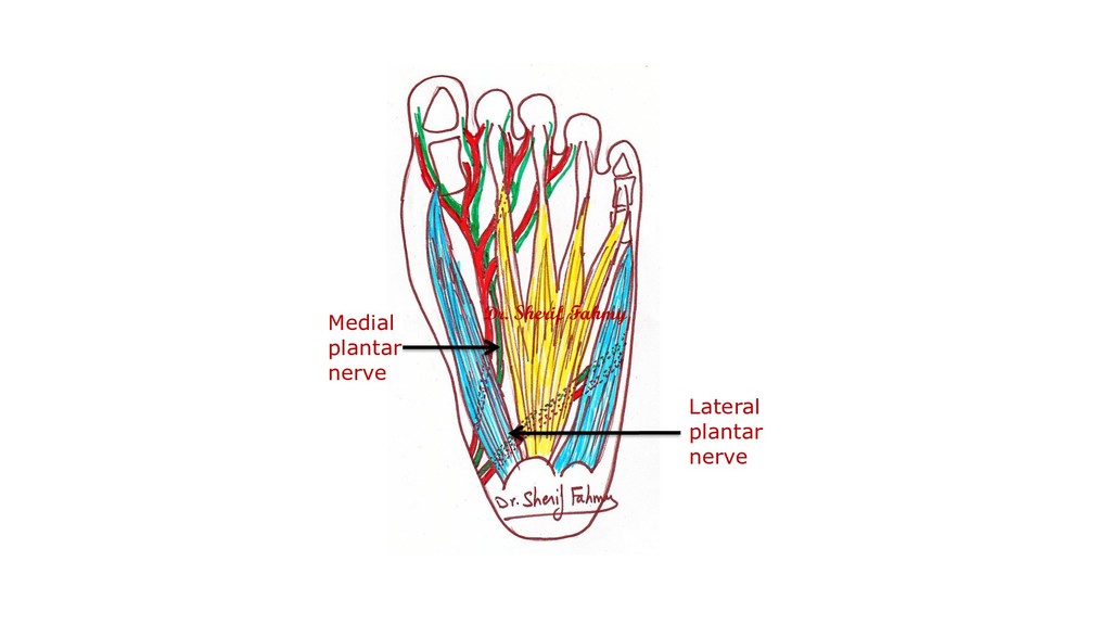
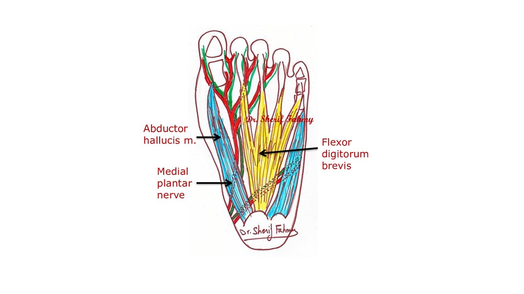
1- Superficial peroneal (Musculocutaneous) n. in the loateral compartment of leg. 2- Deep peroneal (Anterior tibial) n. in the anterior compartment of leg. B- Posterior tibial n.: In the posterior compartment of leg.
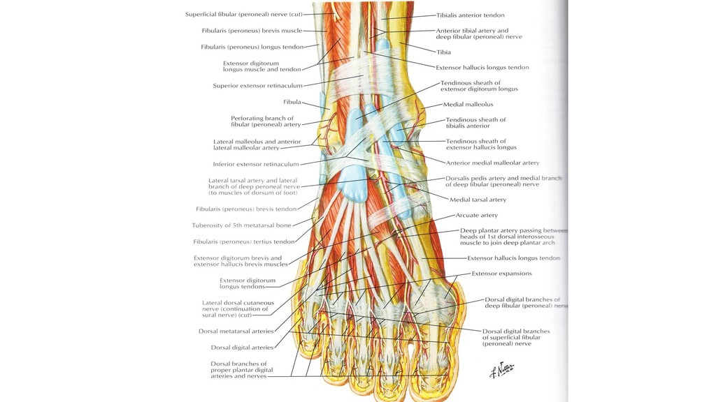
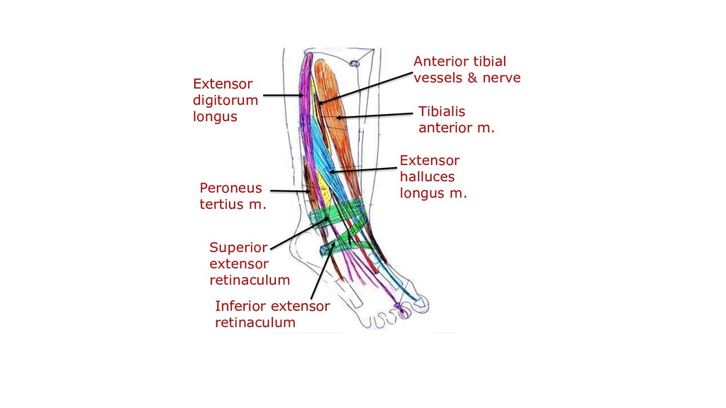
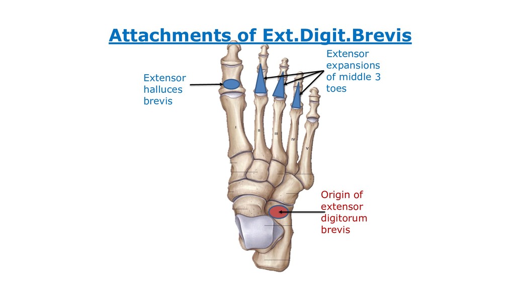
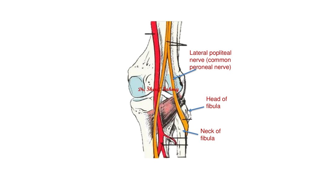
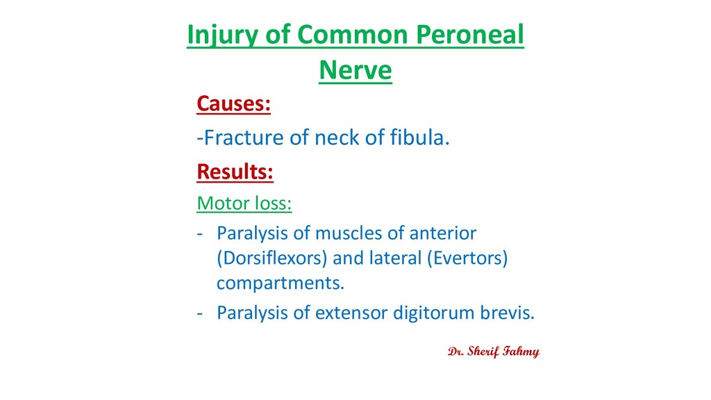
fibula. Results: Motor loss: - Paralysis of muscles of anterior (Dorsiflexors) and lateral (Evertors) compartments. - Paralysis of extensor digitorum brevis. Dr. Sherif Fahmy
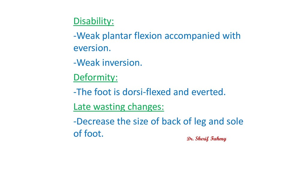
1/3 and front of lower 1/3 and dorsum of foot except borders. Disability: - Loss of dorsi flexion and eversion. - Weak inversion. Deformity: The foot is plantar flexion (foot drop) and inverted (Equinovarus). Late wasting changes: Decrease the size of anterolateral aspect of leg. Dr. Sherif Fahmy
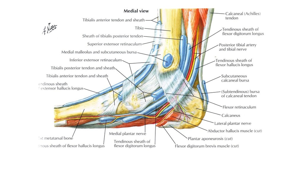
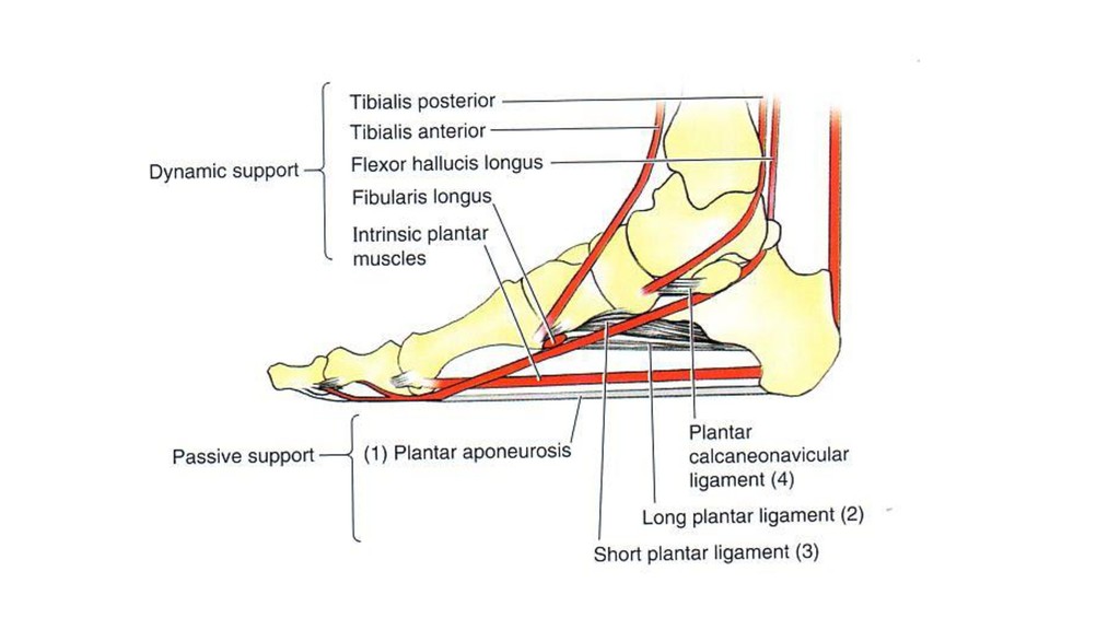
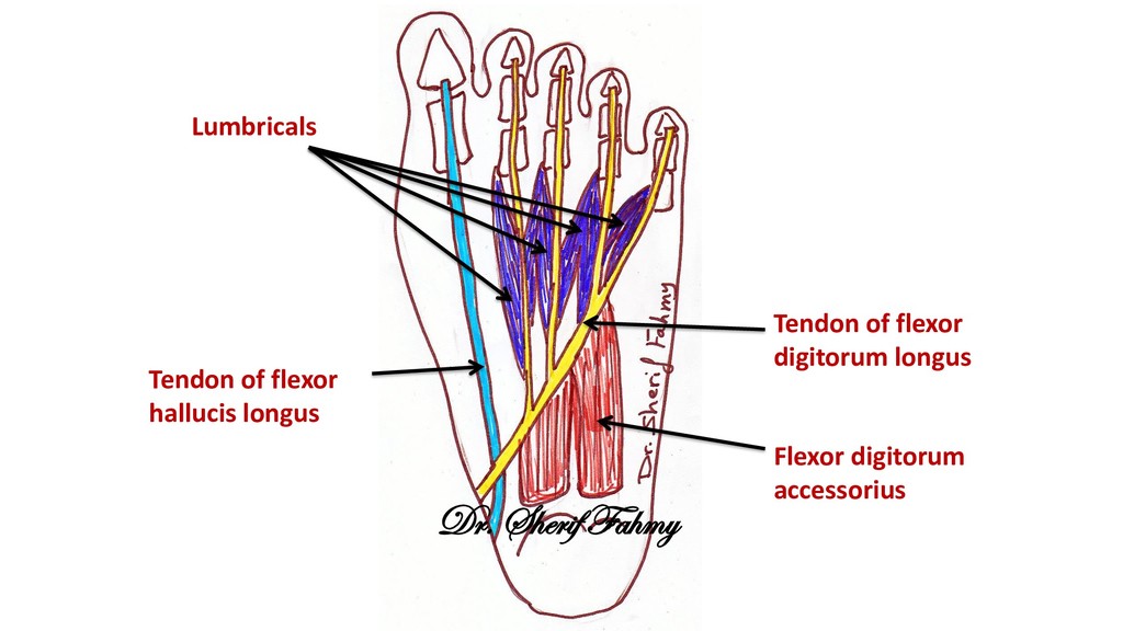
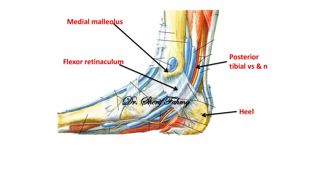
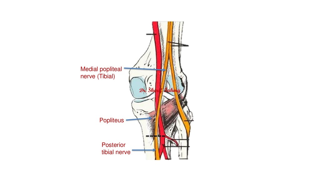
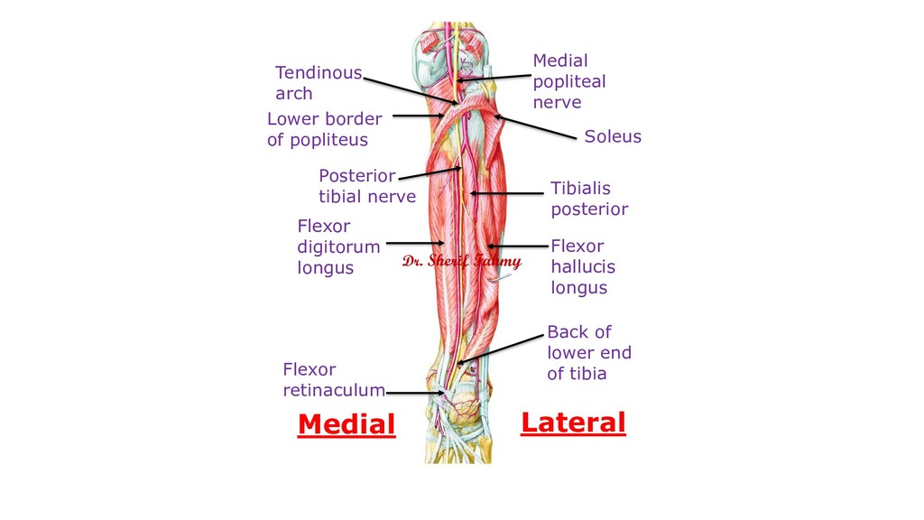
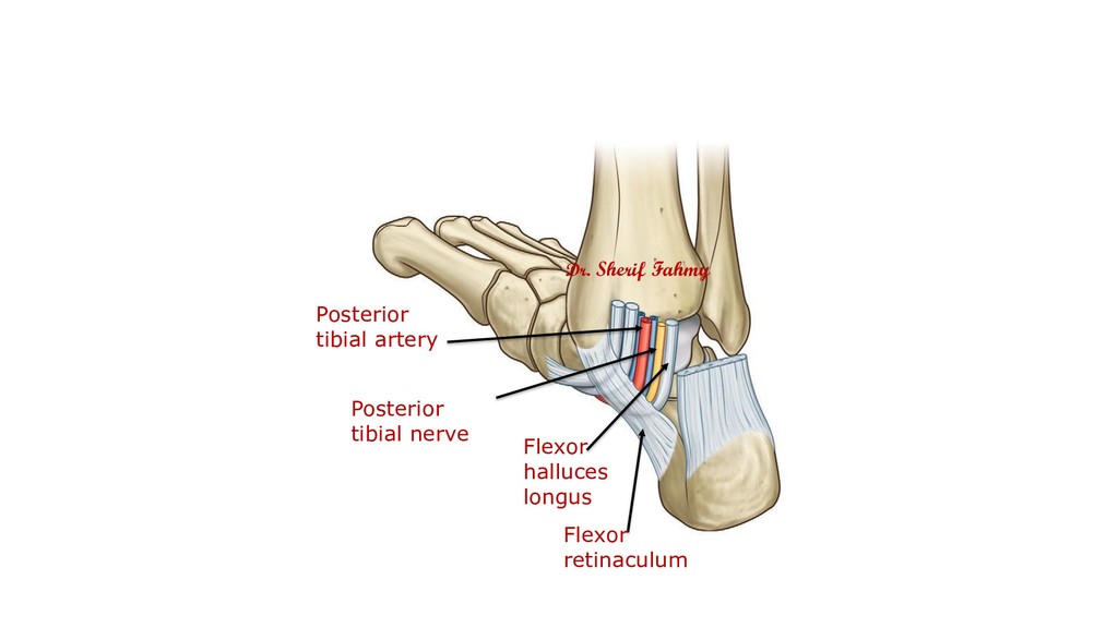
Posterior tibial nerve Flexor digitorum longus Tibialis posterior Flexor hallucis longus Back of lower end of tibia Flexor retinaculum Lateral Medial Dr. Sherif Fahmy