Lock in $30 Savings on PRO—Offer Ends Soon! ⏳

FY 2024: MDC 4 - Respiratory System

Avatar for e4health e4health PRO
April 03, 2024
790

FY 2024: MDC 4 - Respiratory System

Avatar for e4health

e4health PRO

April 03, 2024
Tweet

More Decks by e4health

Transcript

  1. H I M | C O D I N G

    & C D I | H E A LT H I T | R E V C Y C L E Empowering Better Health e4health tackles healthcare’s data, quality and revenue challenges empowering your providers to focus on better care.
  2. Objectives • Review MDC 4- Diseases and Disorders of the

    Respiratory System with a focus on selected diagnoses and procedures • Learner will acquire a basic understanding of the diagnoses and procedures included in MDC-4 • Discuss Query opportunities in MDC-4 • Review coding clinics relevant to the chosen topics in each DRG
  3. MDC 4-MS- DRGs (Medical) • 175-176 Pulmonary Embolism with and

    without MCC • 177-178-179 Respiratory Infections and Inflammations with/without CC/MCC • 180-181-182 Respiratory Neoplasms with/without CC/MCC • 183-184-185 Major Chest Trauma with/without CC/MCC • 186-187-188 Pleural Effusion with/without CC/MCC • 189 Pulmonary Edema and Respiratory Failure • 190-191-192 COPD with/without CC/MCC • 193-194-195 Simple Pneumonia and Pleurisy • 196-197-198 Interstitial Lung Disease with/without CC/MCC • 199-200-201 Pneumothorax with/without CC/MCC • 202-203 Bronchitis and Asthma with/without CC/MCC • 204 Respiratory Signs and Symptoms • 205-206 Other Respiratory System Diagnoses with/without CC/MCC • 208-207 Respiratory System Diagnosis with Ventilator Support MV < 96 hrs and MV >96 hrs
  4. MDC 4-MS- DRGs (Surgical)  DRG 163 MAJOR CHEST PROCEDURES

    WITH MCC  DRG 164 MAJOR CHEST PROCEDURES WITH CC  DRG 165 MAJOR CHEST PROCEDURES WITHOUT CC/MCC  DRG 166 OTHER RESPIRATORY SYSTEM O.R. PROCEDURES WITH MCC  DRG 167 OTHER RESPIRATORY SYSTEM O.R. PROCEDURES WITH CC  DRG 168 OTHER RESPIRATORY SYSTEM O.R. PROCEDURES WITHOUT CC/MCC
  5. Pneumonia • Chest imaging (X-Ray or CT scan with infiltrate

    or consolidation) • Clinical symptoms of fever, chills, productive cough or increased sputum production, SOB, pleuritic chest pain • Physical exam typically shows rales, rhonchi, bronchial breath sounds, and pleural rub • Laboratory data may show elevated WBC and inflammatory markers, sputum cultures if applicable Diagnostic Criteria
  6. Pneumonia Treatment Support respiratory function, O2 administration and pulse ox

    monitoring If chronic lung condition is present, bronchodilators, steroids, nebulizers Antibiotic therapy Supportive care- Fluids, incentive spirometry, increased activity
  7. Pneumonia • An inflammatory response, usually a noninfectious cause Pneumonitis

    • A chemical injury caused by inhalation of sterile gastric contents Aspiration pneumonitis • An infectious process caused by the inhalation of oropharyngeal secretions colonized by pathogenic bacteria Aspiration pneumonia • Community, hospital/healthcare acquired; can be superimposed on viral pneumonia Bacterial pneumonia • Immunosuppressed Viral, fungal, atypical • Develops after 48 hrs. or more on ventilator via trach or endotracheal tube VAP (ventilator associated)
  8. Pneumonia Types • Aspiration pneumonia • Often overlooked • Consider

    related diagnoses/conditions (e.g., functional quadriplegia, current or Hx CVA, neurodegenerative disorders, gastroparesis, GERD, morbid obesity, altered LOC, NG tube, Hx vomiting, dysphagia, aspiration on swallow studies) • RLL infiltrate on CXR • Antibiotic choice - Cefepime, Zosyn • Pneumonia due to coronavirus disease 2019 • Code first COVID-19 U07.1 • Review for additional MCC’s such as respiratory failure and sepsis This Photo by Unknown author is licensed under CC BY-NC.
  9. Pneumonia Types • MRSA pneumonia and MSSA pneumonia • Susceptible

    populations-long term care, indwelling lines/drains, open wounds/ulcers, recent surgical procedures, immunosuppression, CPAP/BIPAP, ventilator • Gram negative pneumonia • Klebsiella, Proteus, Serratia, Pseudomonas, and Enterobacter species • Susceptible populations-elderly, long term care, immunosuppressed, chronic respiratory illness, ESRD, CPAP/BIPAP, ventilator, recent hospitalization or broad spectrum Abx therapy • Respiratory Syncytial Virus Pneumonia • RSV usually causes mild cold-like symptoms • Can be severe in children and older adults • Ensure clarification of the type of viral pneumonia in documentation if cultures are done • Susceptible populations-Children, elderly, immunosuppressed, and those with chronic respiratory illness
  10. Pneumonia Coding Instruction • CC 3rd Q 2019, p. 37

    - Only code lobar PNA when specifically documented as such and a causal organism is not specified; not location directed • CC 2nd Q 2019, p. 6 - Assign T86.818, complication of lung transplant, followed by J69.0 when aspiration PNA is present with a transplanted lung • CC 1st Q 2019, p. 10 - Documentation must link PNA to HIV to be HIV-related illness • CC 4th Q 2013, p. 118 - Assign Y95, Nosocomial infection for a healthcare acquired condition
  11. Pneumonitis • Pneumonitis Coding Instruction: • CC 2nd Q 2019,

    p. 31- Aspiration bronchitis is more commonly caused by aspiration of food rather than aspiration of gases. Therefore, code J69.0, Pneumonitis due to inhalation of food and vomit, is a more appropriate code assignment than code J68.0, Bronchitis and pneumonitis due to chemicals, gases, fumes and vapors • CC 2nd Q 2019, p. 28 - If the pneumonitis due to chemotherapy is documented as acute, assign code J70.2, Acute drug-induced interstitial lung disorders, followed by code T45.1X5A, Adverse effect of antineoplastic and immunosuppressive drugs, initial encounter
  12. Ventilator Associated Pneumonia • Assign only when provider directly links

    PNA to ventilator • Assign an additional code for organism • Do not assign codes from J12-18 for type on PNA VAP (Ventilator associated pneumonia) • CC 2nd Q 2020, p. 17-VAP and sepsis both present, sepsis sequenced first • CC 2nd Q 2020, p. 28-Sepsis and aspiration PNA are related and POA, sepsis is sequenced first Coding Clinics
  13. VAP Coding guidelines • Documentation of Ventilator Associated Pneumonia •

    As with all procedural or postprocedural complications, code assignment is based on the provider’s documentation of the relationship between the condition and the procedure. • Code J95.851, Ventilator associated pneumonia, should be assigned only when the provider has documented ventilator associated pneumonia (VAP). An additional code to identify the organism (e.g., Pseudomonas aeruginosa, code B96.5) should also be assigned. Do not assign an additional code from categories J12-J18 to identify the type of pneumonia. • Code J95.851 should not be assigned for cases where the patient has pneumonia and is on a mechanical ventilator and the provider has not specifically stated that the pneumonia is ventilator-associated pneumonia. If the documentation is unclear as to whether the patient has a pneumonia that is a complication attributable to the mechanical ventilator, query the provider.
  14. VAP Coding guidelines • Ventilator Associated Pneumonia Develops after Admission

    • A patient may be admitted with one type of pneumonia (e.g., code J13, Pneumonia due to Streptococcus pneumonia) and subsequently develop VAP. In this instance, the principal diagnosis would be the appropriate code from categories J12-J18 for the pneumonia diagnosed at the time of admission. • Code J95.851, Ventilator associated pneumonia, would be assigned as an additional diagnosis when the provider has also documented the presence of ventilator associated pneumonia.
  15. Sepsis due to Ventilator-Associated Pneumonia - Coding Clinic Second Quarter

    2020 Page 17 • Question: A patient with a complicated past medical history is admitted for severe sepsis due to ventilator- associated pneumonia (VAP). The VAP is due to Escherichia coli and methicillin susceptible Staphylococcus aureus. Would VAP be considered a complication or a localized infection? What are the appropriate code assignments and sequencing of sepsis due to VAP? • Answer: The VAP is the localized infection, which is sequenced after the underlying systemic infection, sepsis. In this case, sequence either code A41.51, Sepsis due to Escherichia coli [E. coli], or A41.01, Sepsis due to Methicillin susceptible Staphylococcus aureus, as the principal diagnosis. Codes J95.851, Ventilator associated pneumonia, and R65.20, Severe sepsis without septic shock, should be assigned as additional diagnoses. • The Official Guidelines for Coding and Reporting, Section I.C.1.d.4, states, "If the reason for admission is sepsis or severe sepsis and a localized infection, such as pneumonia or cellulitis, a code for the underlying systemic infection should be assigned first and the code for the localized infection should be assigned as a secondary diagnosis."
  16. Pneumonia - General Query Opportunity • Review documentation for: •

    Causative or suspected organism based on treatment protocol • Causative or suspected mechanism • Vent-associated • Aspiration • Associated conditions, such as: • Respiratory failure and its acuity/type • Sepsis • Tobacco or vaping use • COPD or asthma exacerbation • Underlying lung disorders
  17. Influenza • Code only confirmed diagnoses of influenza due to

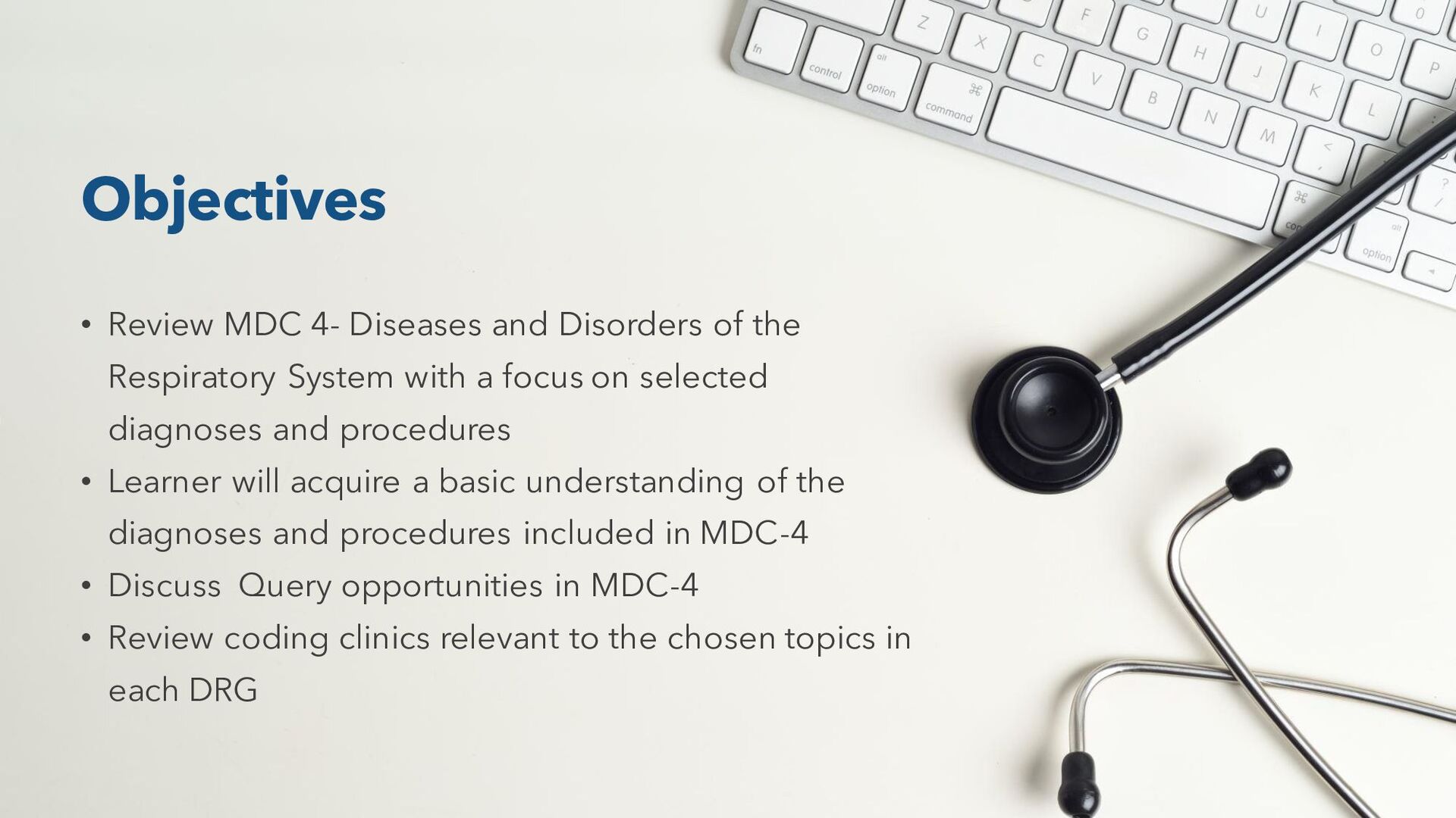
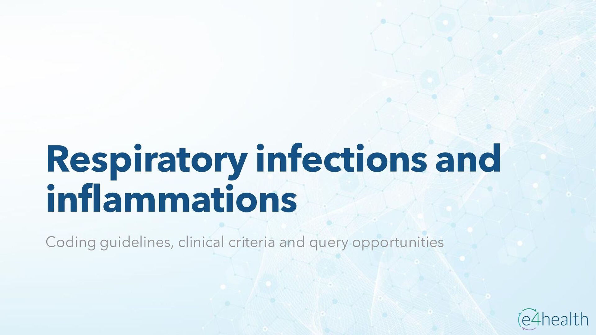
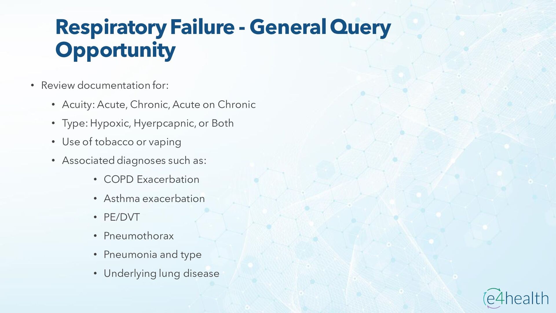
    certain influenza viruses (J09) or influenza due to other identified influenza virus (J10). • J09.X is used to report a specific strain of influenza (such as “novel” influenza A) that includes avian (bird) influenza, influenza A/H5N1, influenza of other animal origin (not bird or swine), and swine influenza virus, • Category J10, Influenza due to other identified influenza virus, is used to report ordinary seasonal influenza A (non-novel), influenza B, and influenza C • If provider documentation indicates “suspected,” “possible, “probable,” “likely,” utilize category J11 is used for influenza due to unidentified influenza virus • Influenza with any form of pneumonia or bronchopneumonia is assigned to influenza with pneumonia combination code (J09.X1, J10.00–J10.08, and J11.00–J11.08) followed by a code for the specified type of pneumonia • Influenza with other types of respiratory manifestations such as upper respiratory infection, laryngitis, pharyngitis, and pleural effusion are classifiable to J09.X2, J10.1, and J11.1. • Influenza may also involve body systems other than the respiratory system, such as the gastrointestinal tract (J09.X3, J10.2, and J11.2), and other manifestations such as encephalopathy, myocarditis, and otitis media (J09.X9, J10.81–J10.89, and J11.81–J11.89).
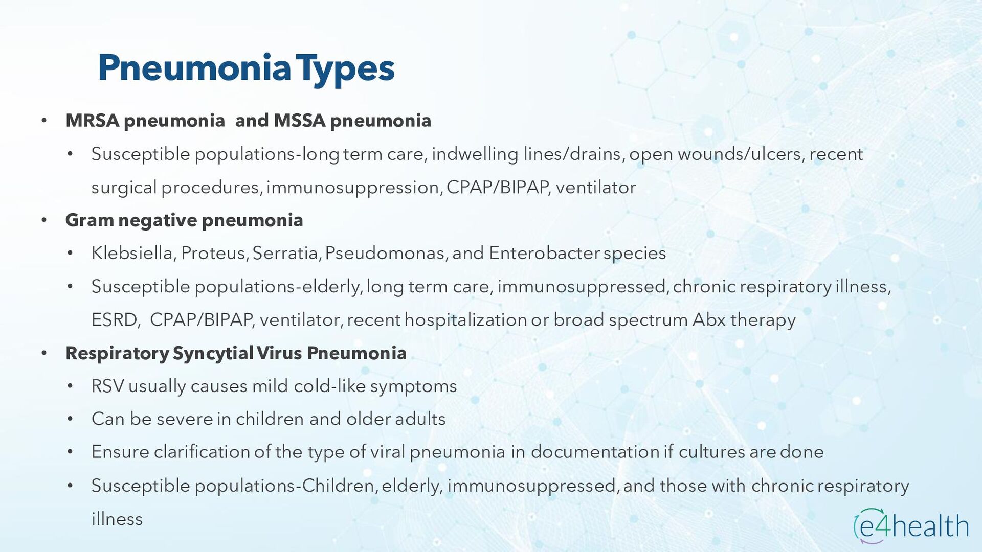
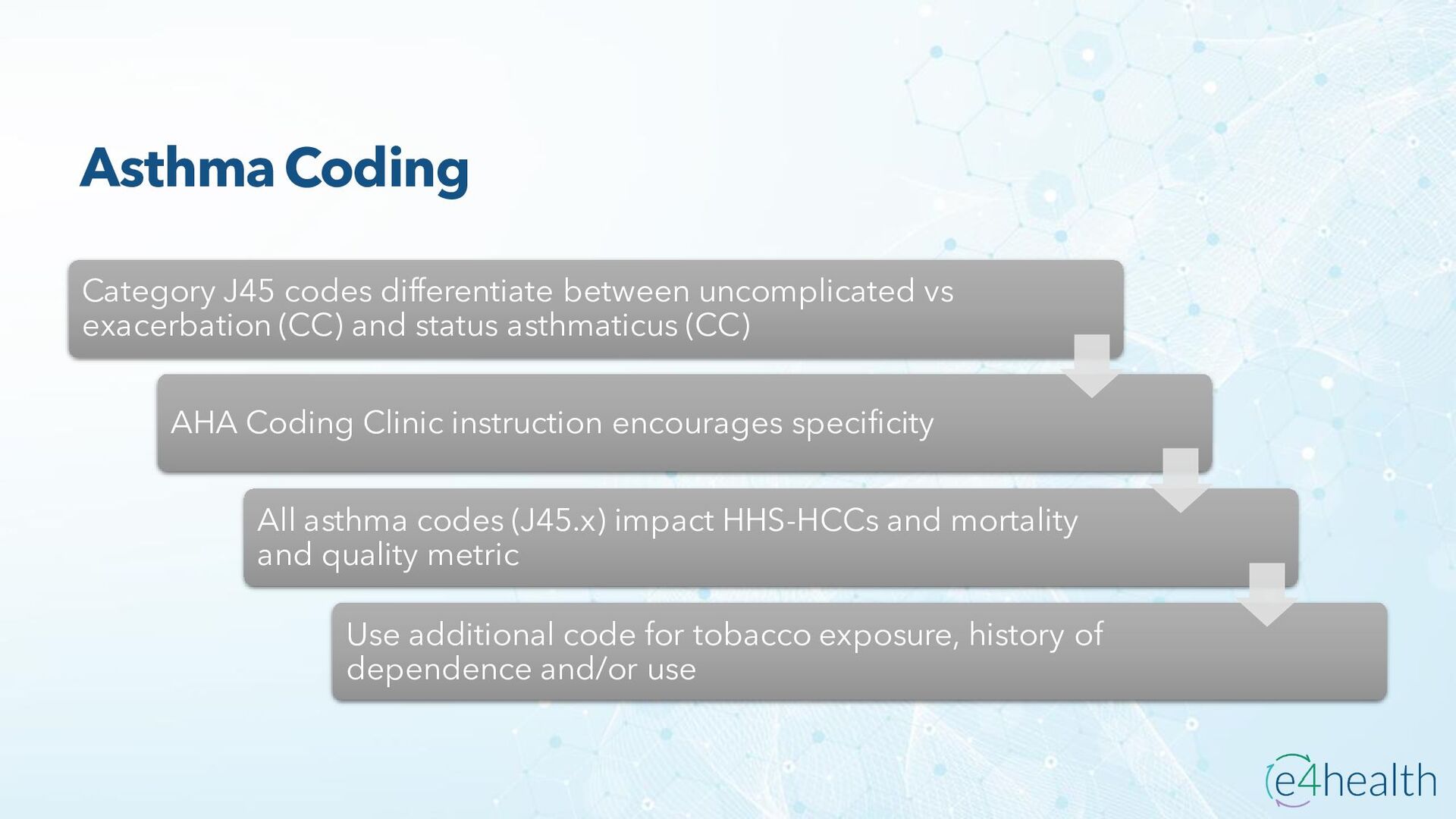
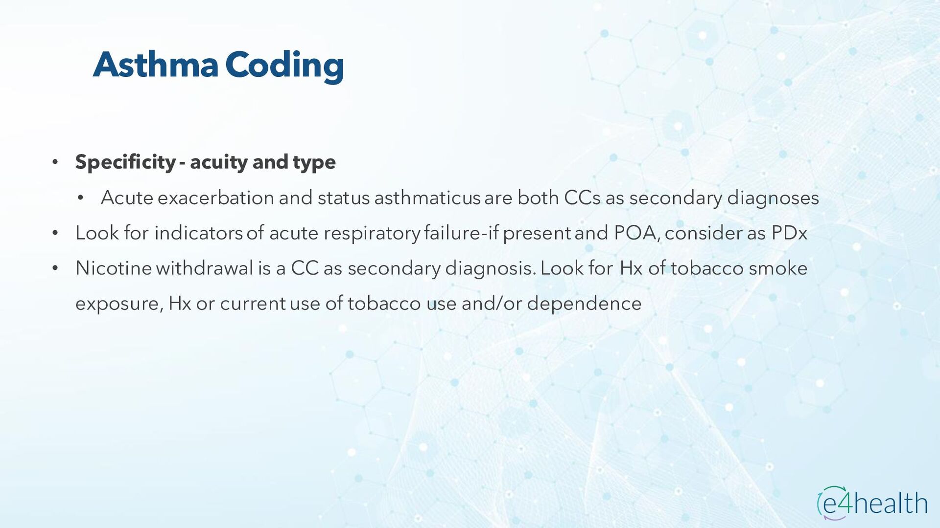
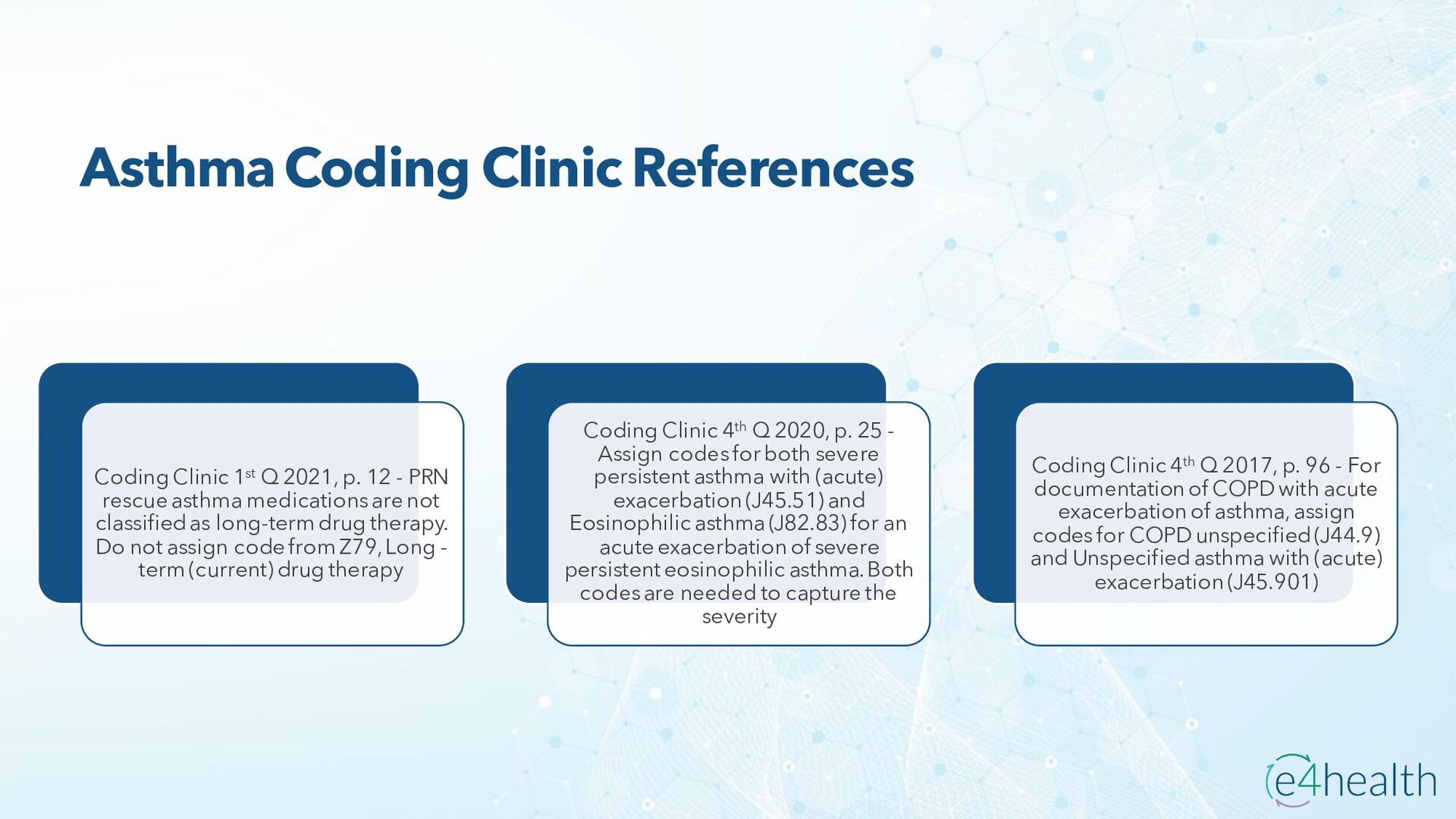
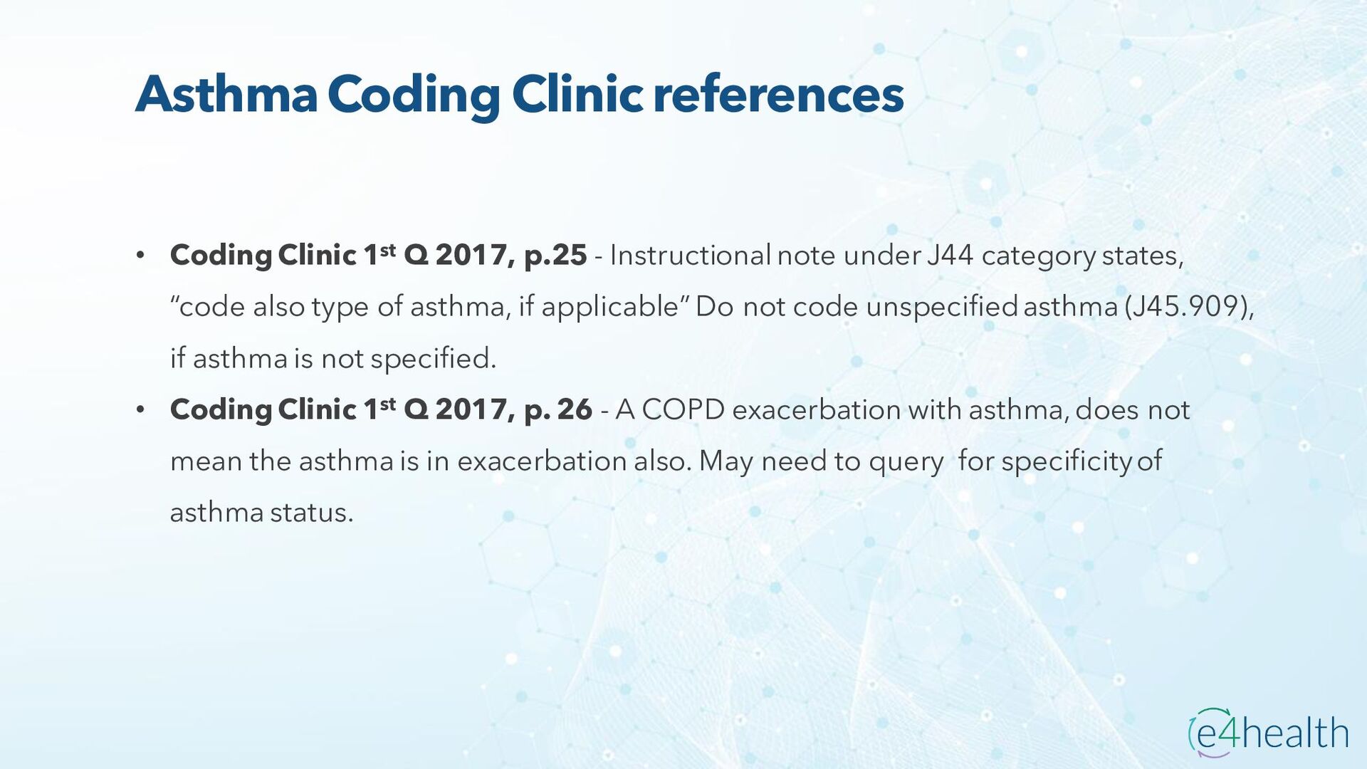
  18. Sepsis due to bronchitis - Coding Clinic Third Quarter 2016

    Page 8 • Question: What are the correct ICD-10 CM codes for a provider's diagnostic statement of "viral sepsis due to acute viral bronchitis due to influenza A?" The sepsis was present on admission. • Answer: Assign code A41.89, Other specified sepsis, for a diagnosis of sepsis due to influenza A. Although codes A30-A49 classify bacterial illnesses, there is no specific code for viral sepsis. Code A41.89 is the best available option to capture the concept of sepsis when no specific code exists. "Sepsis, specified organism NEC" is indexed to code A41.89. Assign also code J10.1, Influenza due to other identified influenza virus with other respiratory manifestations, and code J20.8, Acute bronchitis due to other specified organisms. • Codes from subcategory J09.X-, Influenza due to identified novel influenza A virus, are intended for a specific strain of influenza A, such as "novel" influenza A, and not the ordinary seasonal influenza A.
  19. Sepsis secondary to influenza - Coding Clinic Third Quarter 2016

    Page 8 • Question: What are the appropriate ICD-10-CM codes for a diagnosis of sepsis secondary to influenza B? • Answer: Assign code A41.89, Other specified sepsis, for a diagnosis of sepsis due to influenza B. Although codes A30-A49 classify bacterial illnesses, there is no specific code for viral sepsis. Code A41.89 is the best available option to capture the concept of sepsis when no specific code exists. "Sepsis, specified organism NEC" is indexed to code A41.89. Assign also code J10.1, Influenza due to other identified influenza virus with other respiratory manifestations, for the influenza B. • Question: How do you code a patient who is admitted with sepsis from influenza with pneumonia? • Answer: Assign code A41.89, Other specified sepsis, for a diagnosis of sepsis due to influenza. Although codes A30-A49 classify bacterial illnesses, there is no specific code for viral sepsis. Code A41.89 is the best available option to capture the concept of sepsis when no specific code exists. "Sepsis, specified organism NEC" is indexed to code A41.89. In addition, assign code J11.00, Influenza due to unidentified influenza virus with unspecified type of pneumonia, for the influenza with pneumonia.
  20. Influenza- GENERAL QUERY OPPORTUNITY • Review documentation for: • Causative

    or suspected organism for influenza based on treatment protocol • Associated conditions, such as: • Pneumonia and its type • Respiratory failure and its acuity/type • Sepsis • Tobacco or vaping use • COPD or asthma exacerbation • Underlying lung disorders
  21. General COVID Sequencing • U07.1, COVID-19, is assigned only if

    the provider documentation confirms the diagnosis OR there is documentation of a positive COVID-19 test result (an Exception to the ICD-10-CM Coding Guidelines, Section II.H) • If provider documentation states “suspected,” “possible,” “probable,” or “inconclusive” COVID-19, assign any presenting signs and symptoms only. Do not assign U07.1. • Additionally, provider documentation does not need to link a positive COVID-19 test to a respiratory condition. • Sequencing COVID-19, U07.1, only if it meets the definition of principal diagnosis • Followed by the appropriate codes for associated COVID-19 manifestations • Non-respiratory manifestations, such as diarrhea, need to be linked to COVID-19 in order for U07.1 to be principal diagnosis • For COVID-19 with respiratory signs and symptoms which progresses to sepsis or severe sepsis, circumstance of admission determines if sepsis or U07.1 meets the definition of principal diagnosis
  22. COVID & Obstetrics Sequencing Codes from ICD-10-CM Chapter 15, Pregnancy,

    Childbirth and the Puerperium, take precedence over codes from other chapters, including code U07.1. For COVID-19 admission that is related to pregnancy or childbirth, the appropriate code from subcategory O98.5, other viral diseases complicating pregnancy, childbirth, and the puerperium, will be sequenced as principal diagnosis Followed by the code U07.1 and associated manifestations If the reason for admission during pregnancy or childbirth is unrelated to COVID-19 and the patient tests COVID-19 positive, the principal diagnosis is the reason for admission The appropriate code from O98.5, followed by U071. and associated manifestation codes for COVID-19 will be secondary diagnoses
  23. Contact and Exposure of COVID-19 • If the exposed individual,

    whether symptomatic or asymptomatic, tests positive for the COVID-19 virus, assign code U07.1. Although the individual is asymptomatic, the individual has tested positive and is considered to have COVID-19 infection. • For asymptomatic individuals with actual or suspected exposure, assign code Z20.822, Contact with and (suspected) exposure to COVID-19. • For symptomatic individuals where there is an actual or suspected exposure to COVID-19, and infection has been ruled out, or the test results are inconclusive or unknown, assign code Z20.822, Contact with and (suspected) exposure to COVID-19. • For a patient who receives an organ transplant that came from a donor who was COVID-19 positive, assign code Z20.822, Contact with and (suspected) exposure to COVID-19.
  24. Testing for COVID-19 For encounters for COVID-19 testing, including preoperative

    testing, code as exposure to COVID-19, using code Z20.822, Contact with and (suspected) exposure to COVID-19 For an encounter for COVID-19 antibody testing that is not being performed to confirm a current COVID-19 infection AND is not a follow-up test after resolution of COVID-19, assign code Z01.84, Encounter for antibody response examination. Antibody testing is a blood test performed to determine whether an individual has developed antibodies to a disease
  25. Personal History of COVID-19 • If a patient has a

    history of COVID-19, without residual symptom(s) or condition(s), code Z86.16, Personal history of COVID-19, is assigned. This Photo by Unknown author is licensed under CC BY-SA.
  26. Multisystem Inflammatory Syndrome (MIS) • M35.81, Multisystem Inflammatory syndrome (MIS)

    is utilized for both adults and children, MIS-A and MIS-C, respectively • Serious condition associated with COVID-19 where different internal and external body parts become inflamed • Symptoms include fever or chills, tachycardia, gastrointestinal symptoms, rash, conjunctival injection, and mucosal changes with a relative lack of severe respiratory disease as well as elevated inflammatory markers • Treatment includes supportive care to reduce inflammation, prevent clots, treat/prevent bacterial infections, hydration, and oxygen support
  27. Coding Multisystem Inflammatory Syndrome (MIS) • Assign code U07.1, COVID-19,

    as the principal/first-listed diagnosis • Followed by code M35.81, Multisystem inflammatory syndrome, as an additional diagnosis • Additional codes should be assigned for any associated complications of MIS For individuals with multisystem inflammatory syndrome (MIS) and COVID-19: • Assign codes M35.81, Multisystem inflammatory syndrome • Followed by U09.9, Post COVID-19 condition, unspecified • Additional codes should be assigned for any associated complications of MIS If an individual with a history of COVID- 19 develops MIS: • Assign codes M35.81, Multisystem inflammatory syndrome • Followed by Z20.822, Contact with and (suspected) exposure to COVID-19 • Additional codes should be assigned for any associated complications of MIS If an individual with a known or suspected exposure to COVID-19, and no current COVID-19 infection or history of COVID-19, develops MIS:
  28. Post COVID-19 condition • Code U09.9, Post COVID-19 condition, unspecified,

    is utilized for conditions or symptoms related to a previous COVID-19 infection • Provider documentation should state that the condition presented is due to a previous COVID-19 infection • Code U09.9 should not be assigned for manifestations of an active (current) COVID-19 infection • For sequela of COVID-19, or associated symptoms or conditions that develop following a previous COVID-19 infection: • Assign a code(s) for the specific symptom or condition related to the previous COVID-19 infection, if known, • Followed by the code U09.9 • If a patient has a condition associated with a previous COVID-19 infection and develops a new active (current) COVID-19 infection: • code U09.9 may be assigned in conjunction with code U07.1, COVID-19, to identify that the patient also has a condition(s) associated with a previous COVID-19 infection • Followed by Code(s) for the specific condition(s) associated with the previous COVID-19 infection and code(s) for manifestation(s) of the new active (current) COVID-19 infection should also be assigned
  29. COVID-19 - GENERAL QUERY OPPORTUNITY • Review documentation for: •

    Active or history or previous COVID-19 condition • If applicable, review documentation if non-respiratory symptoms are potentially linked to COVID-19 • Associated conditions, such as: • Sepsis • MIS in both adults or children • Acute kidney injury or acute tubular necrosis • Encephalopathy and type • DIC of intubation
  30. Respiratory Neoplasms • Benign - localized, does not spread •

    In-situ - “in its original place”, does not spread • Malignant - cancerous growths that may metastasize • Uncertain behavior - unpredictable behavior of tumor on path, currently benign may become malignant • Primary - origin or Secondary - metastasis Neoplasm - abnormal, usually rapid new cell growth, differentiated by behavior
  31. Respiratory Neoplasms Lung cancer Leading cause cancer deaths Smokers at

    greatest risk Symptoms occur in advanced disease- persistent new cough, hemoptysis, SOB, chest pain, weight loss
  32. Respiratory Neoplasms • Types • Small cell lung cancer -

    heavy smokers, less common • Non-small cell lung cancer - umbrella term • Squamous cell - very slow growing, often in airways • Large cell carcinoma - uncommon, rapid, more likely to spread • Adenocarcinoma - slow growing, outer regions
  33. Respiratory Neoplasms - General Query Opportunity • Review documentation for:

    • Neoplasm behavior and any secondary sites • Laterality and location of neoplasms • Associated diagnoses, such as: • Malnutrition • Anemia and its type and due to cause • Pancytopenia or Neutropenia • Complications of chemotherapy or radiation therapy • Tumor lysis syndrome This Photo by Unknown author is licensed under CC BY-SA-NC.
  34. PULMONARY EMBOLISM vs ACUTE COR PULMONALE • I26.90 - I26.99

    - without acute cor pulmonale • I27.82 Chronic pulmonary embolism • T790XXA and T791XXA Air embolism and Fat embolism initial encounter • T80.0XXA Air embolism following infusion, transfusion and therapeutic injection initial encounter Pulmonary embolism • I26.01 Septic pulmonary embolism with acute cor pulmonale • I26.02 Saddle embolus pulmonary artery with acute cor pulmonale • I26.09 Other pulmonary embolus with acute cor pulmonale Acute Cor Pulmonale
  35. Acute Cor Pulmonale Cor pulmonale is Latin for pulmonary heart

    Alteration in the structure and function of the right ventricle of the heart caused by a primary disorder of the respiratory system resulting in pulmonary hypertension Most common cause of acute cor pulmonale is massive pulmonary embolism Most common cause of chronic cor pulmonale is COPD *Acute Cor Pulmonale can only be coded when the patient has a pulmonary embolus
  36. Acute Cor Pulmonale • Pathophysiology • Increased right-sided filling pressure

    from pulmonary hypertension from disease of the lung • Signs and Symptoms • Right sided heart failure not related to left sided heart failure • Massive pulmonary embolism • Pulmonary hypertension on echocardiogram
  37. Deep vein thrombosis • DVT • Afib When patient has

    a pulmonary embolism, it is likely it comes from one of two sources: • Pain in calf, redness, swelling • History of travel • Ultrasound/doppler of the legs When reviewing a patient who has a pulmonary embolism, look for: DVT is code-able along with pulmonary embolism and impacts MS-DRG
  38. Subsegmental Pulmonary Embolism - Coding Clinic Fourth Quarter 2019 pages

    6-7 • New ICD-10-CM codes have been created to identify single subsegmental pulmonary embolism without acute cor pulmonale (I26.93) and multiple subsegmental pulmonary emboli without acute cor pulmonale (I26.94). • The use of advanced imaging techniques has increased the detection of small subsegmental pulmonary emboli (SSPE) in asymptomatic patients that may not be clinically significant. • These SSPEs are often isolated to distal (subsegmental) branches of the pulmonary artery, without coexisting deep venous thrombosis, and are usually too small to cause any major problems. Previously, subsegmental pulmonary emboli were treated with anticoagulation for months or years. However, it is unknown whether these emboli are in fact an indication for future thromboembolic events, and there is no consistent evidence that patients with SSPE benefit from short- and long-term anticoagulation therapy. • The most recent guidelines from the American College of Chest Physicians (ACCP) recommend that patients with isolated SSPE and no proximal DVT undergo surveillance rather than anticoagulation. • These new codes will enable important clinical differentiation, and will be beneficial for quality measures for hospitals, as well as for research and evaluation of treatment efficacy.
  39. Segmental and Subsegmental Pulmonary Emboli - Coding Clinic Second Quarter

    2022 page 13 Question: The patient had a chest computerized tomography angiography (CTA) which revealed pulmonary emboli in the segmental branches of the right middle and lower lobes of the lung. The radiologist also noted emboli in the subsegmental branches of the right and left lobes of the lung. The provider's final diagnostic statement listed "Segmental and subsegmental pulmonary emboli." How should segmental and subsegmental pulmonary emboli be coded? Answer: Assign codes I26.99, Other pulmonary embolism without acute cor pulmonale and I26.94, Multiple subsegmental pulmonary emboli without acute cor pulmonale, for documentation of segmental and multiple subsegmental pulmonary emboli. In this case, the patient had segmental emboli in the proximal branches of the right middle and lower lobes as well as bilateral subsegmental emboli. Two codes are needed to fully capture the patient's condition.
  40. Pulmonary Embolism - General Query Opportunity • Review documentation for:

    • Laterality, location, and acuity • Associated diagnoses, such as: • Respiratory failure • Presence of Cor Pulmonale and its acuity • Presence of DVT and laterality • Associated mechanical device, such as prosthesis • Stroke This Photo by Unknown author is licensed under CC BY-NC-ND.
  41. Major Chest Trauma Types • Rib fractures most common, usually

    due to blunt trauma • Complications-Pneumonia, flail chest, pneumothorax, contusions • Local tenderness, chest pain on inspiration or with coughing, hematoma, ecchymosis • Sternal fractures most common in MVC • Can be associated with rib fractures and hemothorax/hemopneumothorax • Local pain and hypersensitivity, confirmed on CXR, CT scan • Flail chest uncommon • 3 or more rib fractures in 2 or more sites w/wo sternal lesion and paradoxical respiratory movement and dysfunction-frequent respiratory failure • 75% associated with pulmonary contusion, high morbidity and mortality
  42. Major Chest Trauma Types cont. • Pneumothorax - hemothorax, hemopneumothorax,

    tension, open, simple (Different MS-DRG if PDx) • Simple - air in pleural cavity, non-complex • Tension - air cannot flow out of pleural space, hemithorax hyper expands, deviation of mediastinal organs, decreased inspiration, extreme risk of death without immediate chest tube insertion • Open - entry of air into the pleural cavity from penetrating chest wound >70% tracheal diameter, “sucking and bubbling” can be seen in inspiration/expiration • Hemothorax - blood in pleural space usually due to rib fractures causing venous vessel damage, pulmonary trauma, arterial injury is rare • Hemopneumothorax - air and blood in pleural space • Pulmonary contusion - life-threatening, suspected in blunt trauma with hypoxia, respiratory failure may be immediate, rib fractures may or may not be present
  43. Major Chest Trauma Types cont. • Pneumomediastinum - air in

    mediastinum (space between lungs) • Common in blunt chest trauma, clinical significance not well defined • May be associated with esophageal and/or tracheobronchial injuries • Diaphragm injury • Very rare, high morbidity and mortality • 90% due to blunt force trauma related to car accidents • Tracheobronchial injuries - Damage to the tracheal and/or bronchial cartilage from cricoid to bronchial division • Majority are fatal • Cardiac injuries as PDx fall under different MS-DRGs
  44. Major Chest Trauma • Continuous assessment and support of the

    respiratory system • Imaging with serial CXR or CT scan • Chest tube insertion • Supplemental O2/mechanical ventilation • Pain control • Pulmonary hygiene • Bronchoscopy • Tracheostomy Treatment
  45. Major Chest Trauma • Acute respiratory failure • ARDS •

    Shock • Ventilator associated pneumonia • Pulmonary embolism • Other organ failure, such as acute kidney Injury, liver laceration Complications & Potential Query Opportunity
  46. Coding Guidelines • Acute respiratory failure as principal diagnosis •

    A code from subcategory J96.0, Acute respiratory failure, or subcategory J96.2, Acute and chronic respiratory failure, may be assigned as a principal diagnosis when it is the condition established after study to be chiefly responsible for occasioning the admission to the hospital, and the selection is supported by the Alphabetic Index and Tabular List. However, chapter- specific coding guidelines (such as obstetrics, poisoning, HIV, newborn) that provide sequencing direction take precedence.
  47. Coding Guidelines • Acute respiratory failure as secondary diagnosis •

    Respiratory failure may be listed as a secondary diagnosis if it occurs after admission, or if it is present on admission, but does not meet the definition of principal diagnosis.
  48. Coding Guidelines • Sequencing of acute respiratory failure and another

    acute condition • When a patient is admitted with respiratory failure and another acute condition, (e.g., myocardial infarction, cerebrovascular accident, aspiration pneumonia), the principal diagnosis will not be the same in every situation. This applies whether the other acute condition is a respiratory or non- respiratory condition. Selection of the principal diagnosis will be dependent on the circumstances of admission. If both the respiratory failure and the other acute condition are equally responsible for occasioning the admission to the hospital, and there are no chapter-specific sequencing rules, the guideline regarding two or more diagnoses that equally meet the definition for principal diagnosis (Section II, C.) may be applied in these situations. • If the documentation is not clear as to whether acute respiratory failure and another condition are equally responsible for occasioning the admission, query the provider for clarification.
  49. Respiratory failure • Hypoxic - low arterial oxygen levels •

    Hypercapnic - elevate carbon dioxide levels • Combination of both hypoxic and hypercapnic Types • Acute • Acute on chronic • Chronic Acuity
  50. Acute Hypoxemic Respiratory Failure Definitions Abnormal arterial oxygen and or

    carbon dioxide Failure of one or both functions, oxygenation and carbon dioxide elimination The inability of the lungs to perform their basic task of gas exchange
  51. Acute Hypoxemic Respiratory Failure pO2 < 60 mmHg on room

    air measured by ABG or pO2 decrease by 10 mmHg from baseline < 91% on room air measured by pulse oximetry, or P/F ratio of < 300 on oxygen Room air values are not available Calculate P/F ratio PO2/FIO2% Clinical Indicators
  52. Acute Hypercapnic Respiratory Failure Definitions Elevation in arterial carbon dioxide

    levels -pCO2 > 50 mmHg with a pH < 7.35, or pCO2 increase by 10 mmHg from known baseline function Requires an arterial or venous blood gas demonstrating elevated pCO2 levels
  53. Acute Hypercapnic Respiratory Failure • Adverse reaction to sedatives or

    narcotics • COPD • Hypoventilation/Obesity hypoventilation syndrome • Obstructive sleep apnea Etiologies include:
  54. Causes: COPD, pulmonary fibrosis, lung cancer, interstitial lung disease Criteria:

    • Hypoxemia • Elevated pCO2 • Elevated bicarb (or CO2 on Chemistry Panel) • Normal pH (7.35-7.45) • Often do not have ABG’s in a stable outpatient • Worsening dyspnea Treatment: Continuous home oxygen at a minimum of 2L/min Chronic Respiratory Failure
  55. Chronic Respiratory failure Definitions SpO2 < 91% on room air

    pCO2 > 50 on room air Requires continuous oxygen supplementation
  56. Chronic Respiratory failure Hypoxic (J96.11) SpO2 < 91% on room

    air Hypercapnic (J96.12) Elevated pCO2 > 50 with normal pH (7.35-7.45)
  57. Respiratory Failure - General Query Opportunity • Review documentation for:

    • Acuity: Acute, Chronic, Acute on Chronic • Type: Hypoxic, Hyerpcapnic, or Both • Use of tobacco or vaping • Associated diagnoses such as: • COPD Exacerbation • Asthma exacerbation • PE/DVT • Pneumothorax • Pneumonia and type • Underlying lung disease
  58. Coding Guidelines • Chronic Obstructive Pulmonary Disease [COPD] and Asthma

    • Acute exacerbation of chronic obstructive bronchitis and asthma • The codes in categories J44 and J45 distinguish between uncomplicated cases and those in acute exacerbation. An acute exacerbation is a worsening or a decompensation of a chronic condition. An acute exacerbation is not equivalent to an infection superimposed on a chronic condition, though an exacerbation may be triggered by an infection.
  59. Chronic Obstructive Pulmonary Disease (COPD) COPD With or without exacerbation

    Emphysema Interstitial, Panlobar, Unilateral Chronic Bronchitis Simple, Mucopurulent, Unsp. COPD Medications • Theophylline • Advair • Ventolin (Albuterol) • Spiriva (Tiotropium) • ProAir HFA (Albuterol) • Pulmicort (Budesonide) • Singular (Montelukast) • Dulera (Formoterol/Mometasone) • Breo (Fluticasone/Vilanterol) • Symbicort (Budesonide/Formoderol) • Xopenex (Levalbuterol) • Incruse Ellipta (Umeclidinium)
  60. Bronchitis A common lung disorder in which inflammation causes the

    bronchi to swell narrowing the airways and causing airflow obstruction Bronchospasm, edema, copious secretions create SOB, cough, wheezing, and chest tightness Symptoms may overlap with COPD, wheezing suggests asthma, chronic cough and sputum production suggest COPD
  61. Asthma Asthma exacerbation-worsening or decompensation of chronic asthma evidenced by

    decreases in expiratory airflow compared to baseline Acute severe asthma or status asthmaticus-severe asthma unresponsive to repeated courses of therapy. Medical emergency requiring immediate treatment Eosinophilic- subtype, usually more severe, typical for patients who develop asthma as adults, immune mechanisms contribute to asthma, characterized by presence of eosinophils in circulation and airways
  62. Asthma Treatment • Varies depending severity of asthma and acuity

    of situation • Long - term control medications • Inhaled corticosteroids • Leukotriene modifiers • Long-acting beta agonists (LABA) • Theophylline • Combination inhalers - Corticosteroid and LABA • Immediate relief “Rescue medications” • Short - acting beta agonists - Albuterol and ipratropium (Atrovent) • Oral/IV corticosteroids
  63. Asthma Treatment • Others • Allergy medications • Medications to

    assist with biologic responses that increase inflammation in the lung. • Supportive care • Supplemental O2 • Pulse ox monitoring • Emotional support for anxiety • Hydration • Antibiotics if bacterial infection suspected • Status asthmaticus - Emergent intervention • Combination of beta2-agonists, anticholinergics, glucocorticoids, and bronchodilators.
  64. Asthma Coding Category J45 codes differentiate between uncomplicated vs exacerbation

    (CC) and status asthmaticus (CC) AHA Coding Clinic instruction encourages specificity All asthma codes (J45.x) impact HHS-HCCs and mortality and quality metric Use additional code for tobacco exposure, history of dependence and/or use
  65. Asthma Coding • Specificity - acuity and type • Acute

    exacerbation and status asthmaticus are both CCs as secondary diagnoses • Look for indicators of acute respiratory failure-if present and POA, consider as PDx • Nicotine withdrawal is a CC as secondary diagnosis. Look for Hx of tobacco smoke exposure, Hx or current use of tobacco use and/or dependence
  66. Asthma Coding Clinic References Coding Clinic 1st Q 2021, p.

    12 - PRN rescue asthma medications are not classified as long-term drug therapy. Do not assign code from Z79, Long - term (current) drug therapy Coding Clinic 4th Q 2020, p. 25 - Assign codes for both severe persistent asthma with (acute) exacerbation (J45.51) and Eosinophilic asthma (J82.83) for an acute exacerbation of severe persistent eosinophilic asthma. Both codes are needed to capture the severity Coding Clinic 4th Q 2017, p. 96 - For documentation of COPD with acute exacerbation of asthma, assign codes for COPD unspecified (J44.9) and Unspecified asthma with (acute) exacerbation (J45.901)
  67. Asthma Coding Clinic references • Coding Clinic 1st Q 2017,

    p.25 - Instructional note under J44 category states, “code also type of asthma, if applicable” Do not code unspecified asthma (J45.909), if asthma is not specified. • Coding Clinic 1st Q 2017, p. 26 - A COPD exacerbation with asthma, does not mean the asthma is in exacerbation also. May need to query for specificity of asthma status.
  68. Acute vs chronic bronchitis • Acute bronchitis (J20.9) - Often

    related to viral infection, resolves in a few weeks • Treatment - O2 therapy with saturation monitoring, ABG, CPAP or vent if severe, antibiotics for identified infection or prophylaxis, pulmonary consult • Chronic bronchitis (J42) - Form of COPD, chronic obstruction of airflow, symptoms for at least 3 months of 2 consecutive years • Treatment - Combo of oral/inhaled steroids and bronchodilators, home O2 for advanced stages Inflammation of the bronchial tubes leading to cough and mucus production, possible SOB with acute presentation
  69. bronchitis • Include specificity in coding • Use additional code

    for tobacco use/dependence • “Smoker’s cough” codes to J41.0. simple chronic bronchitis • Impacts CMS and HHS HCC risk adjustment, hospital mortality and quality metrics • Review for clinical indicators of acute and or chronic respiratory failure • Consider if respiratory infection also present (such as pneumonia)
  70. COPD, asthma, or bronchitis - General Query Opportunity • Review

    documentation for: • If COPD, presence of COPD exacerbation • If Asthma: • Presence of asthma exacerbation or status asthmaticus • Severity of asthma: Mild intermittent, mild/moderate/severe persistent • Due to cause of asthma • If Bronchitis, suspected or causative organism • With tobacco use or exposure • Associated diagnoses, such as: • PE or pneumothorax • Respiratory Failure • Medication non-compliance
  71. Interstitial Lung Disease Blanket term covering over 200 diagnoses addressing

    scarring in the lung Cleveland Clinic study identified significant importance of identifying underlying connective tissue diseases Causes: Connective tissue disorders, Drugs, Idiopathic interstitial pneumonias, Exposures, Vasculitis
  72. Interstitial Lung Disease • Labs - autoimmune indicators, chest imaging,

    PFTs, bronchoscopy, lung biopsy Diagnosis • DOE, dry cough, crackles, chest discomfort, fatigue, clubbing of nails, imaging with GGO, reticulations, honeycombing, cysts, or nodules Assessment • Depends on underlying cause, not curative but prevents further damage, avoidance of causative substances, bronchodilators, corticosteroids, antifibrotics, oxygen, lung transplant Treatment
  73. Interstitial Lung Disease General Query Opportunity • Review documentation for:

    • Specified causative nature, such as lupus or rheumatoid arthritis, neoplasms • Associated conditions, such as: • Lung infections and its type • Obstructive sleep apnea • Pulmonary hypertension • GERD • Pulmonary fibrosis
  74. Pneumothorax • A collapsed lung due to air leakage into

    the pleural Definition • Primary or secondary spontaneous • Traumatic • Hemopneumothorax • Iatrogenic • Tension Types
  75. Pneumothorax • Chest imaging (CXR, CT scan) showing air between

    the lung and chest wall Diagnosis • SOB, pleuritic chest pain, tachypnea, tachycardia, cyanosis Assessment • Close monitoring of respiratory status, pulse oximetry, serial CXRs, O2 therapy, thoracostomy (chest tube), pleurodesis Treatment
  76. Pneumothorax • Coding Considerations • Excludes note for category J93,

    Pneumothorax and air leak • Secondary spontaneous pneumothorax (J93.12) - “code first” underlying condition • Careful coding iatrogenic or postprocedural pneumothorax (J95.811) – this is PSI 06 • Pneumothorax is a CC as a secondary diagnosis • Bump to Hemopneumothorax (J94.2), spontaneous tension pneumothorax (J93.0) or traumatic hemopneumothorax (S27.2xx) provides an MCC • Query Opportunities • Seek specificity of type/etiology for accurate code capture • Establish accurate POA status
  77. Pneumothorax - General Query Opportunity • Review documentation for: •

    Acuity • Causative nature, such as tension, trauma, complication of procedure • Associated conditions, such as: • Respiratory Failure and it’s acuity/type • COPD exacerbation • Diaphragm injuries • Neoplasms • Pulmonary hypertension • Ventilator associated pneumonia
  78. Vaping • Electronic cigarette devices resemble traditional cigarettes and produce

    an inhalable aerosol that may contain nicotine, tetrahydrocannabinol (TCH) and cannabinoid (CBD) oils, flavoring, and other chemicals. • Code U07.0 is utilized for identifying respiratory illnesses associated with the use of electronic nicotine delivery systems. • Patients presenting with Vaping related disorders: • U07.0 would be assigned as principal diagnosis • Followed by additional codes for other manifestations, such as acute respiratory failure or pneumonitis • Do not assign respiratory symptoms due to vaping, such as short of breath or cough, since U07.0, Vaping-related disorder, is a definitive diagnosis and is inherent to the diagnosis. • However, if there are non-respiratory symptoms due to vaping present, a secondary code for these symptoms (such as diarrhea or abdominal pain) can be added • Associated respiratory signs and symptoms due to vaping, such as cough or shortness of breath, are not coded separately, because a definitive diagnosis has been established. However, it would be appropriate to code separately any gastrointestinal symptoms, such as diarrhea and abdominal pain.
  79. Vaping-Related Disorders - General Query Opportunity • Review documentation for:

    • Non-respiratory symptoms that are potentially related to vaping • Ensure consistency and clinical indicators of respiratory symptoms are due to vaping • Associated conditions, such as: • Respiratory Failure and it’s acuity/type • COPD exacerbation • Non-compliance This Photo by Unknown author is licensed under CC BY.
  80. ARDS Direct and Indirect lung injury Direct-acid aspiration, pneumonia, near-

    drowning Indirect - Sepsis, trauma, massive blood transfusion Pulmonary or systemic inflammation Release of cytokines and other proinflammatory molecules Damage to capillary and alveolar endothelium Fluid build up in alveoli causing hypoxia Surfactant disruption Airspace collapse Ventilation-perfusion mismatch
  81. ARDS Treatment • Maintain oxygenation to support organ perfusion •

    Address the precipitating incident • Supportive care-prone position if able, fluid management, nutrition • Ventilator, ECMO, tracheostomy, bronchoscopy • Continued assessments of CXR, ABG, labs, cultures This Photo by Unknown author is licensed under CC BY-SA.
  82. ARDS Coding Clinic reference • CC 4th Q 2020, p.

    96 instructs the coder to assign code J80, ARDS, for acute hypoxic respiratory failure that progresses to ARDS. Per the Excludes 1 note under category J96, only code J80 should be assigned when both respiratory failure and ARDS are documented. Assign the POA indicator “Y” for the ARDS (even if not identified until after admission) because the patient experienced deterioration and worsening of their respiratory condition.
  83. ARDS • Coding Considerations • MCC as secondary diagnosis •

    Not assigned for documentation of acute respiratory distress (R06.03), CC 4th Q 2017 states “Respiratory distress refers to difficulty breathing that may be due to conditions such as asthma, aspiration, trauma, heart disease, pneumonia, etc. Respiratory distress is not associated with a respiratory system inability to supply adequate oxygen and/or eliminate carbon dioxide to maintain metabolism.” • Excludes 1 note - Codes from J96 cannot be coded with ARDS (J80), acute respiratory failure is inherent to ARDS.
  84. ARDS • Query Opportunities • Clear documentation differentiating respiratory distress

    from respiratory failure • Documentation of ALI (Acute lung injury) and/or respiratory failure does not capture ARDS • Documentation of the etiology or precipitating event in record • Identify complications of ARDS - infections, skin breakdown, pneumothorax, DVT/PE, malnutrition, organ failure • May need to clarify if ARDS present in cases with rapid recovery, consider the underlying etiology and treatment course
  85. Do not code ‘Ventilator dependence’ (Z99.11) unless the provider has

    documented ‘dependence’ or ‘dependent’ Placement on a vent during an acute phase illness does not necessarily determine ventilator dependence A patient simply being ‘on a ventilator’ is not enough to code Z99.11 Patient on CPAP or oxygen also does not code to Z99.11 Most likely will only be coded on an IP record; however, is often over-coded Respirator Dependence/Tracheostomy Status maps to an HCC Ventilator Dependence
  86. Anatomy review- respiratory system • Tracheobronchial tree • Trachea •

    Carina • Separates into the right and left bronchi • Bronchus • Bronchioles • Alveoli • Lung • Pleura • Diaphragm
  87. Anatomy review- Lungs and pleura • Right Lung • Upper

    (superior) lobe • Middle (intermediate) lobe • Lower (inferior) lobe • Left Lung • Upper lobe (superior) • Lower lobe (inferior) • Lingula in the middle • Parts of the pleura • Visceral • Parietal • Diaphragmatic • Pleura vs. pleural cavity • If procedure is on the pleural cavity — Use Anatomical Regions body system
  88. Common approaches 0—Open (mini- thoracotomy) 3—Percutaneous 4—Percutaneous endoscopic (thoracoscopic) 7—Via

    natural or artificial opening 8—Via natural or artificial opening endoscopic (bronchoscopic) X—External
  89. Devices • Endotracheal tube • Additional code if ET tube

    is inserted for mechanical ventilation procedures • 0BH17EZ as inserted with laryngoscope, not via the laryngoscope for endoscopic visualization • Diaphragmatic pacemaker leads • Implanted laparoscopically Source: Bigomar2. 2007 (May). “Sonde d’intubation tracheale.” Digital Image. Wikimedia Commons. https://en.wikipedia.org/wiki/File:Sondeintubation.jpg.
  90. Bronchoscopy • Common procedures • Endobronchial Biopsy • Brush biopsy

    • Extraction is root operation with Diagnostic qualifier • Drainage • Extirpation of formed or inhaled foreign bodies • Mucous plug • Bronchial thermoplasty • Endobronchial valve insertion • Tracheal or bronchial dilation
  91. Bronchoscopy Whole lung lavage Therapeutic Irrigation in Administration section Bronchoalveolar

    Lavage (BAL) Drainage is root operation, Diagnostic qualifier Lung tissue—Outside bronchial wall and in alveoli Transbronchial biopsy—Excision is root operation with Diagnostic qualifier Common procedures
  92. Thoracoscopy • Common procedures • Pleura or Pleural Cavity •

    Through the chest wall • Wedge resection—Excision • Resection—Entire lobe or lung • Destruction—Heat or cold cautery • Release—Decortication
  93. External Approach • External approach (X) • Used for Mouth

    and Throat procedures that are visible without the aid of any instrumentation are coded to the approach External
  94. Excision & resection • Definitions: • Excision: Cutting out or

    off (with a sharp instrument), without replacement, a portion of a body part • Resection: Cutting out or off, without replacement, all of a body part • Query opportunity: • Review pathology and documentation of all metastatic sites • Review documentation for the clarity of the intent of excision or resection • If intent is the removal of the whole respiratory organ, it is Resection • If intent is the removal of only part of the respiratory organ, it is Excision • If intent is the destruction of all or portion of the respiratory organ with direct use of energy, force, or a destructive agent, it is Destruction
  95. Video-assisted thoracoscopic surgery (VATS) • Minimally invasive procedure to treat

    diseases of the lung, pleura and mediastinum • A small camera is delivered through small chest incisions • One method of talc pleurodesis performed via VATS delivers talc into the pleural cavity to cause adhesions between the pleura and the chest wall. This seals the space and prevents recurrence of pneumothorax • Assign code 3E0L3GC, Introduction of other therapeutic substance into pleural cavity, percutaneous approach • Assign code 0BJQ4ZZ, Inspection pleura, percutaneous endoscopic approach, to capture the endoscopic component of the procedure • Destruction is not an appropriate root operation for this procedure.
  96. Destruction • Ablation of lung • Alternative to surgical removal

    of lung lesions • Can be achieved using extreme heat, freezing chemicals (cryoablation), focused ultrasound, microwaves, or radiofrequency • Typically performed by interventional radiologist using imaging guidance • Root operation is Destruction, meaning the “physical eradication of all or a portion of a body part by the direct use of energy, force, or a destructive agent” • ICD-10-PCS codes do NOT distinguish between the different energy sources used to ablate the tumor
  97. Pleurodesis Prevents recurrent pneumothorax or recurrent pleural effusion by artificially

    obliterating the pleural space: Surgical (mechanical) Chemical (substance) Chemical pleurodesis uses substances such as bleomycin, povidone-iodine, tetracycline and nitrogen mustard, and talc Coding example: 0B5N0ZZ, Destruction of right pleura, open approach, for right mechanical pleurodesis via thoracotomy
  98. BYPASS • Tracheostomy • Surgical procedure in which an opening

    is made in the neck and into the trachea • Indications: • Prolonged dependence on a ventilator for breathing • To bypass an obstructed upper airway • To clean and remove secretions from the airway • To deliver oxygen to the lungs more easily or safely
  99. Noninvasive mechanical ventilation • Mechanical ventilation is a process by

    which the patient’s own effort to breathe is augmented or replaced by the use of a mechanical device • Mechanical ventilation is considered noninvasive when delivered via a noninvasive interface such as a face mask, nasal mask, nasal pillow, oral mouthpiece, or oronasal mask • ICD-10-PCS classifies noninvasive mechanical ventilation to the Extracorporeal or Systemic Assistance and Performance section (first character 5) • Root operation is “Assistance” because it is “taking over a portion of a physiological function by extracorporeal means” This Photo by Unknown author is licensed under CC BY-NC-ND.
  100. Noninvasive mechanical ventilation • Character 5 is for the duration

    of the ventilation: • 3, Less than 24 consecutive hours • 4, 24–96 consecutive hours • 5, Greater than 96 consecutive hours • Character 7 is for the type of ventilation: • 7, Continuous positive airway pressure (CPAP) • 8, Intermittent positive airway pressure • 9, Continuous negative airway pressure • A, High nasal flow/velocity • B, Intermittent negative airway pressure • Z, No qualifier • Coding example: • 5A09357, Assistance with respiratory ventilation, less than 24 consecutive hours, continuous positive airway pressure
  101. Invasive Mechanical Ventilation • Mechanical ventilation is considered invasive when

    the ventilatory assistance is provided by an invasive interface, such as endotracheal intubation or tracheostomy, and the patient receives mechanical ventilation in an uninterrupted fashion • Endotracheal intubation requires nonsurgical placement of the tracheal tube, placed either orally or nasally • Intubation or tracheostomy carried out elsewhere prior to admission, or in an ambulance prior to arrival at the hospital, cannot be reported even though the ambulance may be operated by the same facility
  102. Invasive Mechanical Ventilation • ICD-10-PCS classifies invasive mechanical ventilation to

    the root operation “Performance” because it is “completely taking over the physiological function by extracorporeal means” • Character 5 provides values for duration: • 3, Less than 24 consecutive hours • 4, 24–96 consecutive hours • 5, Greater than 96 consecutive hours • Coding example: 5A1955Z, Respiratory ventilation, greater than 96 consecutive hours
  103. Starting Time- mechanical ventilation Starting time begins with one of

    these events: • Endotracheal intubation performed in the hospital or hospital emergency room, followed by initiation of mechanical ventilation • Initiation of mechanical ventilation through tracheostomy performed in the hospital or emergency room • Admission of a previously intubated patient or a patient with a tracheostomy who is on mechanical ventilation Start counting hours on ventilation only after mechanical ventilation has actually been initiated
  104. Counting mechanical ventilation hours • Removal endotracheal tube followed with

    immediate replacement counts as part of the duration of mechanical ventilation • If patient is started on mechanical ventilation by means of an endotracheal tube and later receives a tracheostomy through which the ventilation continues, count the number of hours the patient is on ventilation from the time of original initiation • Count the entire period of weaning during the process of withdrawing the patient from ventilatory support. • The duration ends when the patient is extubated and the mechanical ventilation is turned off (after the weaning period) • When the patient is on mechanical ventilation for overnight use (and not being weaned), count the duration that the patient is actually on the ventilator
  105. Ending time- mechanical ventilation Duration of mechanical ventilation ends with

    one of the following events: Removal of the endotracheal tube (extubation) Discontinuance of ventilation for patients with tracheostomy after any weaning period is completed Discharge or transfer of patient while still on mechanical ventilation
  106. Mechanical ventilation During surgery • Mechanical ventilation used during surgery

    is not normally coded unless: • The patient has a specific problem and is maintained on the mechanical ventilation longer than expected • The patient requires mechanical ventilation for an extended period of time postoperatively • The postoperative mechanical ventilation continues for more than two days • The physician has clearly documented an unexpected extended period of mechanical ventilation • The hours of mechanical ventilation should be counted starting from the point of intubation
  107. Surgical MS-DRGs MS-DRG 163, 164 and 165 Major Chest Procedures

    with MCC, with CC and without CC or MCC Includes Surgical procedures such as drainage, excision and resection of the bronchus and lung MS-DRG 166, 167 and 168 Other Respiratory System OR procedures with MCC, with CC and without CC or MCC Procedures from many different body systems that can occur frequently in patients Meant to keep the patient with a respiratory system principal diagnosis in MDC-4
  108. References • Hospital-Acquired Pneumonia - Pulmonary Disorders - Merck Manuals

    Professional Edition • Community-Acquired Pneumonia - Pulmonary Disorders - Merck Manuals Professional Edition • Pneumothorax - Pulmonary Disorders - Merck Manuals Professional Edition • Chronic Obstructive Pulmonary Disease (COPD) - Pulmonary Disorders - Merck Manuals Professional Edition • Asthma - Pulmonary Disorders - Merck Manuals Professional Edition • https://en.wikipedia.org/wiki/Video-assisted_thoracoscopic_surgery • AHA ICD-10-CM and ICD-10-PCS Coding Handbook • ICD-10-PCS: An Applied Approach 2023 • Cengage: 3-2-1 CODE IT!