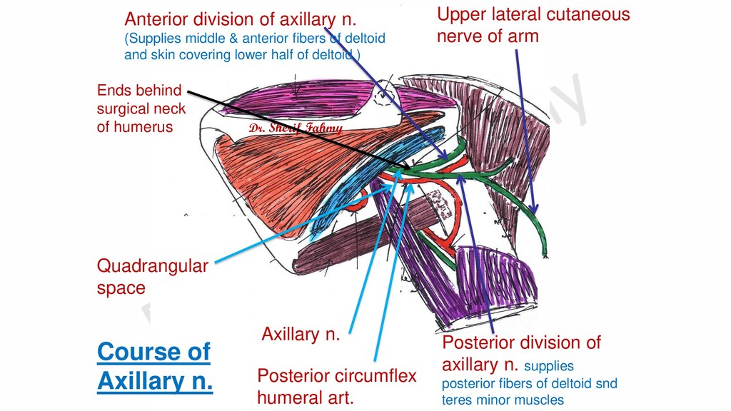
n. (Supplies middle & anterior fibers of deltoid and skin covering lower half of deltoid.) Posterior division of axillary n. supplies posterior fibers of deltoid snd teres minor muscles Upper lateral cutaneous nerve of arm Quadrangular space Course of Axillary n. Dr. Sherif Fahmy Ends behind surgical neck of humerus
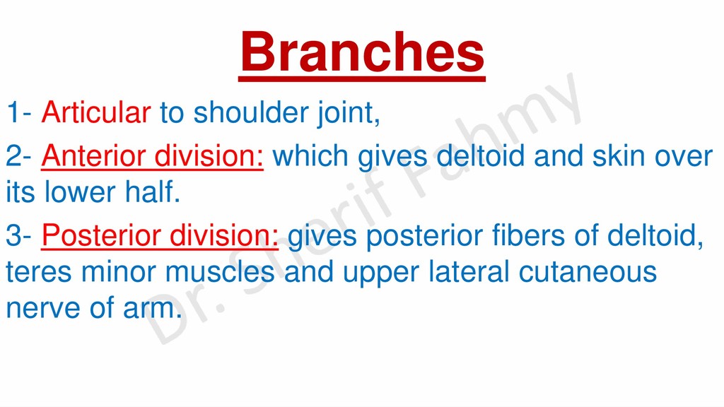
gives deltoid and skin over its lower half. 3- Posterior division: gives posterior fibers of deltoid, teres minor muscles and upper lateral cutaneous nerve of arm.
neck of humerus. 3- Badly adjusted crutch to armpit. Results: Motor loss: -Paralysis of deltoid and teres minor muscles. Sensory loss: -At skin over lower ½ of deltoid muscle. Disability & deformity: -Unable to abduct arm from 15 – 90 Degrees. -Arm is adducted. Late wasting changes: Wasting of deltoid, flat shoulder and prominent acromion.
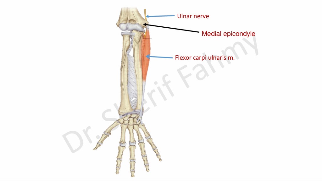
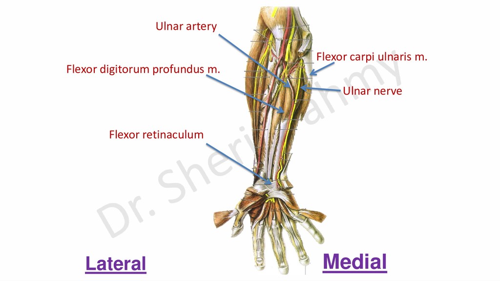
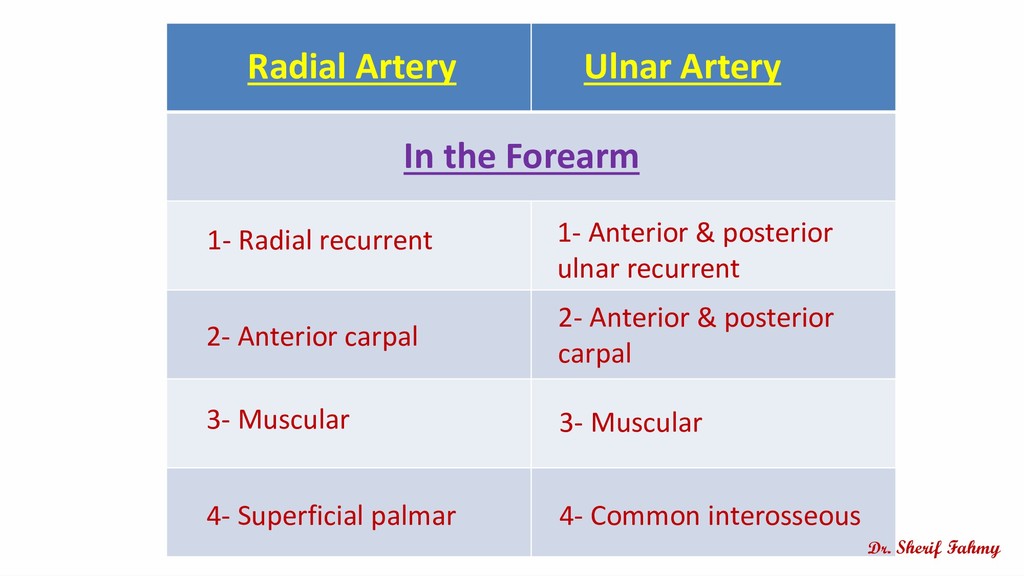
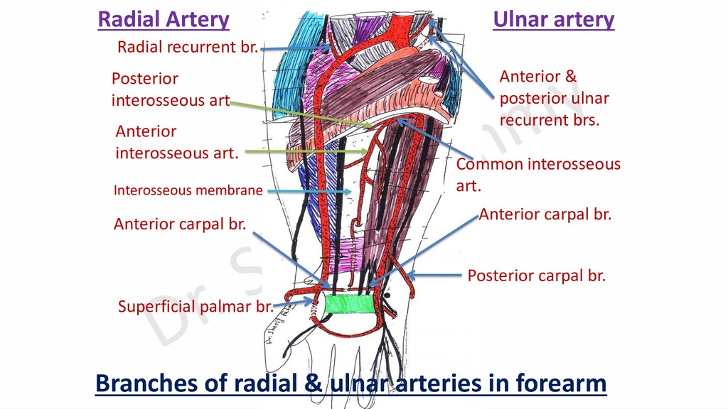
the forearm: Articular: to elbow joint. Muscular: to flexor carpi ulnaris and medial ½ of flexor digitorum profundus m. Cutaneous: Palmar & dorsal cutaneous branches.
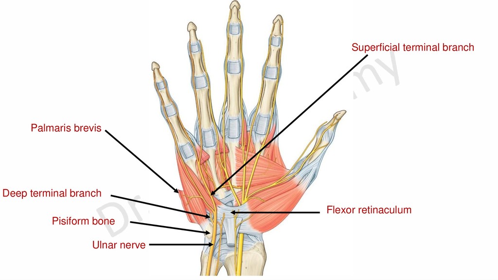
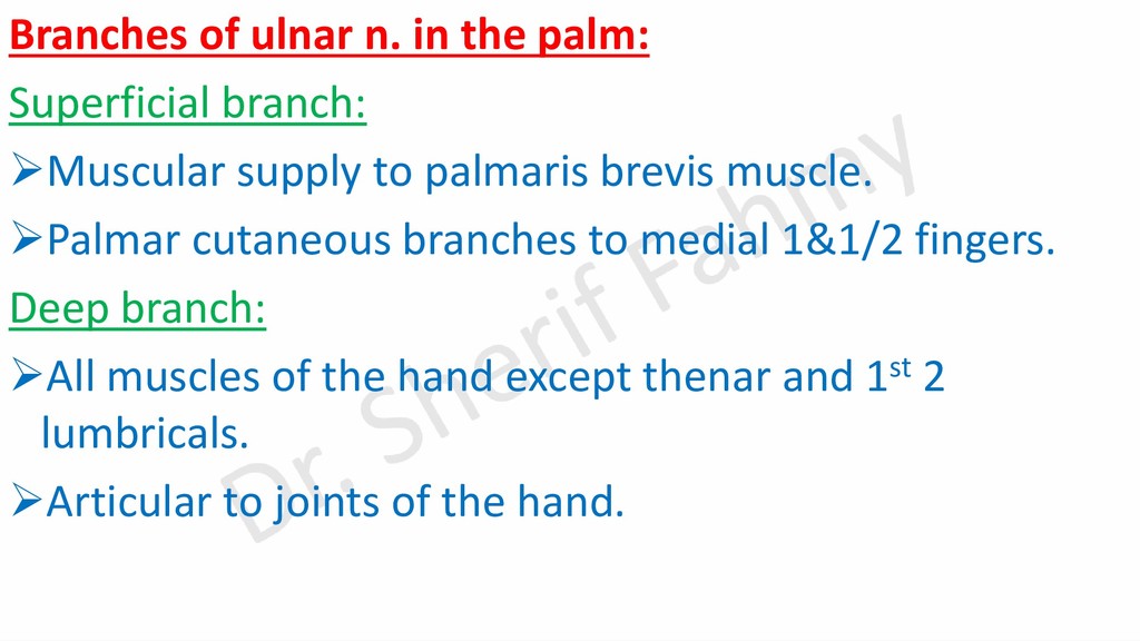
supply to palmaris brevis muscle. ➢Palmar cutaneous branches to medial 1&1/2 fingers. Deep branch: ➢All muscles of the hand except thenar and 1st 2 lumbricals. ➢Articular to joints of the hand.
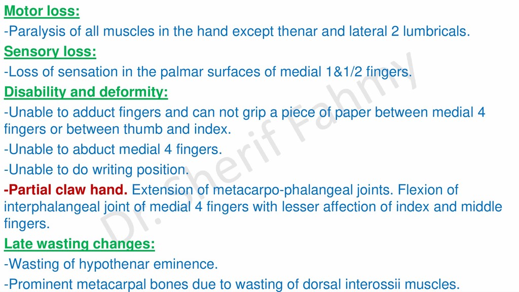
thenar and lateral 2 lumbricals. Sensory loss: -Loss of sensation in the palmar surfaces of medial 1&1/2 fingers. Disability and deformity: -Unable to adduct fingers and can not grip a piece of paper between medial 4 fingers or between thumb and index. -Unable to abduct medial 4 fingers. -Unable to do writing position. -Partial claw hand. Extension of metacarpo-phalangeal joints. Flexion of interphalangeal joint of medial 4 fingers with lesser affection of index and middle fingers. Late wasting changes: -Wasting of hypothenar eminence. -Prominent metacarpal bones due to wasting of dorsal interossii muscles.
of flexor digitorum profundus. -Paralysis of all muscles in the hand except thenar and lateral 2 lumbricals. Sensory loss: -Loss of sensation in medial 1/3 of palm and dorsum of hand. -Loss of sensation in medial 1&1/2 fingers. Disability & Deformity: -As at wrist, loss of adduction of all fingers & loss of abduction of medial 4 fingers. -Weak flexion of wrist with radial deviation due to paralysis of flexor carpi ulnaris muscle. -As in injury at wrist (partial claw hand) but with lesser flexion in ring and little fingers due to paralysis of medial ½ of flexor digitorum profundus muscle (Ulnar paradox). Late wasting changes: -Wasting at the medial side of forearm. -Wasting in muscles of the hand as with injury at wrist.
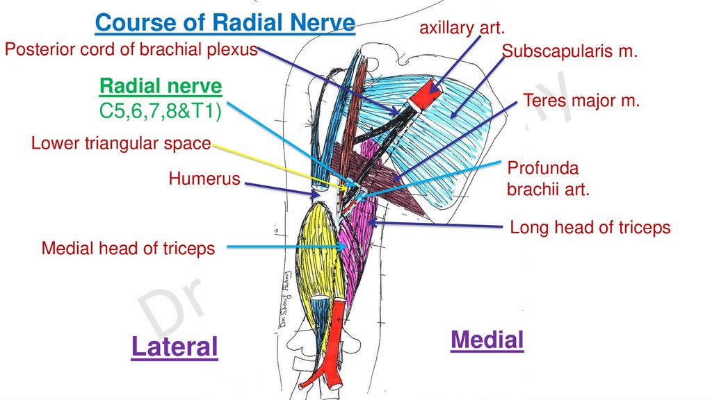
triceps Profunda brachii art. Course of Radial Nerve axillary art. Posterior cord of brachial plexus Humerus Lower triangular space Subscapularis m. Lateral Medial Teres major m.
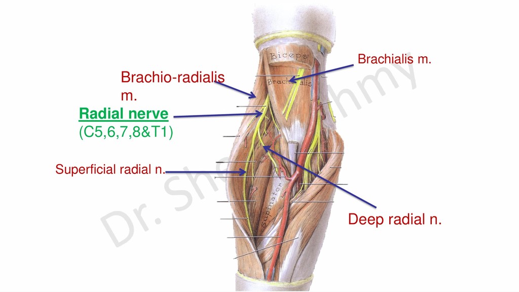
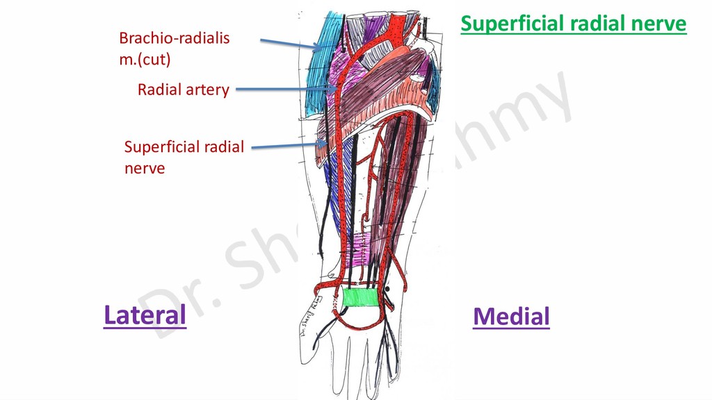
muscles of the back of forearm (extensors of the hand and fingers) except brachioradialis, extensor carpi radialis longus and anconeus ms. Superficial radial nerve: It supplies the skin of -Lateral 2/3 of the dorsum of the hand. -Back of lateral 3 and half fingers except terminal parts (from median nerve).
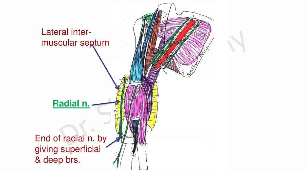
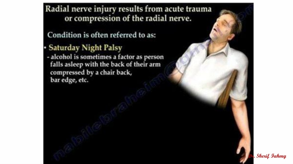
badly adjusted crutch in armpit. B- in upper part of arm: Saturday night palsy. C- In spiral groove: by fracture shaft of humerus. D- At elbow: by supracondylar fracture of humerus. N.B. Main deformity of radial nerve injury is wrist & finger drop (bad hand grip)
loss: Paralysis of triceps and muscles of the back of forearm. Sensory loss: Loss of sensation in skin of: Back of arm, lower lateral of arm, back of forearm and back of lateral 2/3 of dorsum of hand and lateral 3 and half fingers. Disability: Unable to extends forearm against resistance, extends hand against resistance, extends fingers against resistance and supinate extended forearm. Weak hand grip. Deformity: Flexed elbow with wrist and finger drop. Late wasting changes: Wasting of back of arm & forearm.
and part of medial head and muscles of the back of forearm. Sensory loss: Loss of sensation in: lower lateral of arm, back of forearm and back of lateral 2/3 of dorsum of hand and back of lateral 3 and half fingers. Disability: Weak extension of forearm. Unable to extends hand against resistance, extends fingers against resistance and supinate extended forearm. Deformity: • Flexed elbow with wrist and finger drop. Weak hand grip. Late wasting changes: • Partial wasting of back of arm and wasting of back of forearm.
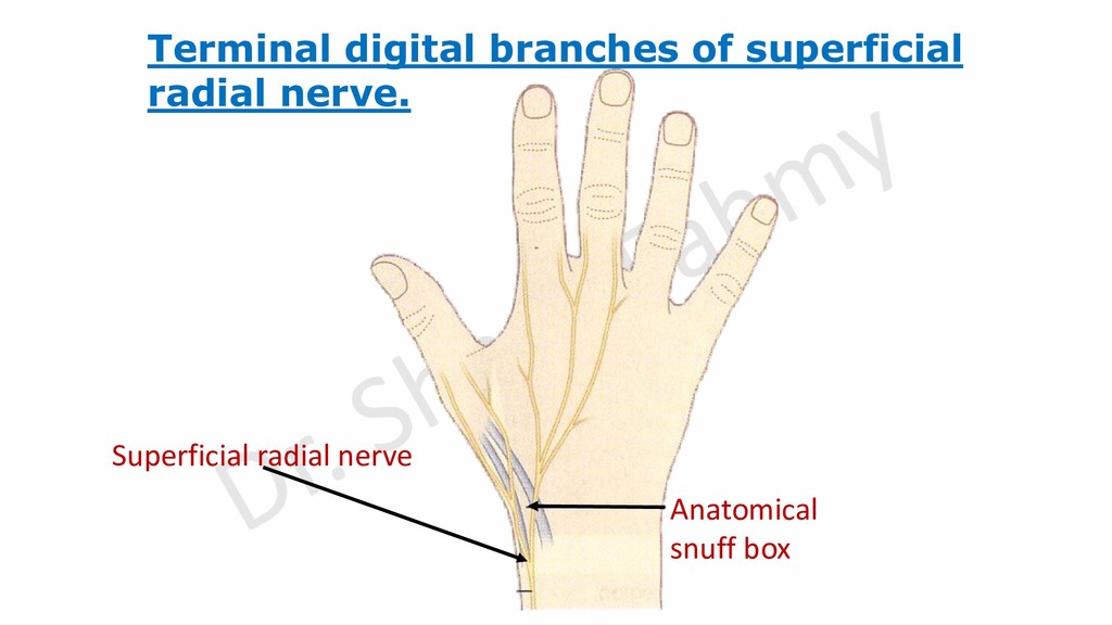
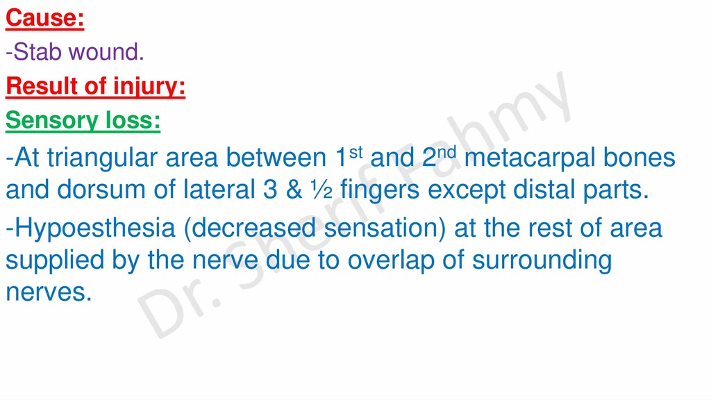
area between 1st and 2nd metacarpal bones and dorsum of lateral 3 & ½ fingers except distal parts. -Hypoesthesia (decreased sensation) at the rest of area supplied by the nerve due to overlap of surrounding nerves.
radius. Results of injury: Motor loss: -Paralysis of muscles of back of forearm except brachio-radialis, extensor carpi radialis longus and anconeus muscles. Disability & deformity: -Weak extension of wrist with abduction of the hand. -Unable to extends fingers. -Finger drop. -No wrist drop due to action of ext.carpi radialis longus. Late wasting changes: -Wasting of back of forearm with lesser affection to lateral side.
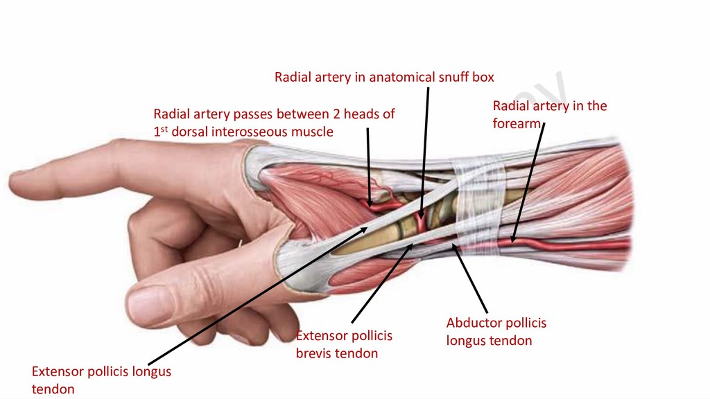
pollicis Deep palmar arch Base of 5th metacarpal bone Deep branch of ulnar artery Radial artery in the palm Radial artery 1st dorsal interosseous m Deep branch of ulnar nerve