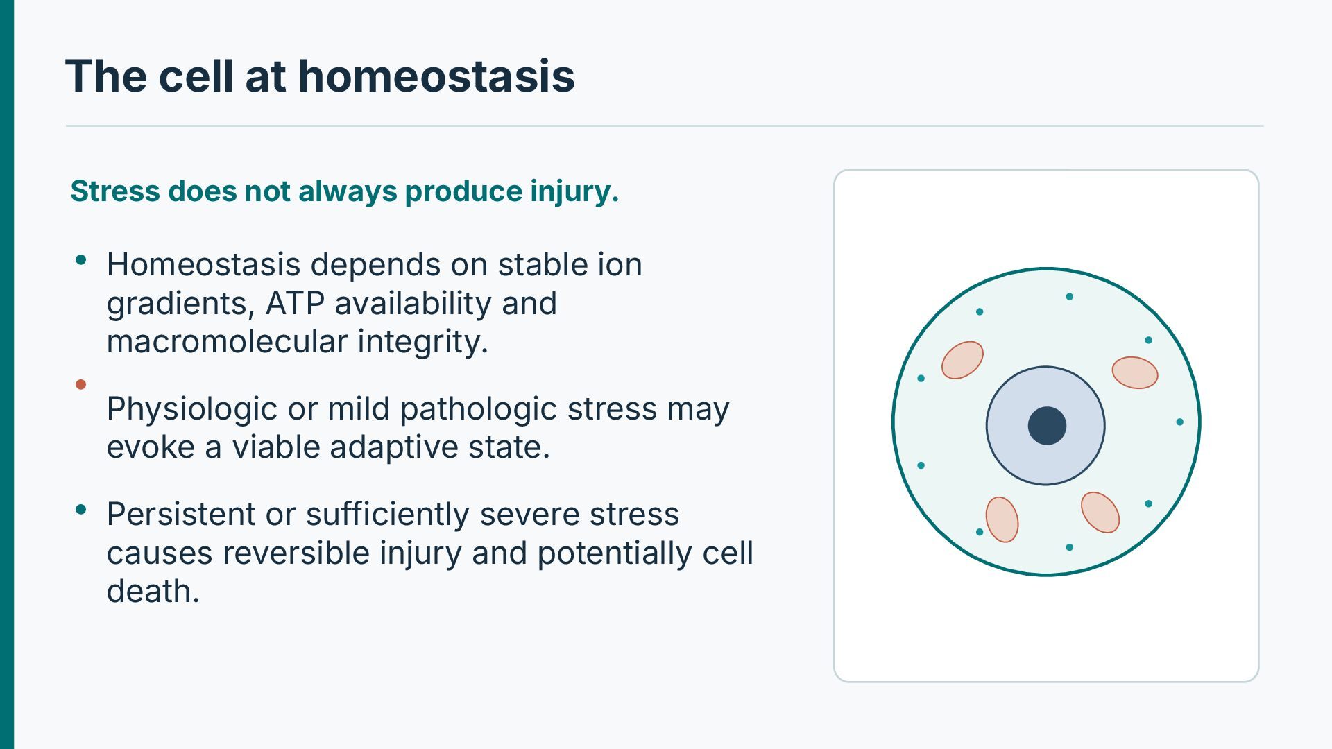
Homeostasis depends on stable ion gradients, ATP availability and macromolecular integrity. Physiologic or mild pathologic stress may evoke a viable adaptive state. Persistent or sufficiently severe stress causes reversible injury and potentially cell death.
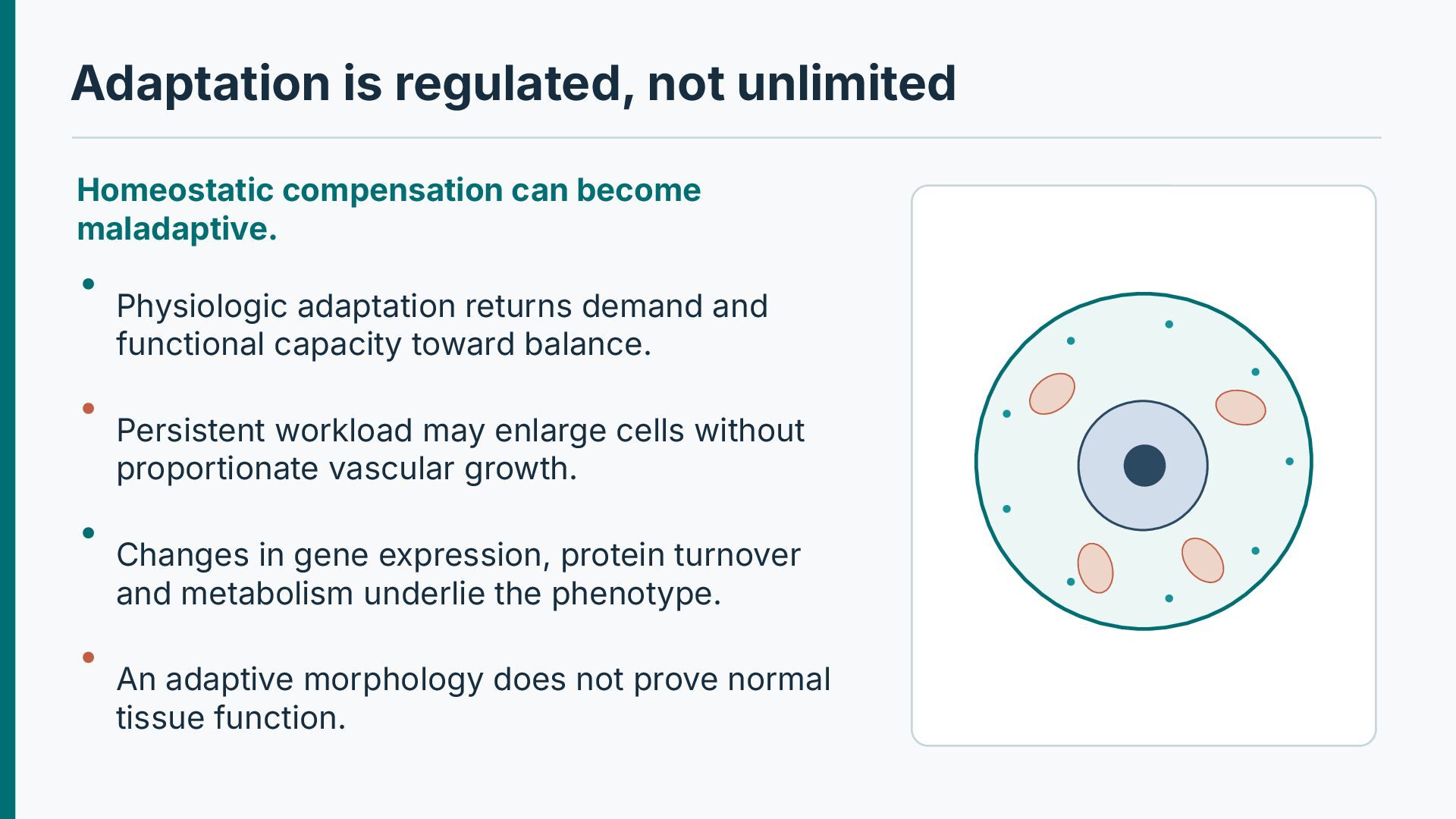
Physiologic adaptation returns demand and functional capacity toward balance. Persistent workload may enlarge cells without proportionate vascular growth. Changes in gene expression, protein turnover and metabolism underlie the phenotype. An adaptive morphology does not prove normal tissue function.
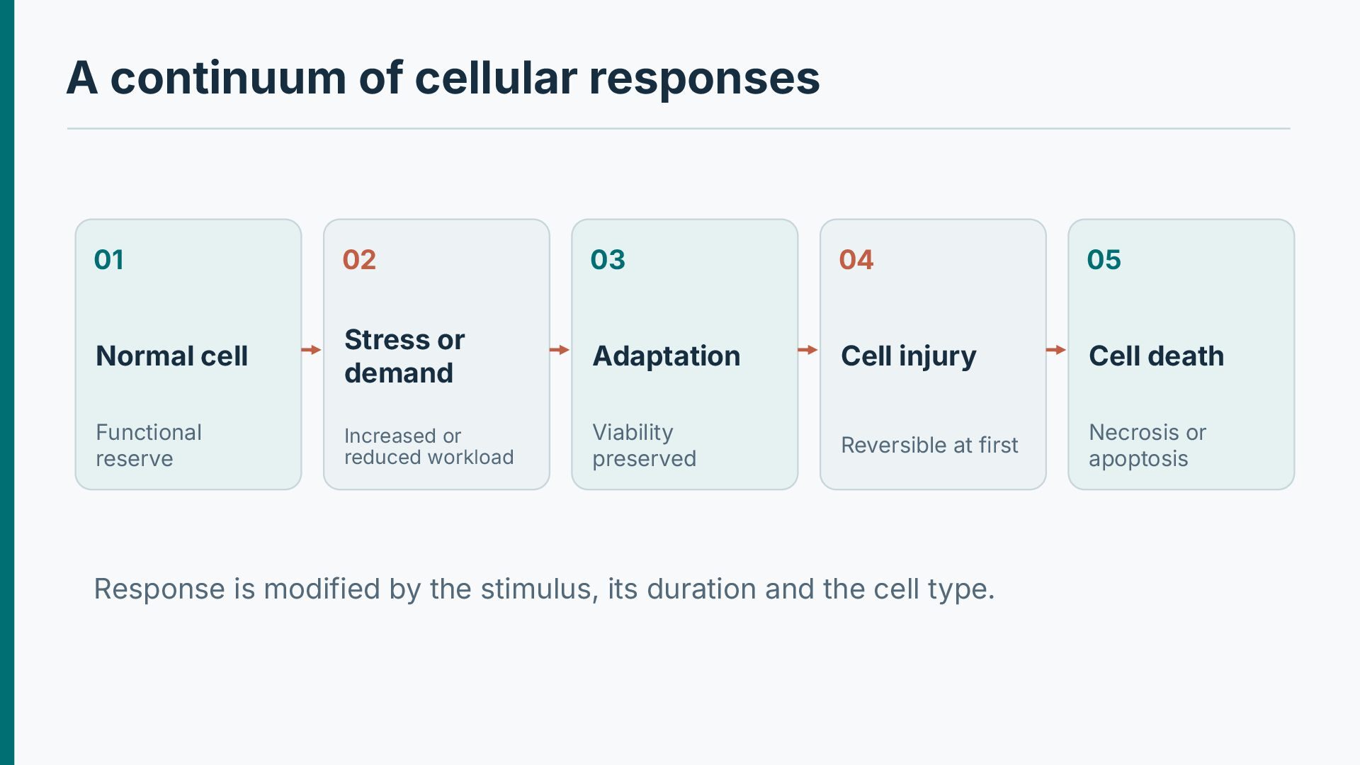
Normal cell Stress or demand Adaptation Cell injury Cell death Functional reserve Increased or reduced workload Viability preserved Reversible at first Necrosis or apoptosis Response is modified by the stimulus, its duration and the cell type.
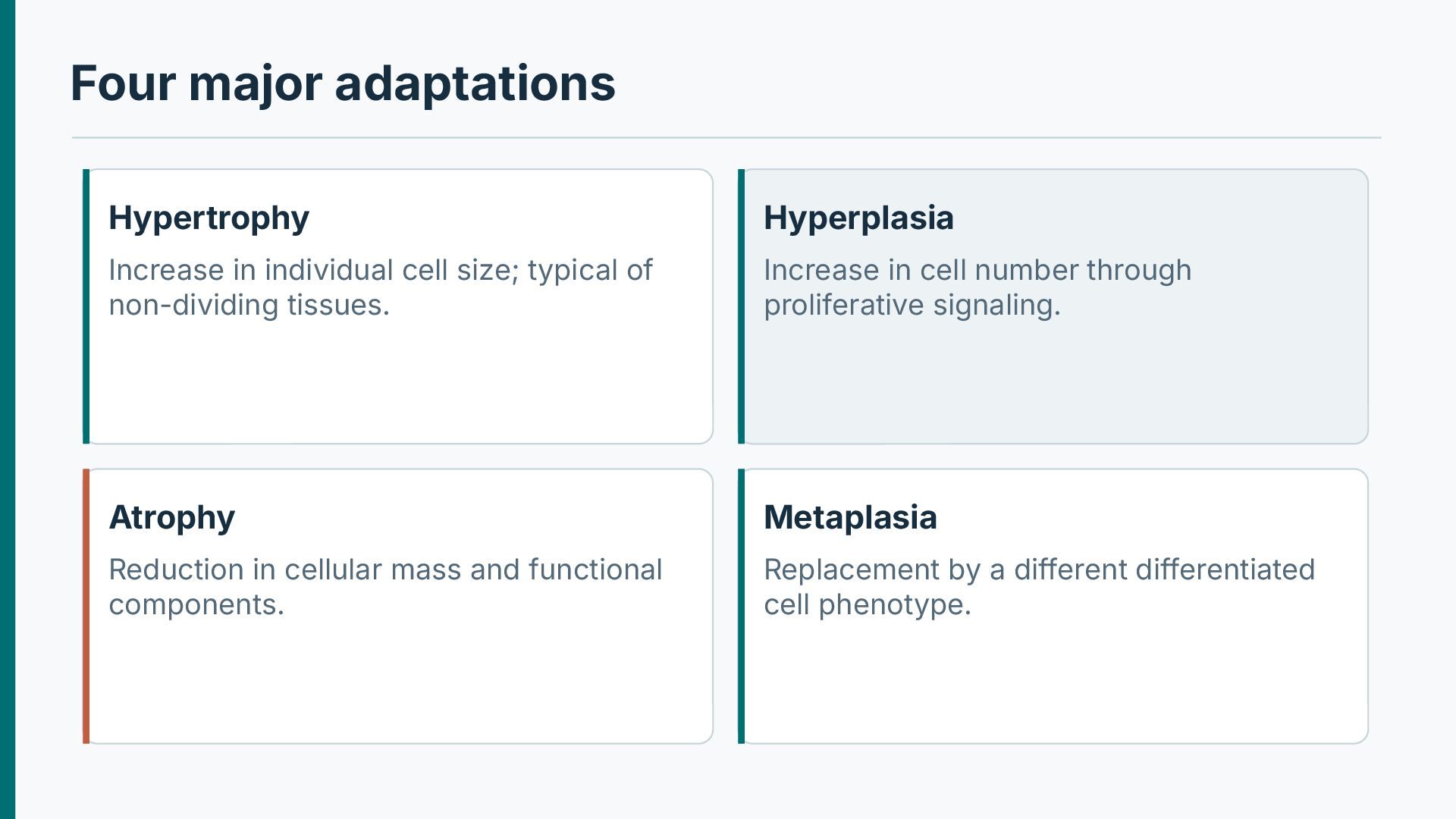
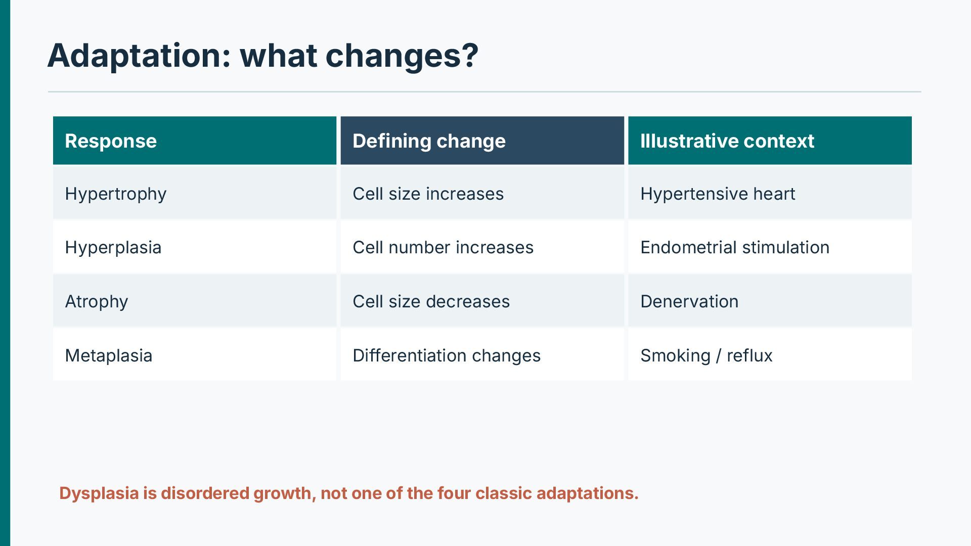
typical of non-dividing tissues. Increase in cell number through proliferative signaling. Atrophy Metaplasia Reduction in cellular mass and functional components. Replacement by a different differentiated cell phenotype.
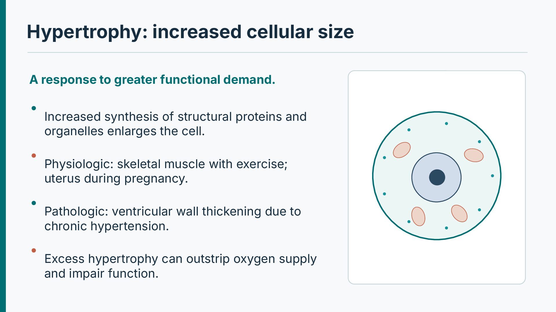
Increased synthesis of structural proteins and organelles enlarges the cell. Physiologic: skeletal muscle with exercise; uterus during pregnancy. Pathologic: ventricular wall thickening due to chronic hypertension. Excess hypertrophy can outstrip oxygen supply and impair function.
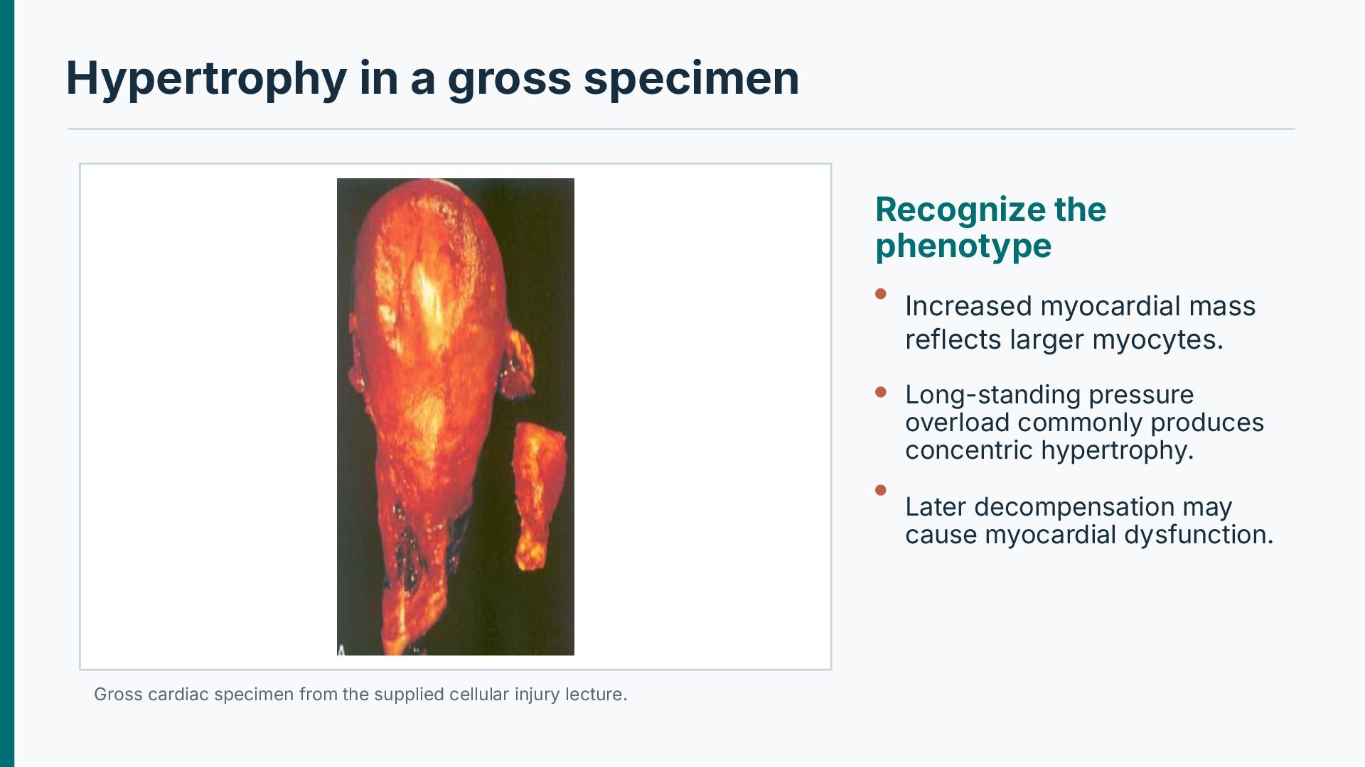
mass reflects larger myocytes. Long-standing pressure overload commonly produces concentric hypertrophy. Later decompensation may cause myocardial dysfunction. Gross cardiac specimen from the supplied cellular injury lecture.
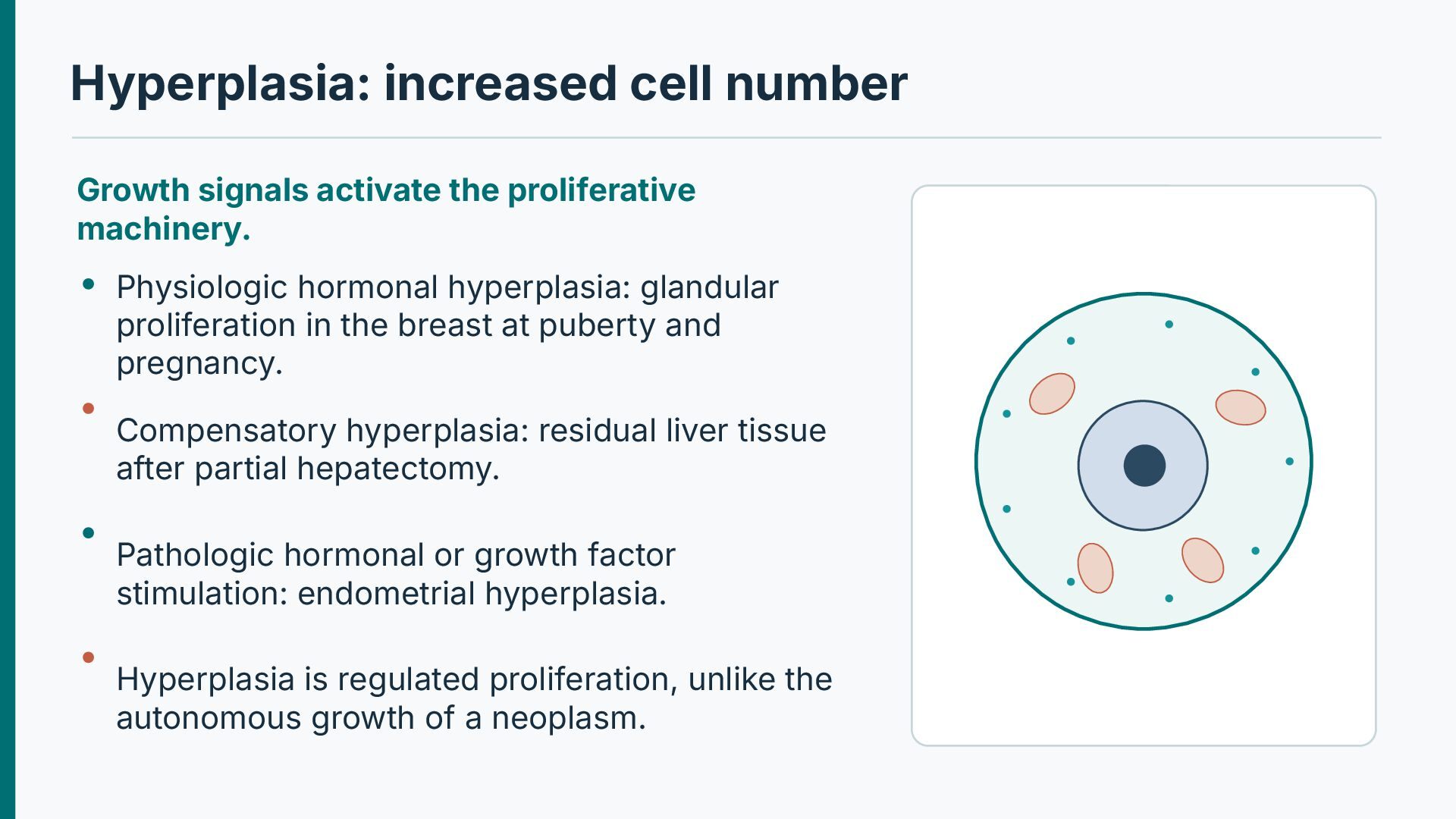
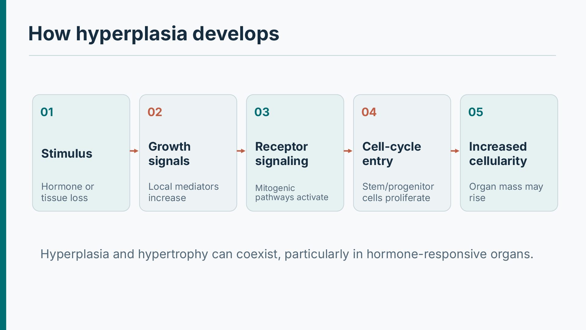
Physiologic hormonal hyperplasia: glandular proliferation in the breast at puberty and pregnancy. Compensatory hyperplasia: residual liver tissue after partial hepatectomy. Pathologic hormonal or growth factor stimulation: endometrial hyperplasia. Hyperplasia is regulated proliferation, unlike the autonomous growth of a neoplasm.
signals Receptor signaling Cell-cycle entry Increased cellularity Hormone or tissue loss Local mediators increase Mitogenic pathways activate Stem/progenitor cells proliferate Organ mass may rise Hyperplasia and hypertrophy can coexist, particularly in hormone-responsive organs.
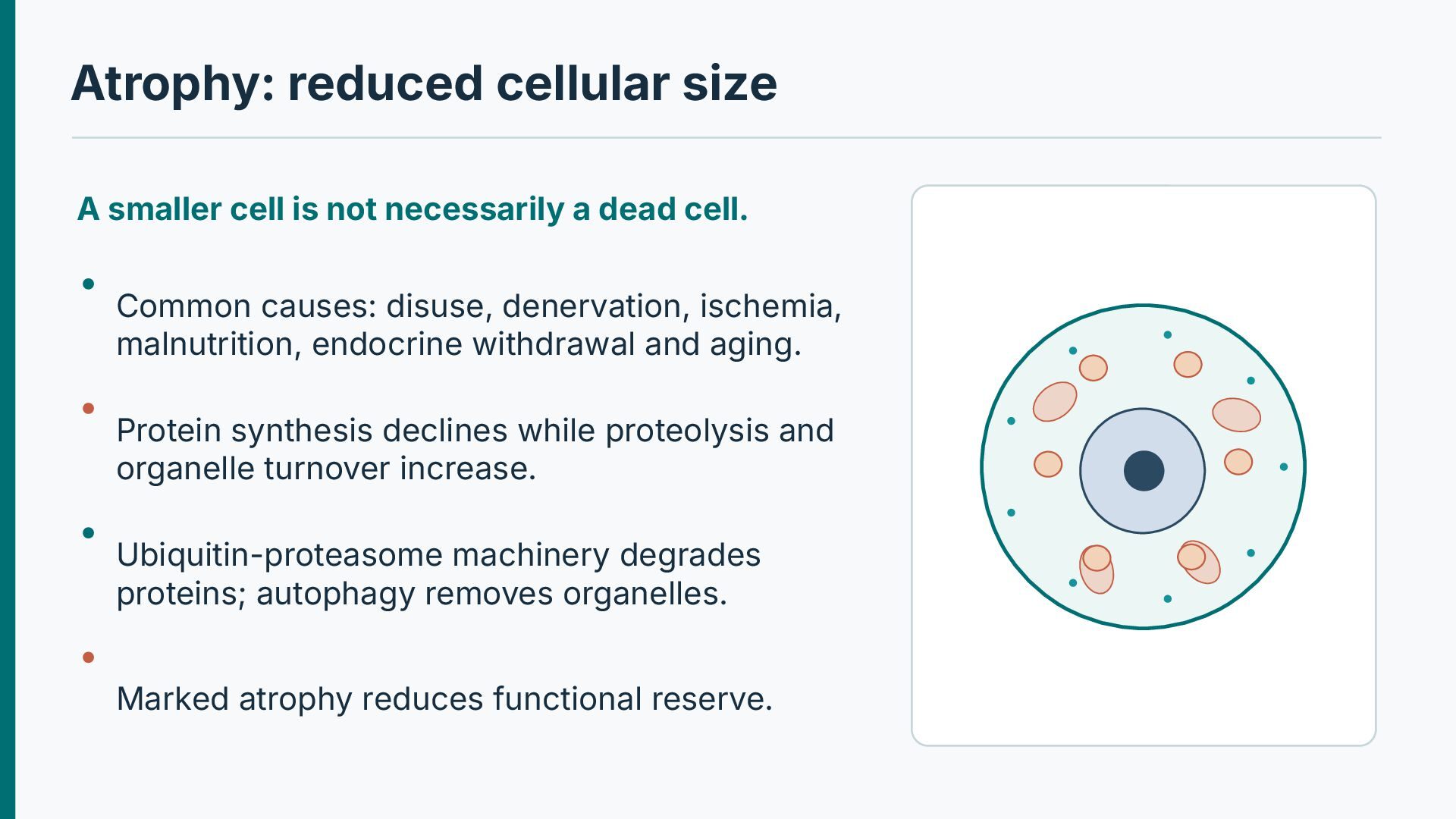
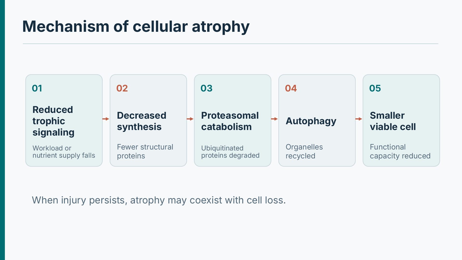
a dead cell. Common causes: disuse, denervation, ischemia, malnutrition, endocrine withdrawal and aging. Protein synthesis declines while proteolysis and organelle turnover increase. Ubiquitin-proteasome machinery degrades proteins; autophagy removes organelles. Marked atrophy reduces functional reserve.
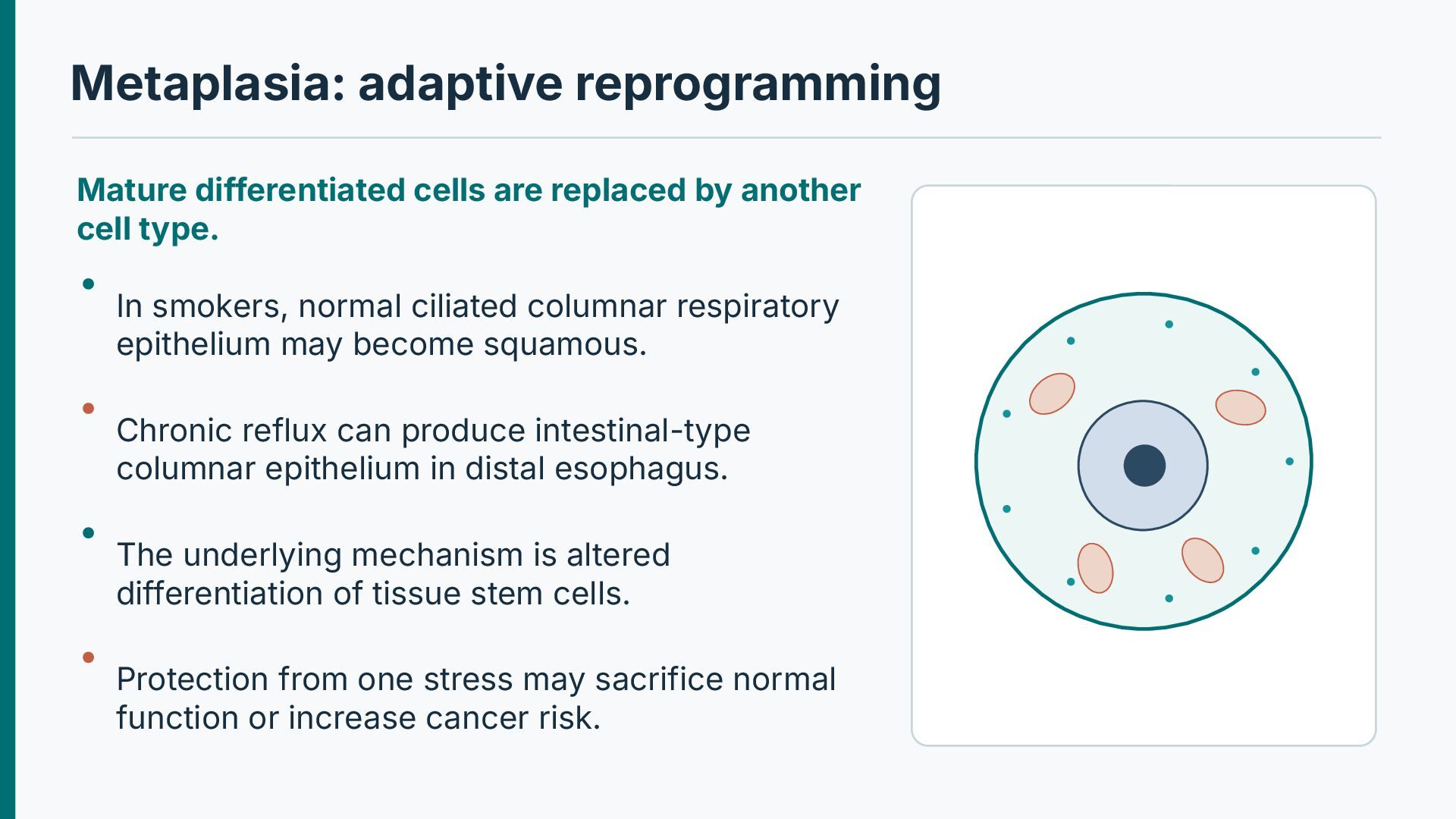
cell type. In smokers, normal ciliated columnar respiratory epithelium may become squamous. Chronic reflux can produce intestinal-type columnar epithelium in distal esophagus. The underlying mechanism is altered differentiation of tissue stem cells. Protection from one stress may sacrifice normal function or increase cancer risk.
size increases Hypertensive heart Hyperplasia Cell number increases Endometrial stimulation Atrophy Cell size decreases Denervation Metaplasia Differentiation changes Smoking / reflux Dysplasia is disordered growth, not one of the four classic adaptations.
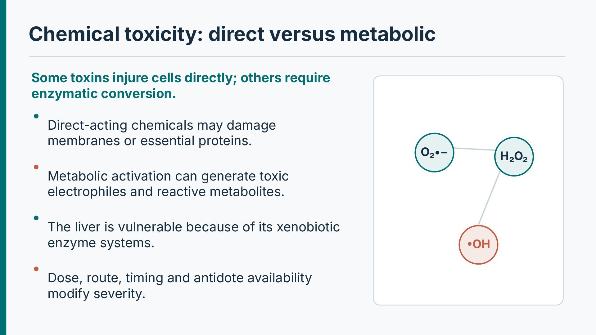
others require enzymatic conversion. Direct-acting chemicals may damage membranes or essential proteins. O₂•− H₂O₂ Metabolic activation can generate toxic electrophiles and reactive metabolites. The liver is vulnerable because of its xenobiotic enzyme systems. Dose, route, timing and antidote availability modify severity. •OH
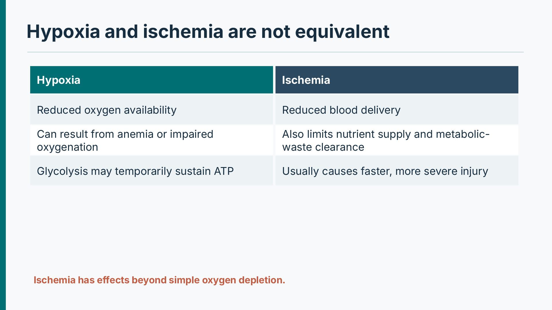
availability Reduced blood delivery Can result from anemia or impaired oxygenation Also limits nutrient supply and metabolicwaste clearance Glycolysis may temporarily sustain ATP Usually causes faster, more severe injury Ischemia has effects beyond simple oxygen depletion.
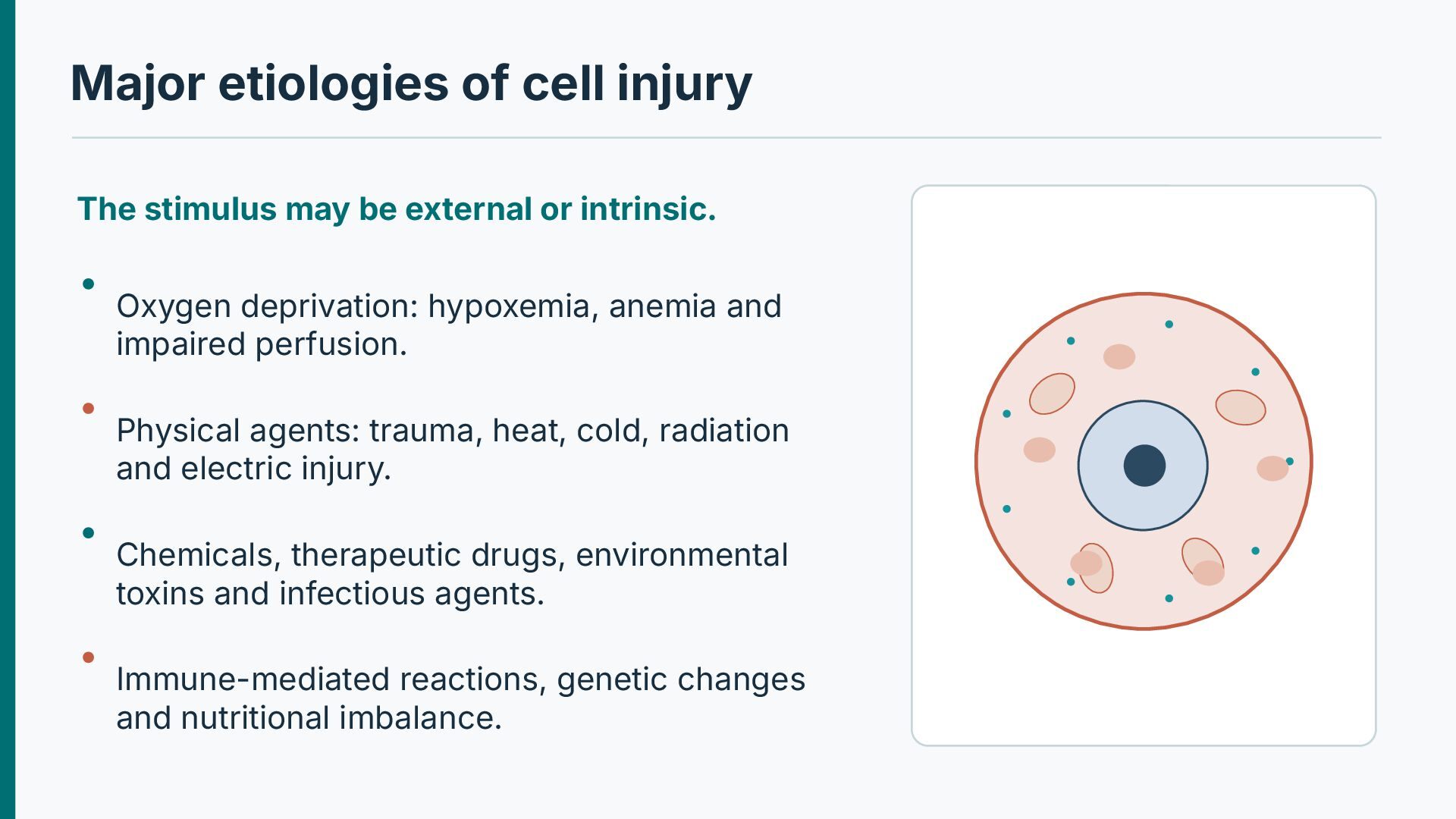
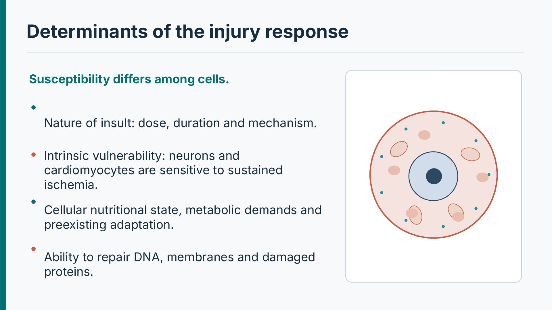
of insult: dose, duration and mechanism. Intrinsic vulnerability: neurons and cardiomyocytes are sensitive to sustained ischemia. Cellular nutritional state, metabolic demands and preexisting adaptation. Ability to repair DNA, membranes and damaged proteins.
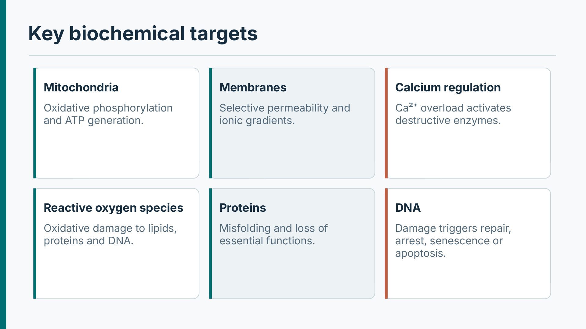
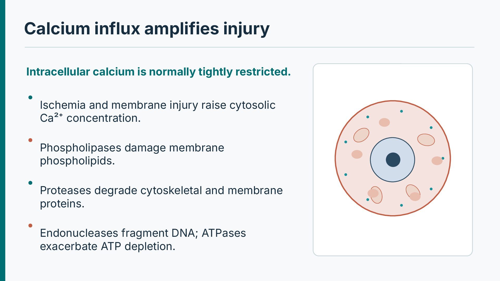
ATP generation. Selective permeability and ionic gradients. Ca²⁺ overload activates destructive enzymes. Reactive oxygen species Proteins DNA Oxidative damage to lipids, proteins and DNA. Misfolding and loss of essential functions. Damage triggers repair, arrest, senescence or apoptosis.
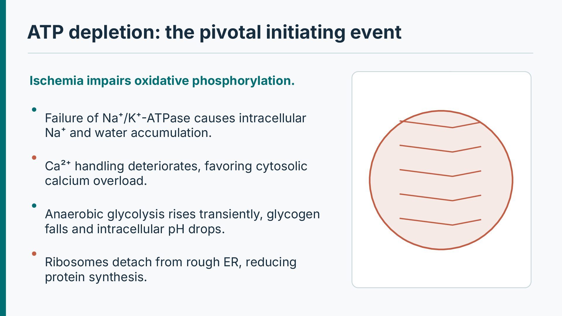
Failure of Na⁺/K⁺-ATPase causes intracellular Na⁺ and water accumulation. Ca²⁺ handling deteriorates, favoring cytosolic calcium overload. Anaerobic glycolysis rises transiently, glycogen falls and intracellular pH drops. Ribosomes detach from rough ER, reducing protein synthesis.
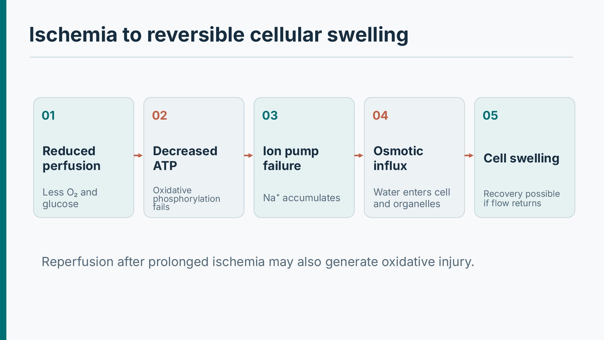
Reduced perfusion Decreased ATP Ion pump failure Osmotic influx Cell swelling Less O₂ and glucose Oxidative phosphorylation fails Na⁺ accumulates Water enters cell and organelles Recovery possible if flow returns Reperfusion after prolonged ischemia may also generate oxidative injury.
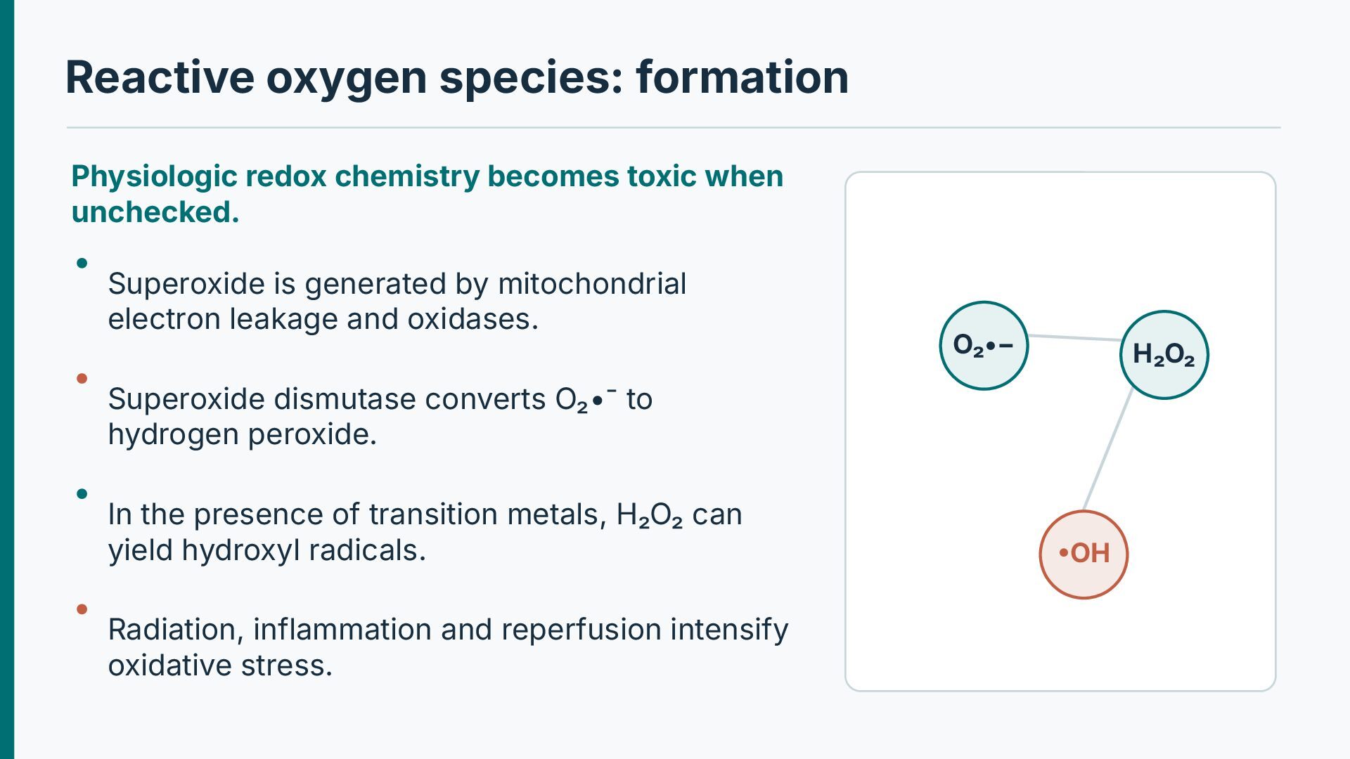
unchecked. Superoxide is generated by mitochondrial electron leakage and oxidases. O₂•− H₂O₂ Superoxide dismutase converts O₂•⁻ to hydrogen peroxide. In the presence of transition metals, H₂O₂ can yield hydroxyl radicals. Radiation, inflammation and reperfusion intensify oxidative stress. •OH
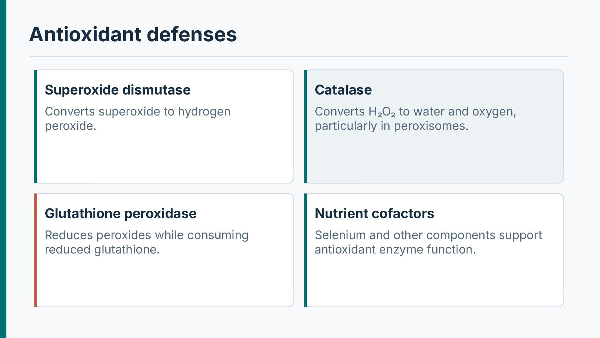
Converts H₂O₂ to water and oxygen, particularly in peroxisomes. Glutathione peroxidase Nutrient cofactors Reduces peroxides while consuming reduced glutathione. Selenium and other components support antioxidant enzyme function.
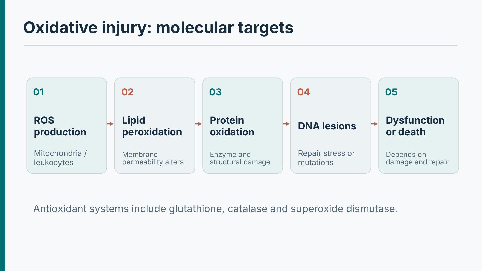
production Lipid peroxidation Protein oxidation DNA lesions Dysfunction or death Mitochondria / leukocytes Membrane permeability alters Enzyme and structural damage Repair stress or mutations Depends on damage and repair Antioxidant systems include glutathione, catalase and superoxide dismutase.
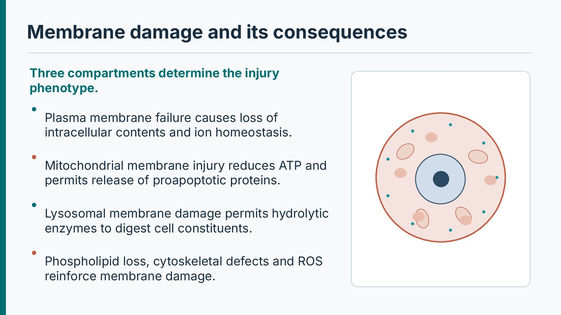
phenotype. Plasma membrane failure causes loss of intracellular contents and ion homeostasis. Mitochondrial membrane injury reduces ATP and permits release of proapoptotic proteins. Lysosomal membrane damage permits hydrolytic enzymes to digest cell constituents. Phospholipid loss, cytoskeletal defects and ROS reinforce membrane damage.
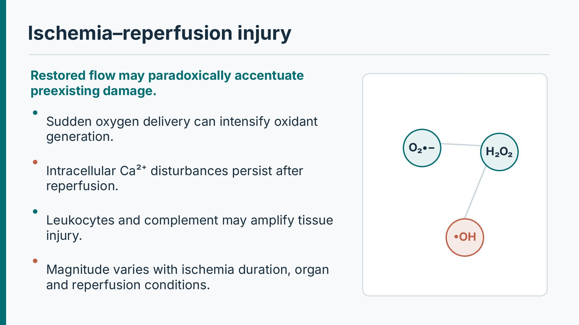
oxygen delivery can intensify oxidant generation. O₂•− H₂O₂ Intracellular Ca²⁺ disturbances persist after reperfusion. Leukocytes and complement may amplify tissue injury. Magnitude varies with ischemia duration, organ and reperfusion conditions. •OH
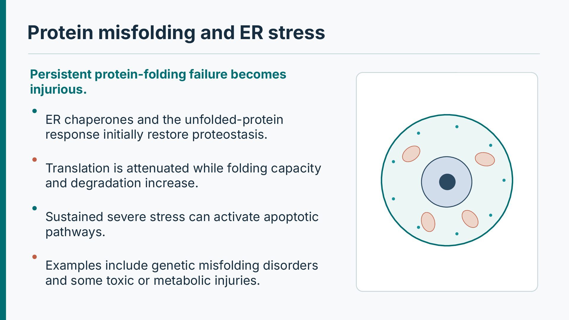
ER chaperones and the unfolded-protein response initially restore proteostasis. Translation is attenuated while folding capacity and degradation increase. Sustained severe stress can activate apoptotic pathways. Examples include genetic misfolding disorders and some toxic or metabolic injuries.
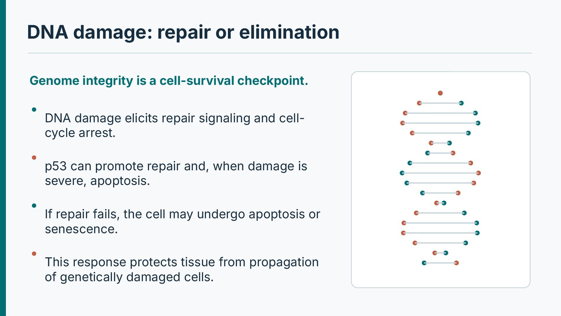
checkpoint. DNA damage elicits repair signaling and cellcycle arrest. p53 can promote repair and, when damage is severe, apoptosis. If repair fails, the cell may undergo apoptosis or senescence. This response protects tissue from propagation of genetically damaged cells.
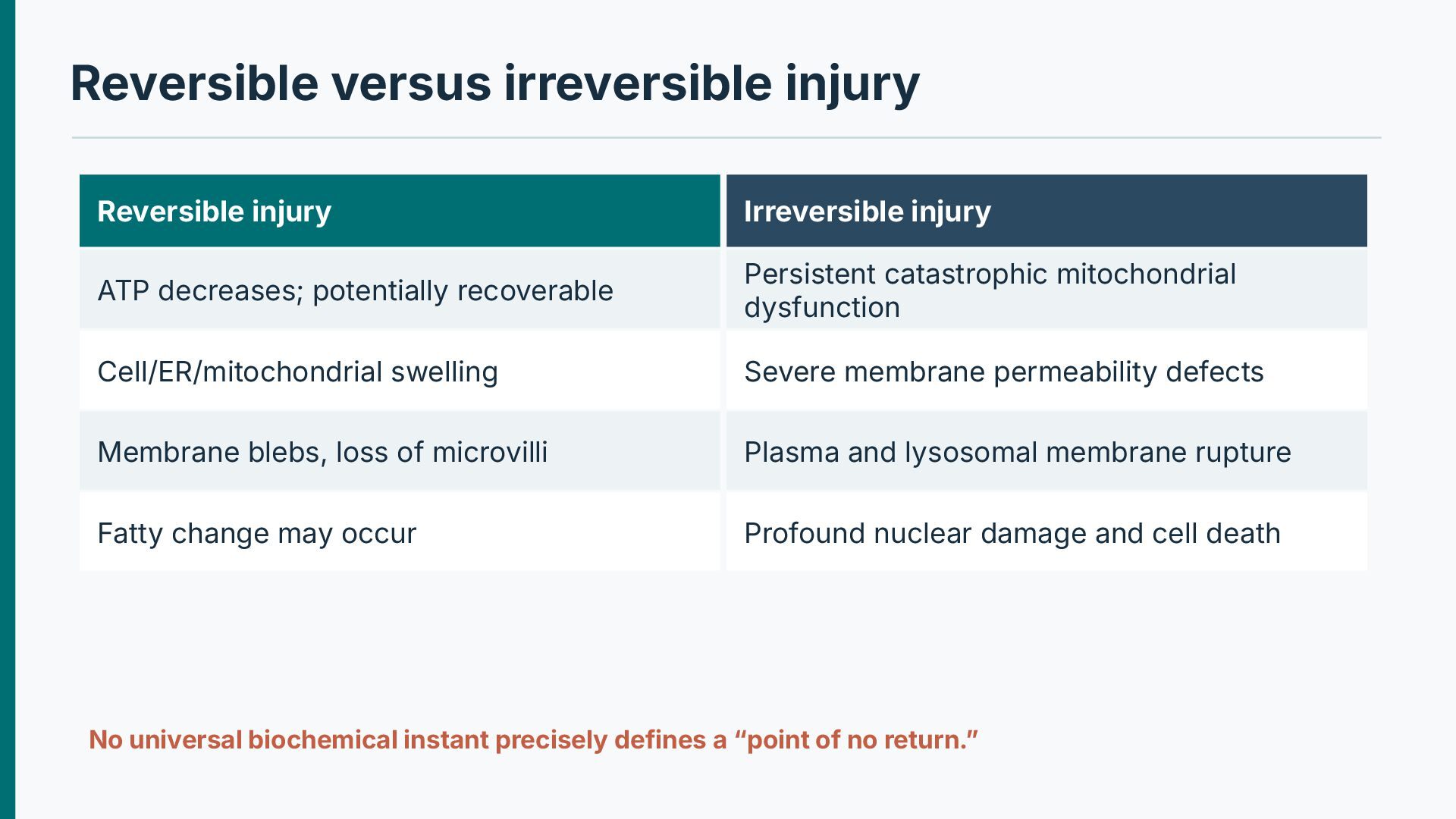
potentially recoverable Persistent catastrophic mitochondrial dysfunction Cell/ER/mitochondrial swelling Severe membrane permeability defects Membrane blebs, loss of microvilli Plasma and lysosomal membrane rupture Fatty change may occur Profound nuclear damage and cell death No universal biochemical instant precisely defines a “point of no return.”
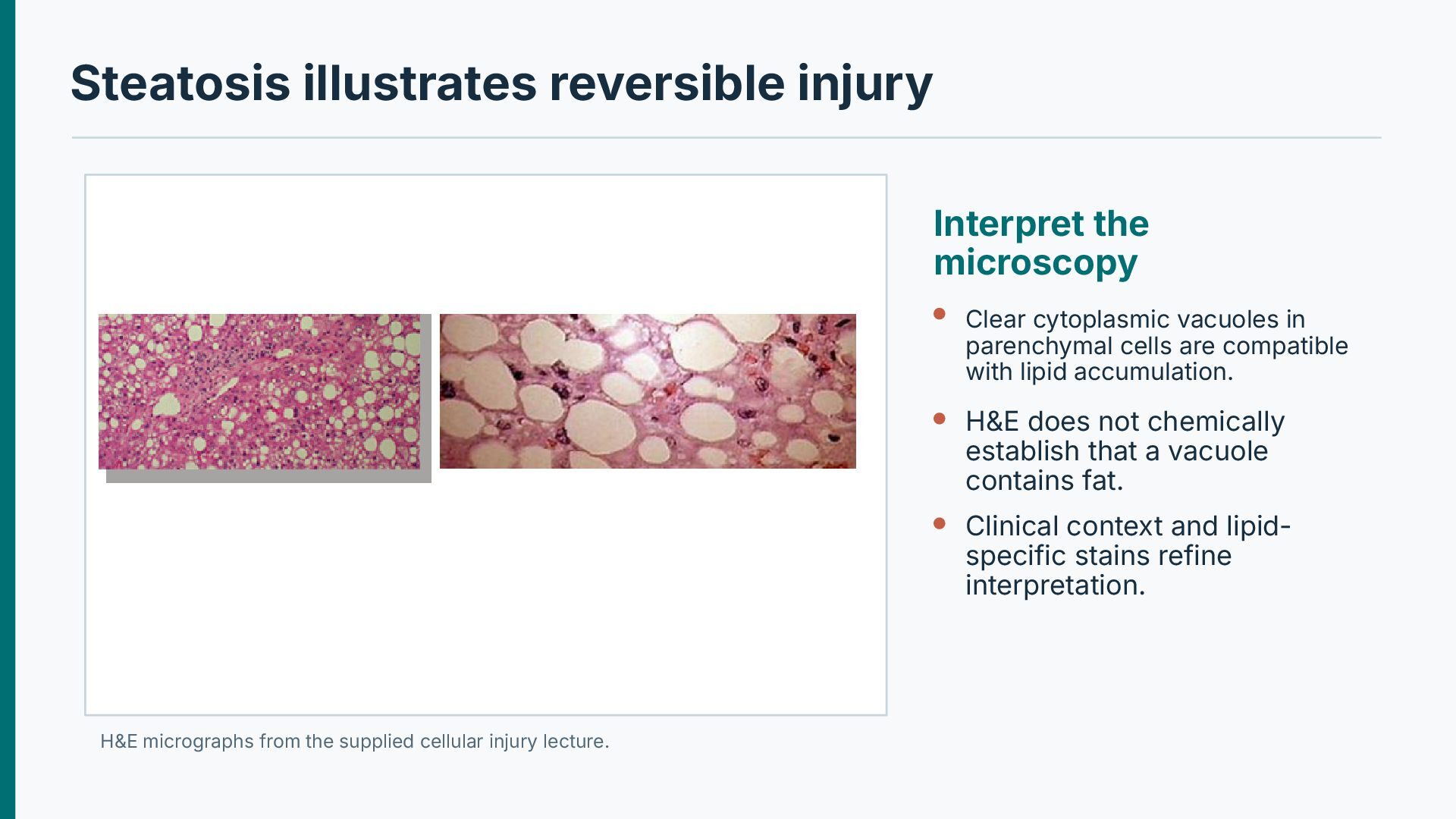
in parenchymal cells are compatible with lipid accumulation. H&E does not chemically establish that a vacuole contains fat. Clinical context and lipidspecific stains refine interpretation. H&E micrographs from the supplied cellular injury lecture.
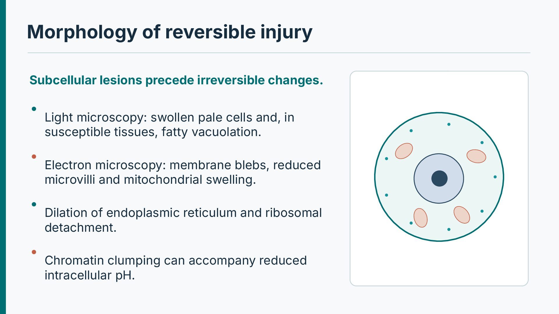
microscopy: swollen pale cells and, in susceptible tissues, fatty vacuolation. Electron microscopy: membrane blebs, reduced microvilli and mitochondrial swelling. Dilation of endoplasmic reticulum and ribosomal detachment. Chromatin clumping can accompany reduced intracellular pH.
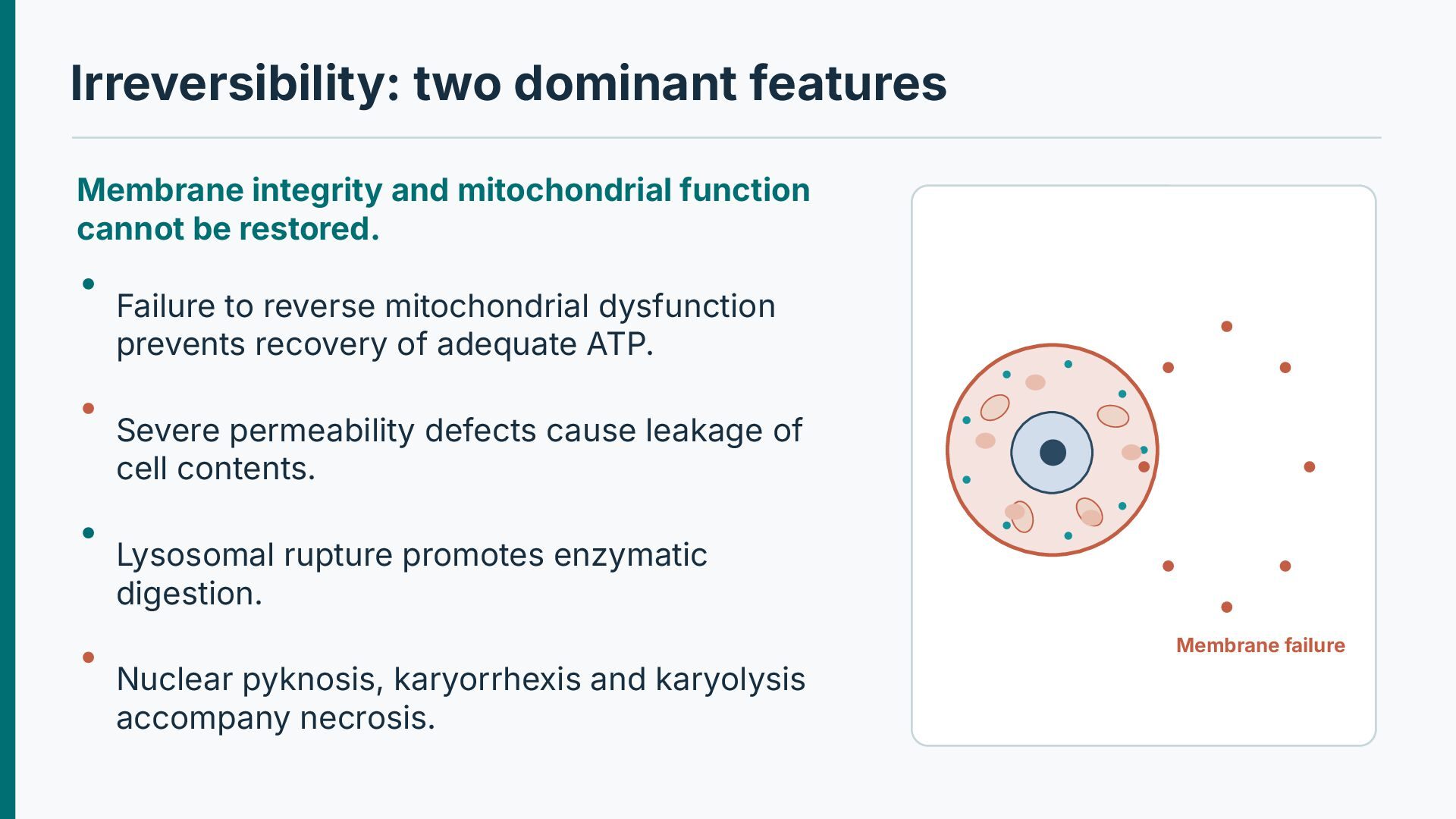
be restored. Failure to reverse mitochondrial dysfunction prevents recovery of adequate ATP. Severe permeability defects cause leakage of cell contents. Lysosomal rupture promotes enzymatic digestion. Membrane failure Nuclear pyknosis, karyorrhexis and karyolysis accompany necrosis.
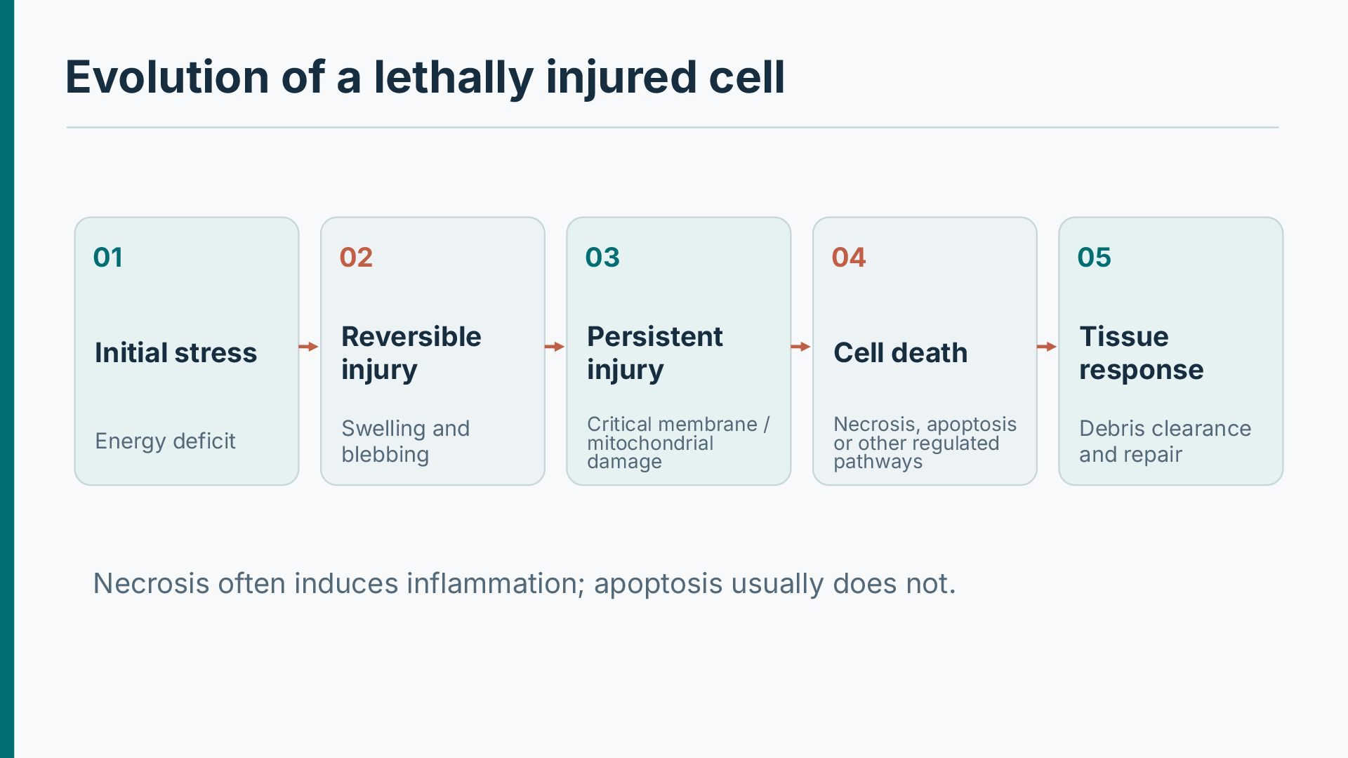
05 Initial stress Reversible injury Persistent injury Cell death Tissue response Energy deficit Swelling and blebbing Critical membrane / mitochondrial damage Necrosis, apoptosis or other regulated pathways Debris clearance and repair Necrosis often induces inflammation; apoptosis usually does not.
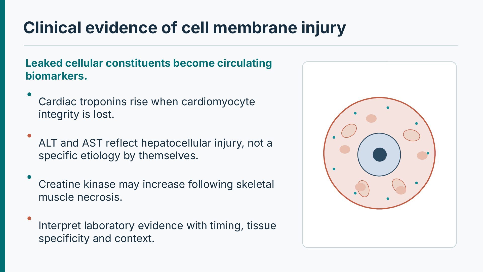
circulating biomarkers. Cardiac troponins rise when cardiomyocyte integrity is lost. ALT and AST reflect hepatocellular injury, not a specific etiology by themselves. Creatine kinase may increase following skeletal muscle necrosis. Interpret laboratory evidence with timing, tissue specificity and context.
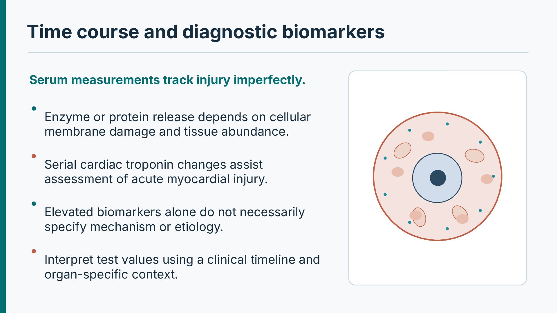
Enzyme or protein release depends on cellular membrane damage and tissue abundance. Serial cardiac troponin changes assist assessment of acute myocardial injury. Elevated biomarkers alone do not necessarily specify mechanism or etiology. Interpret test values using a clinical timeline and organ-specific context.
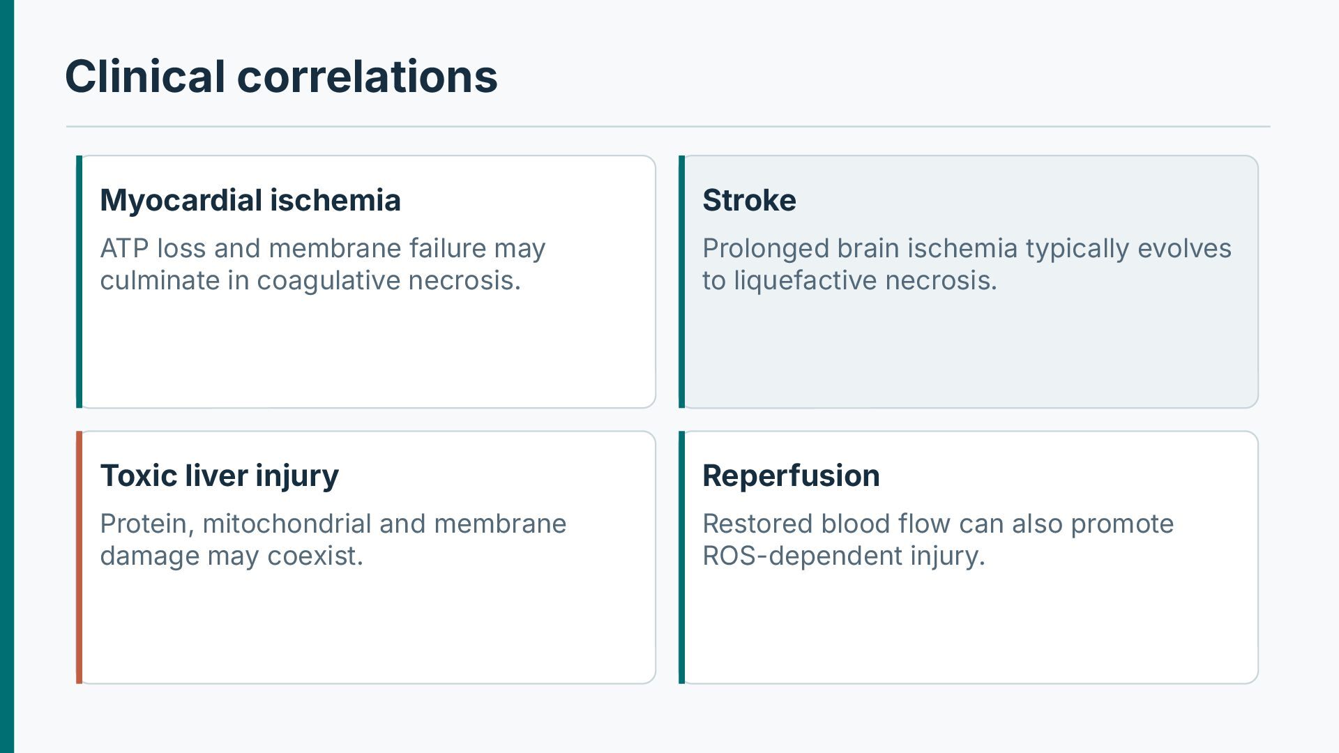
may culminate in coagulative necrosis. Prolonged brain ischemia typically evolves to liquefactive necrosis. Toxic liver injury Reperfusion Protein, mitochondrial and membrane damage may coexist. Restored blood flow can also promote ROS-dependent injury.
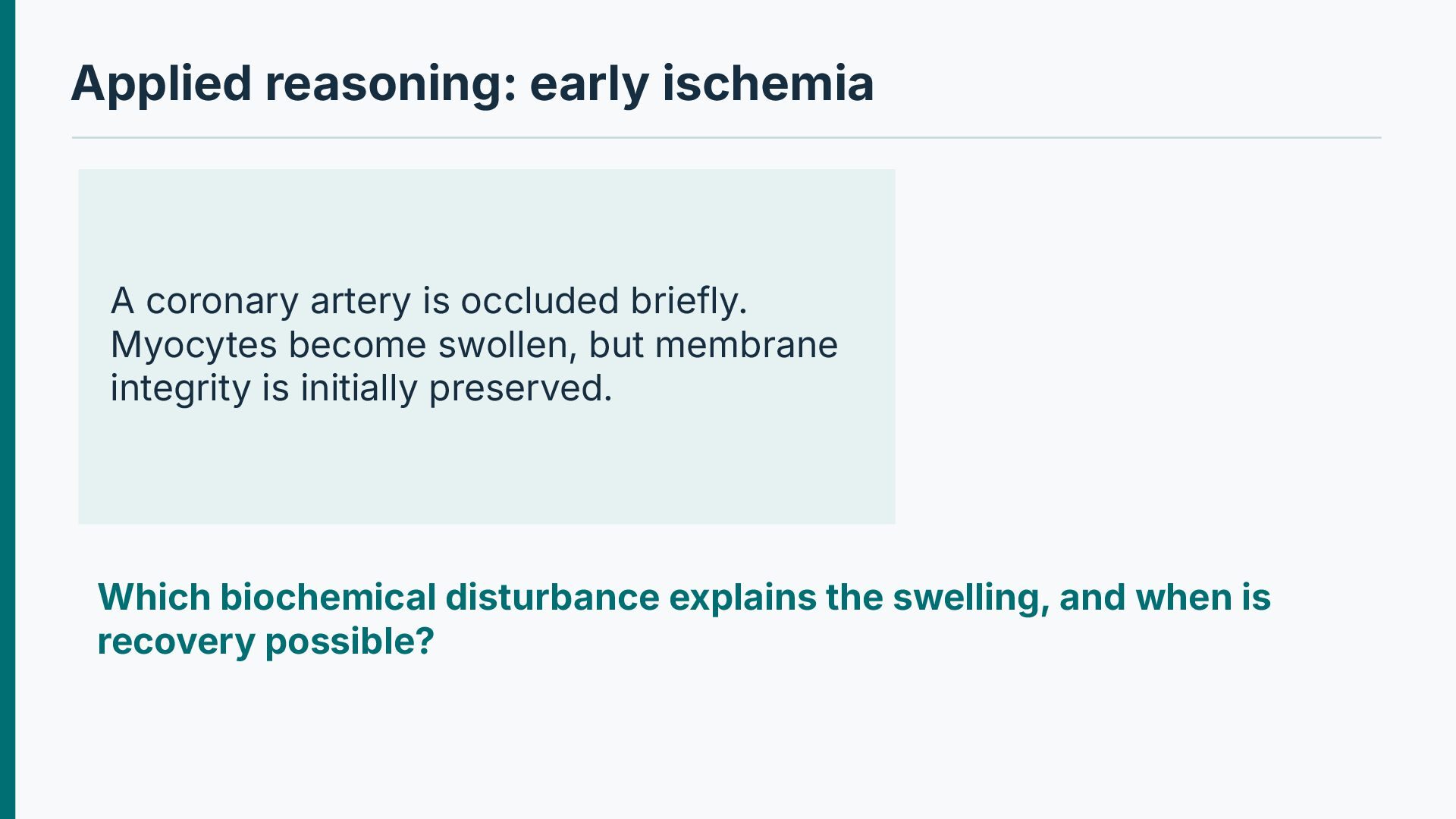
Myocytes become swollen, but membrane integrity is initially preserved. Which biochemical disturbance explains the swelling, and when is recovery possible?
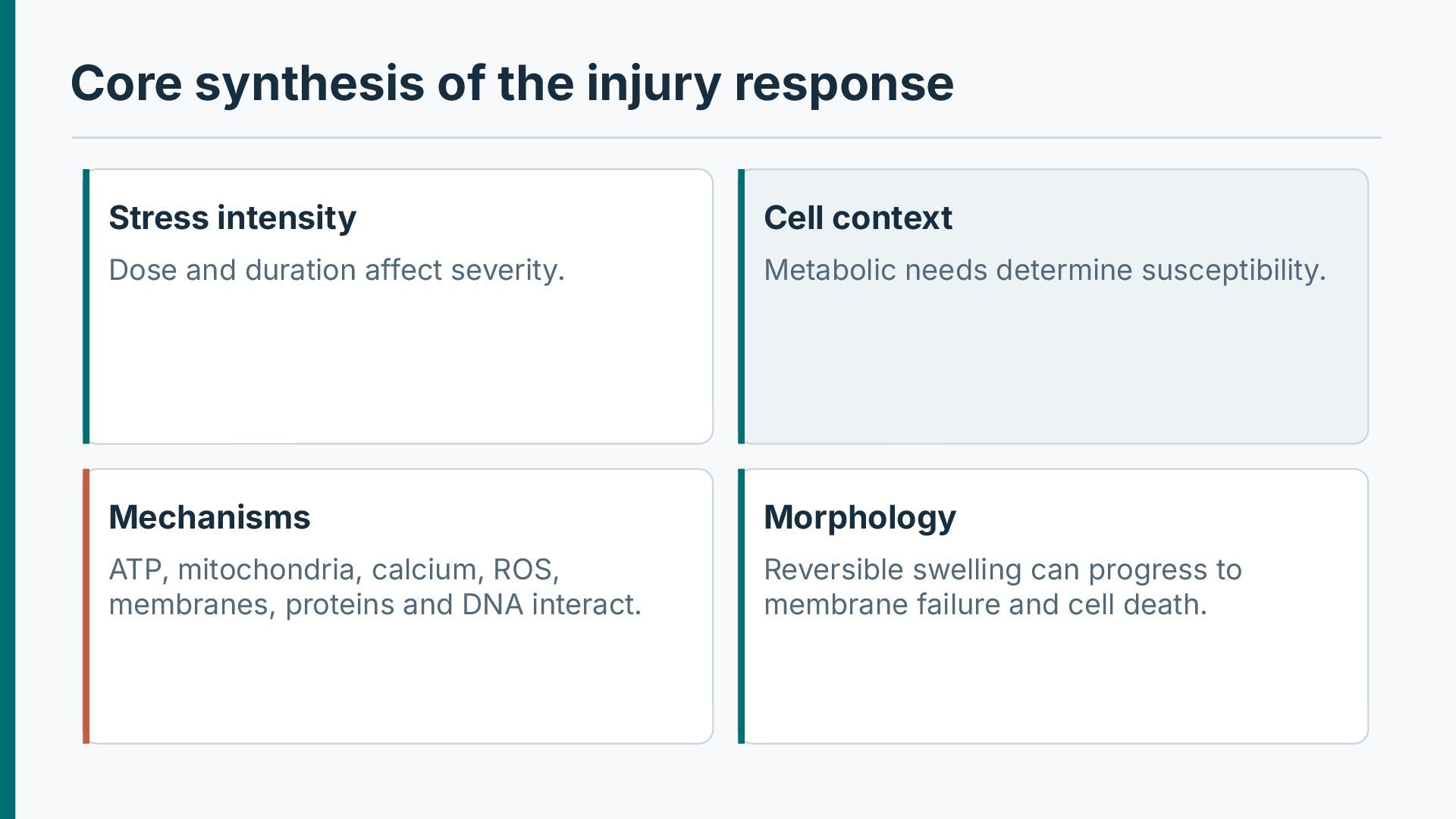
Dose and duration affect severity. Metabolic needs determine susceptibility. Mechanisms Morphology ATP, mitochondria, calcium, ROS, membranes, proteins and DNA interact. Reversible swelling can progress to membrane failure and cell death.
· Chapter: Cell Injury, Cell Death, and Adaptations For further study: correlate the molecular mechanisms with gross and microscopic pathology. Illustrative pathways are original schematics; selected histology images are retained from the supplied teaching material.


 abusuraihsakhri
 institutdelelevage
 ianozsvald
 trycycle
 miyayou
 ykiyota
 shumpei777
 hiranabe
 brainpadpr
 nagano
 martinlippert
 samtorres
 paigeccino
 marketingsoph
 tamaranovitovic
 garrettdimon
 sergeychernyshev
 jeffersonlam
 elsirapls
 jlugia
 baasie
 bethany
 bluesmoon