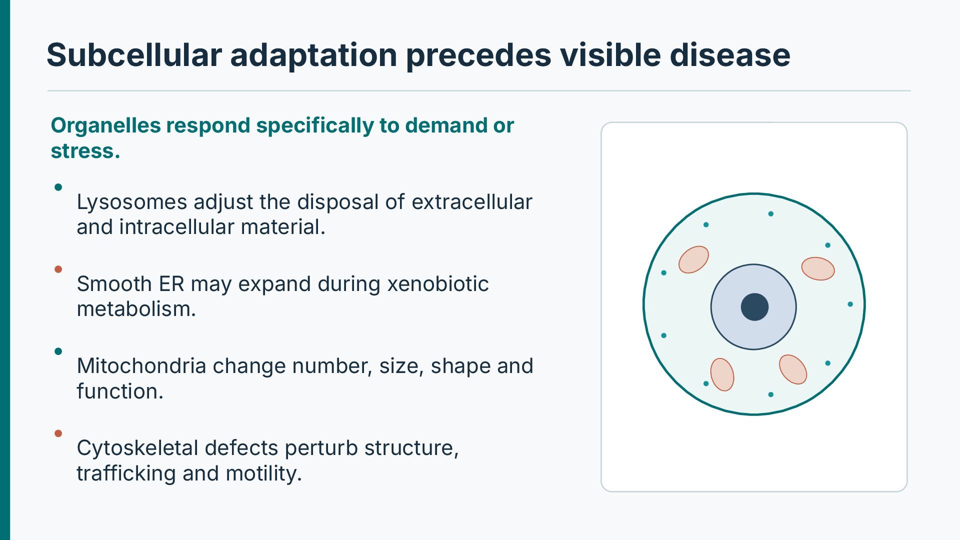
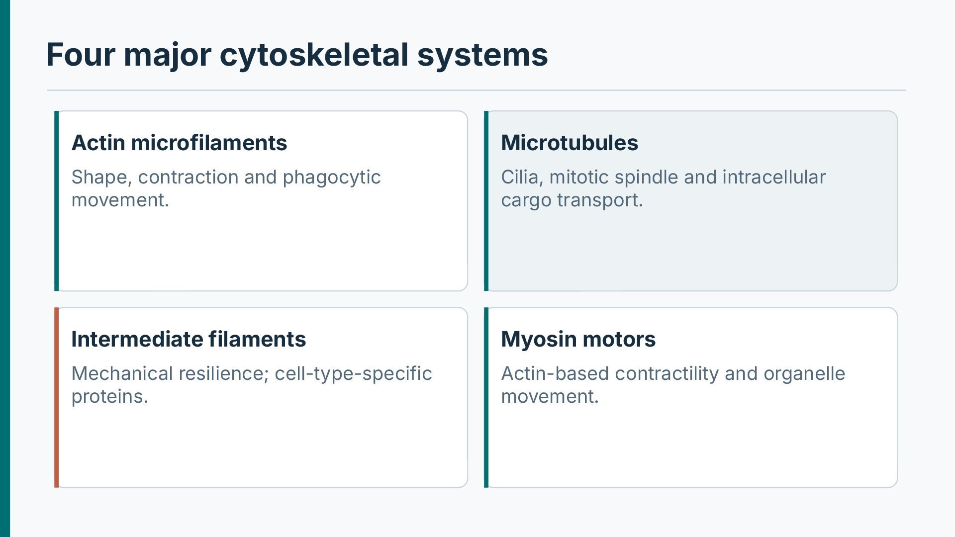
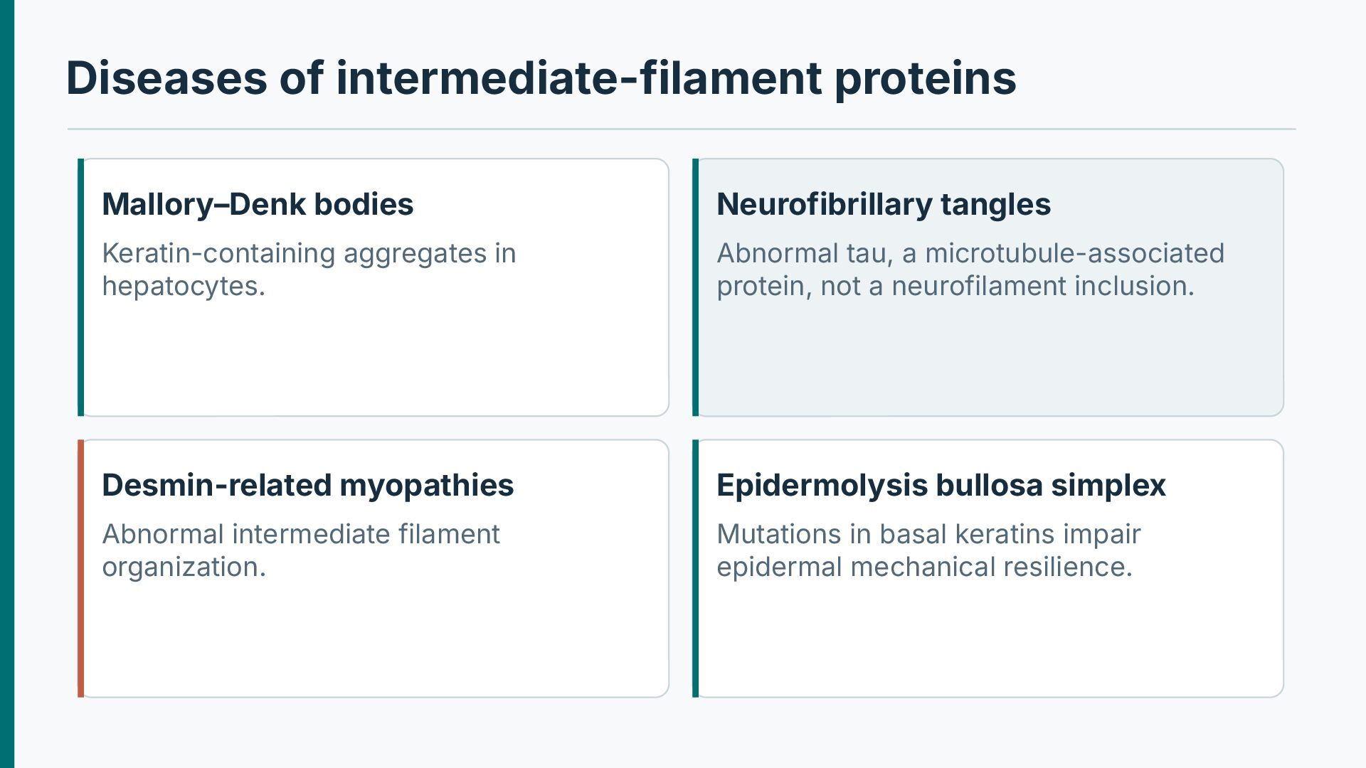
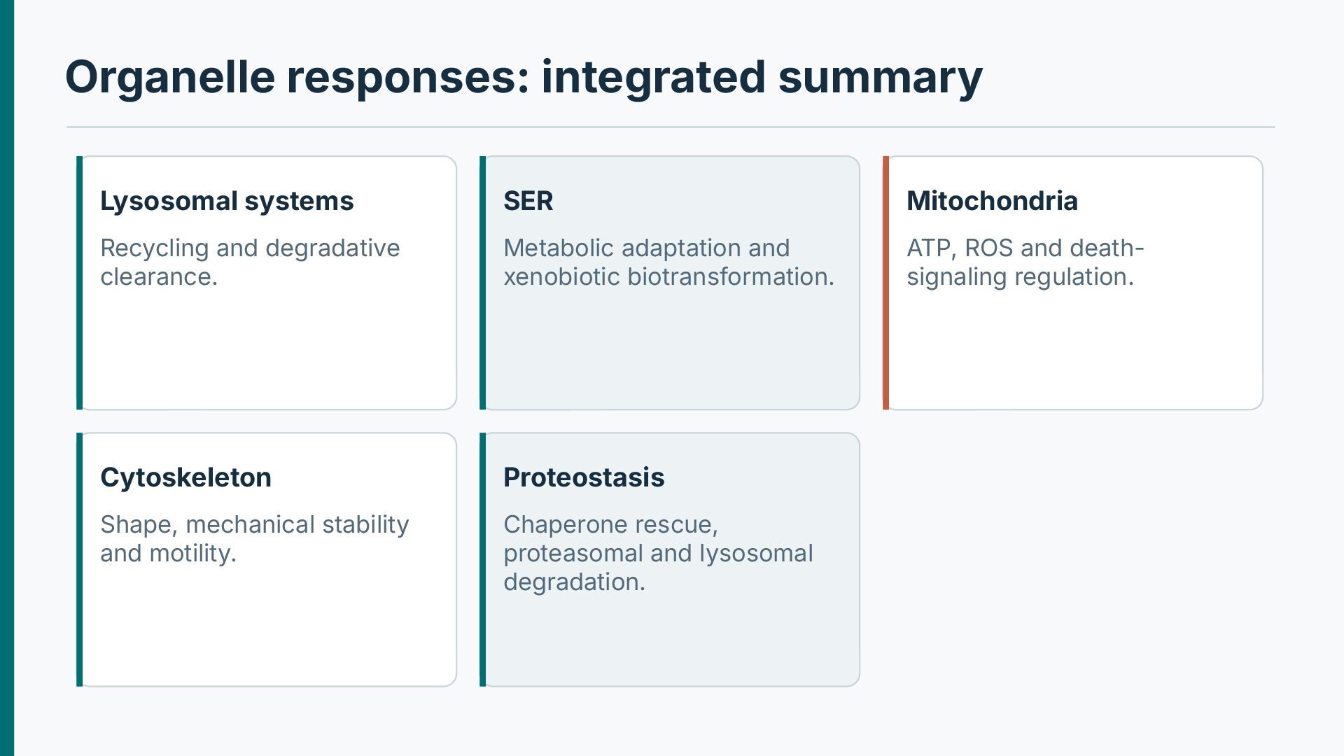
or stress. Lysosomes adjust the disposal of extracellular and intracellular material. Smooth ER may expand during xenobiotic metabolism. Mitochondria change number, size, shape and function. Cytoskeletal defects perturb structure, trafficking and motility.
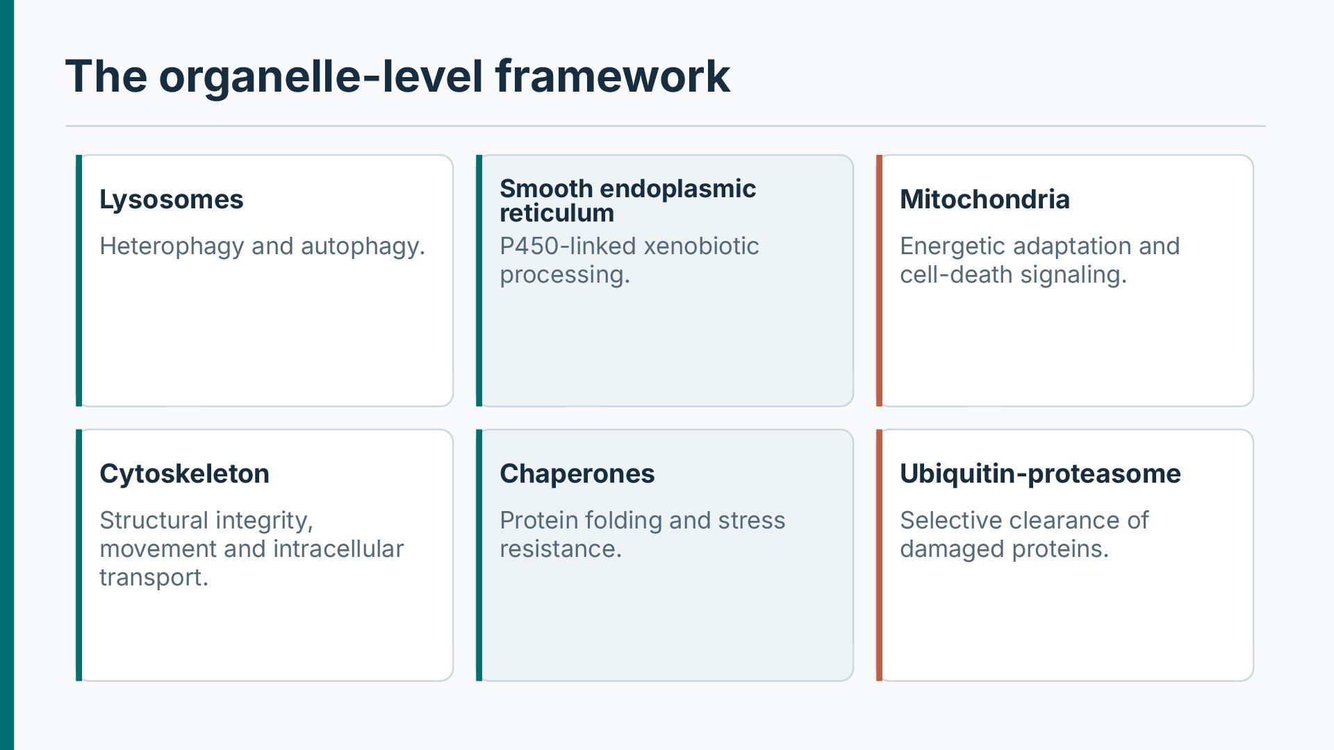
Cytoskeleton Chaperones Ubiquitin-proteasome Structural integrity, movement and intracellular transport. Protein folding and stress resistance. Selective clearance of damaged proteins. Lysosomes Heterophagy and autophagy. Energetic adaptation and cell-death signaling.
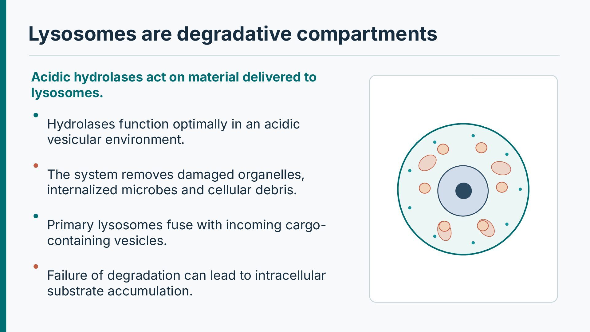
to lysosomes. Hydrolases function optimally in an acidic vesicular environment. The system removes damaged organelles, internalized microbes and cellular debris. Primary lysosomes fuse with incoming cargocontaining vesicles. Failure of degradation can lead to intracellular substrate accumulation.
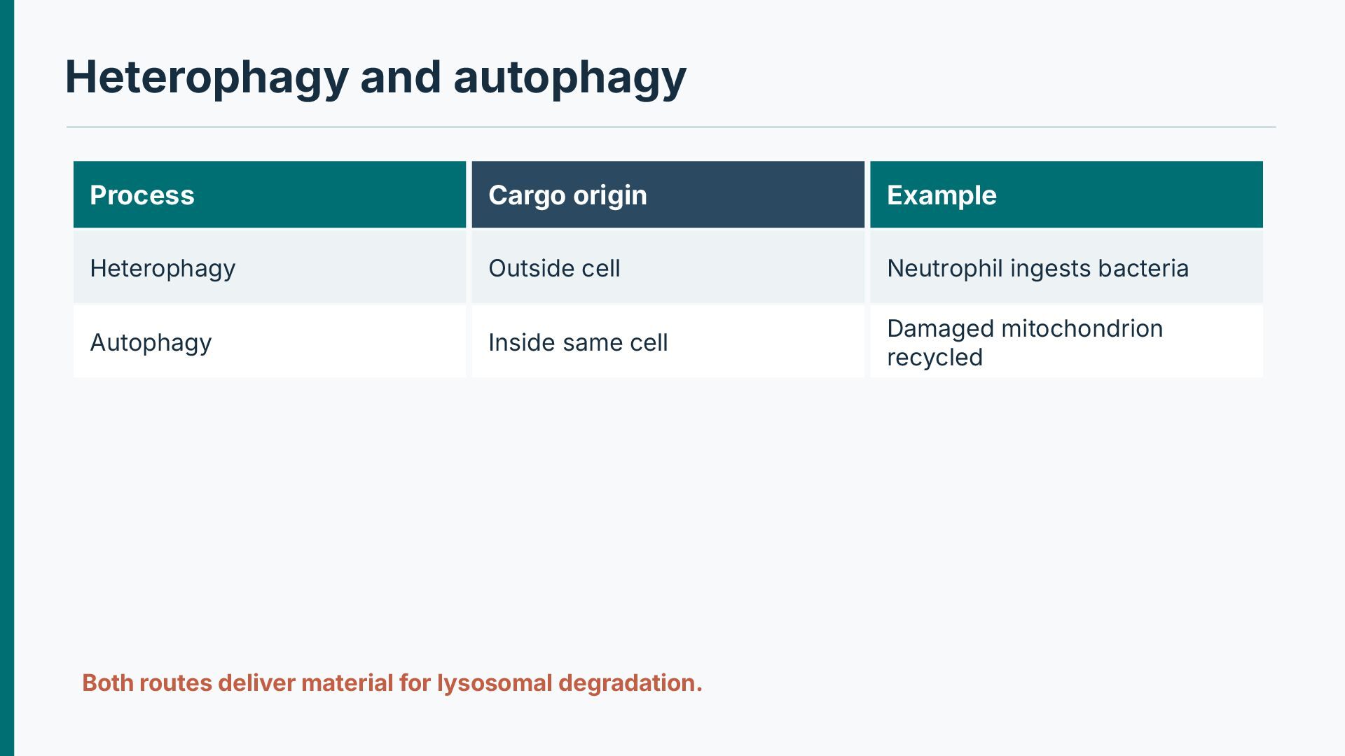
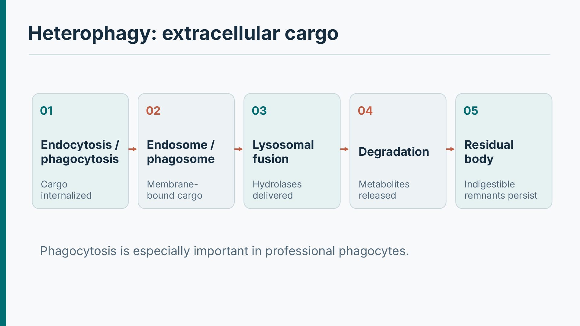
phagocytosis Endosome / phagosome Lysosomal fusion Degradation Residual body Cargo internalized Membranebound cargo Hydrolases delivered Metabolites released Indigestible remnants persist Phagocytosis is especially important in professional phagocytes.
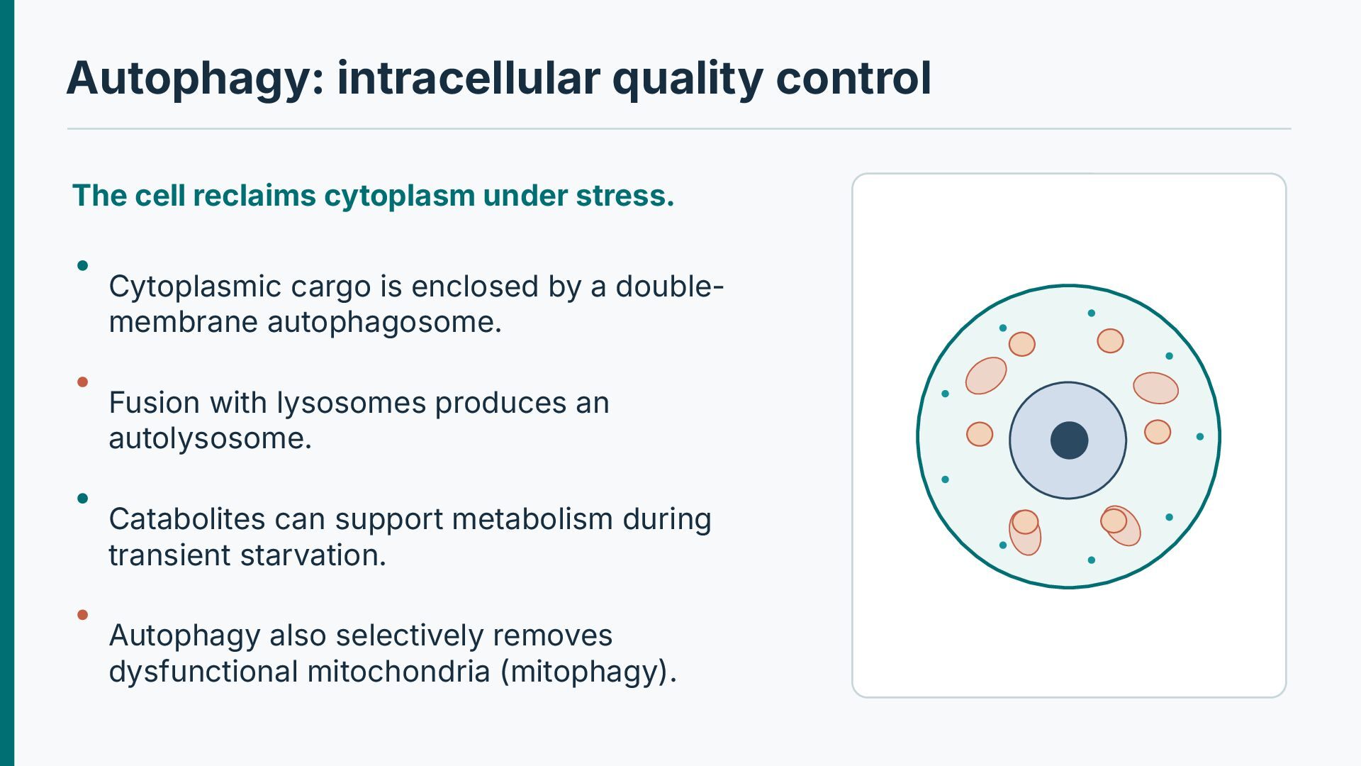
Cytoplasmic cargo is enclosed by a doublemembrane autophagosome. Fusion with lysosomes produces an autolysosome. Catabolites can support metabolism during transient starvation. Autophagy also selectively removes dysfunctional mitochondria (mitophagy).
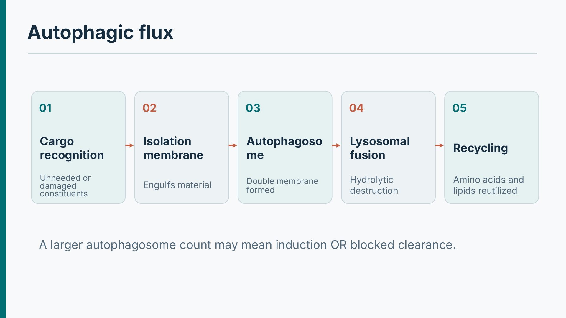
membrane Autophagoso me Lysosomal fusion Recycling Unneeded or damaged constituents Engulfs material Double membrane formed Hydrolytic destruction Amino acids and lipids reutilized A larger autophagosome count may mean induction OR blocked clearance.
the outcome. Starvation activates recycling and conserves cell resources. Defective autophagic flux can aggravate protein and organelle accumulation. Autophagy is implicated in neurodegeneration and some storage disorders. The mere presence of autophagic vacuoles does not prove “autophagic cell death.”
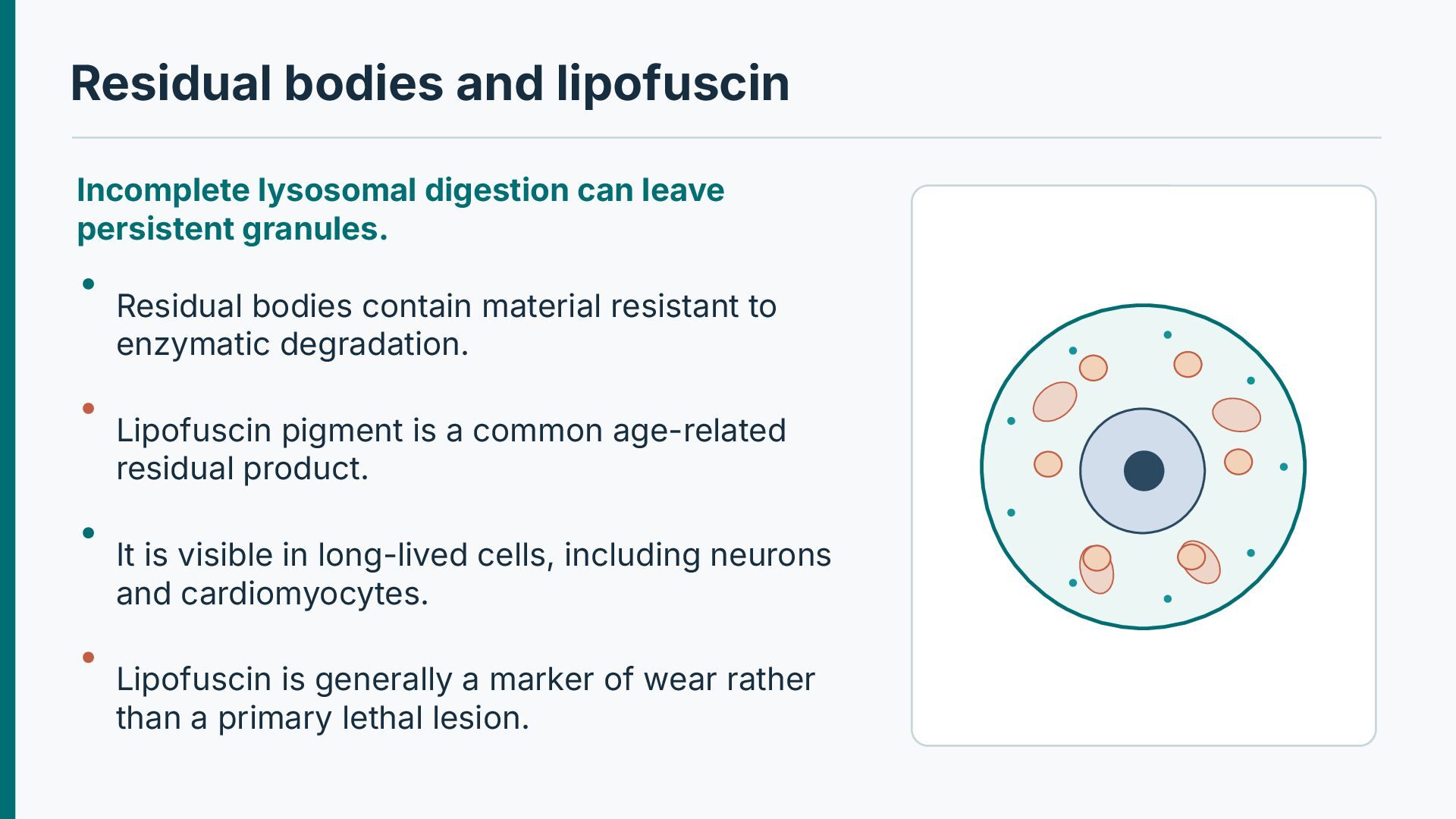
granules. Residual bodies contain material resistant to enzymatic degradation. Lipofuscin pigment is a common age-related residual product. It is visible in long-lived cells, including neurons and cardiomyocytes. Lipofuscin is generally a marker of wear rather than a primary lethal lesion.
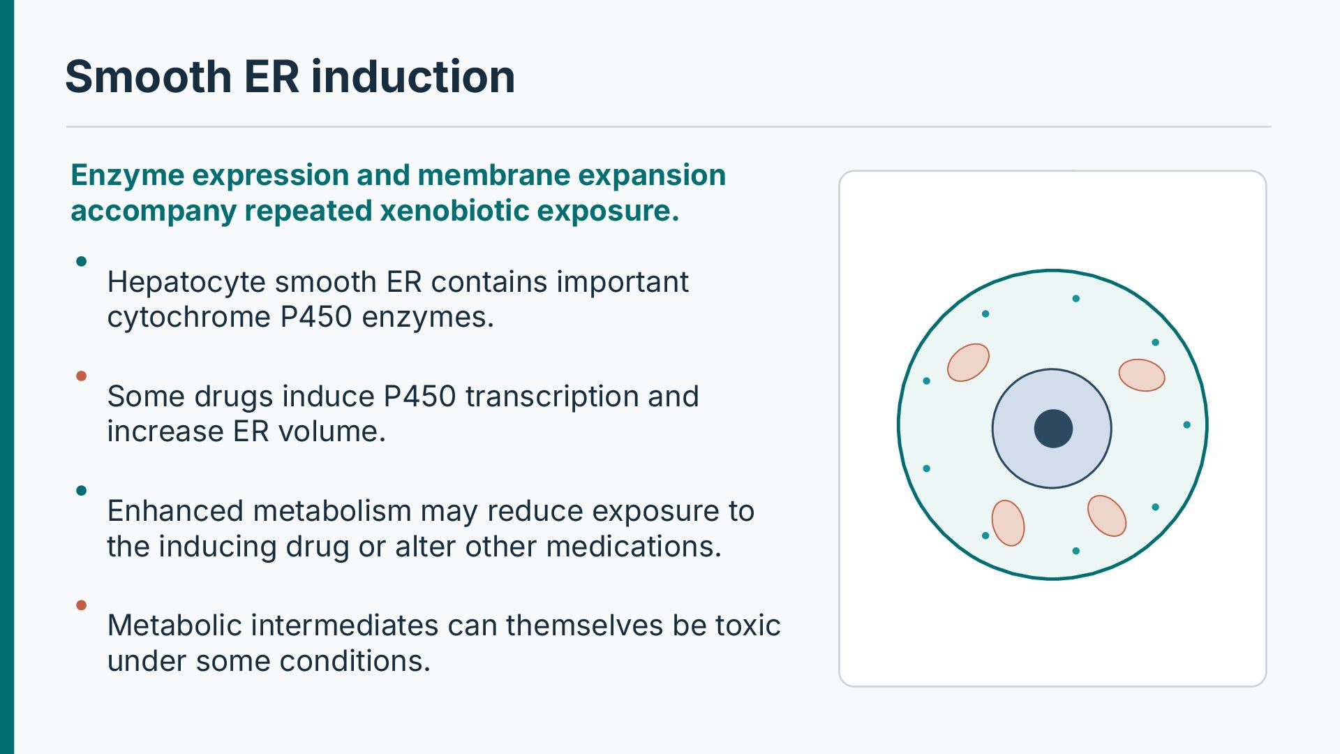
xenobiotic exposure. Hepatocyte smooth ER contains important cytochrome P450 enzymes. Some drugs induce P450 transcription and increase ER volume. Enhanced metabolism may reduce exposure to the inducing drug or alter other medications. Metabolic intermediates can themselves be toxic under some conditions.
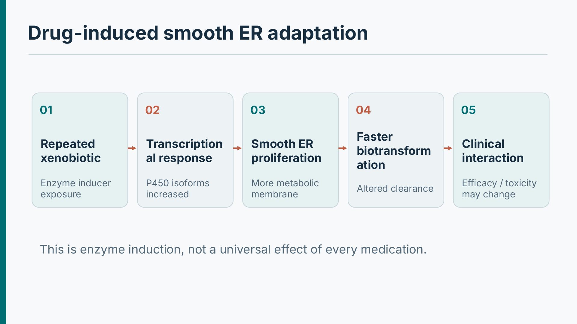
xenobiotic Transcription al response Smooth ER proliferation Faster biotransform ation Clinical interaction Enzyme inducer exposure P450 isoforms increased More metabolic membrane Altered clearance Efficacy / toxicity may change This is enzyme induction, not a universal effect of every medication.
Biogenesis can increase mitochondrial content in energetically active cells. Fission and fusion help maintain quality and distribute mitochondria. Swelling can accompany reversible injury or become severe in lethal injury. Some conditions produce abnormally enlarged mitochondria (“megamitochondria”).
cascade. Loss of membrane potential reduces oxidative ATP generation. Reactive oxygen species can amplify injury. Calcium overload alters permeability and cellular signaling. Outer membrane permeabilization can release cytochrome c and activate apoptosis.
to lysosomes. Loss of mitochondrial membrane potential can stabilize PINK1 on the outer membrane. Parkin-dependent ubiquitination recruits autophagic machinery in relevant contexts. Mitophagy reduces persistence of highly dysfunctional mitochondria. Quality-control failure can worsen energy deficiency and oxidative stress.
genetics. Mitochondrial myopathies may show abnormal mitochondrial proliferation or cristae. Ragged-red fibers can appear with modified Gomori trichrome staining. Oncocytic neoplasms characteristically have abundant mitochondria. Mitochondrial appearance alone rarely specifies a particular genotype.
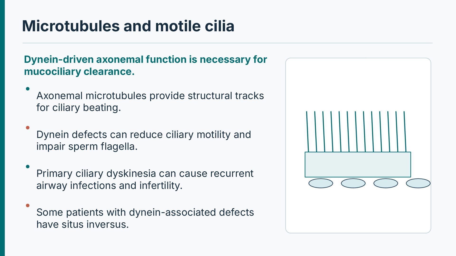
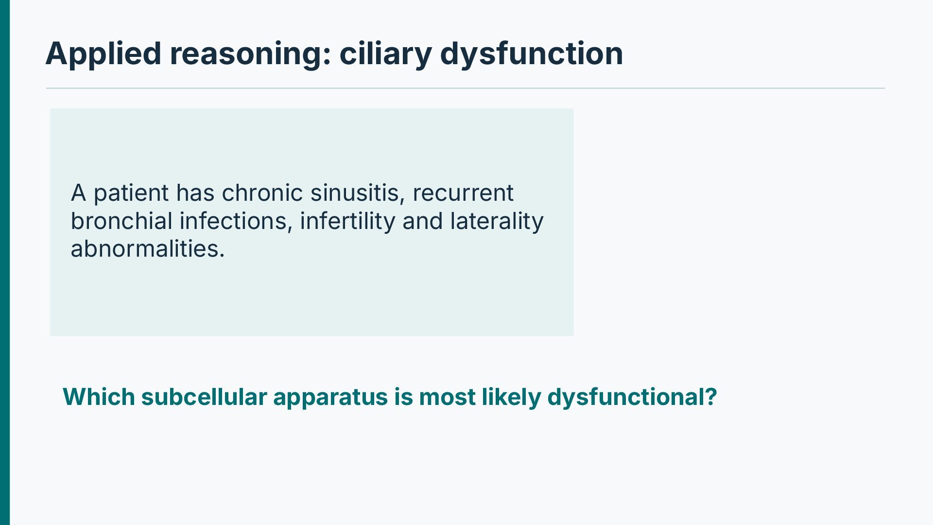
mucociliary clearance. Axonemal microtubules provide structural tracks for ciliary beating. Dynein defects can reduce ciliary motility and impair sperm flagella. Primary ciliary dyskinesia can cause recurrent airway infections and infertility. Some patients with dynein-associated defects have situs inversus.
occur in epithelial cells and many carcinomas. Vimentin labels many mesenchymal cells; desmin characterizes muscle differentiation. Neurofilaments are abundant in neurons; GFAP is a glial intermediate filament. Protein aggregates may reflect abnormal assembly or degradation.
the cell-stress response. Molecular chaperones facilitate correct folding and assembly of proteins. Stress increases production of selected heatshock proteins. Chaperones limit aggregation and can refold damaged proteins. Persistently misfolded proteins are targeted for degradation or stress signaling.
of protein homeostasis. IRE1, PERK and ATF6 are major ER stress sensors. They alter translation, folding capacity and degradation pathways. Acute responses can be protective and reversible. Persistent stress may activate CHOP-linked and other prodeath signaling.
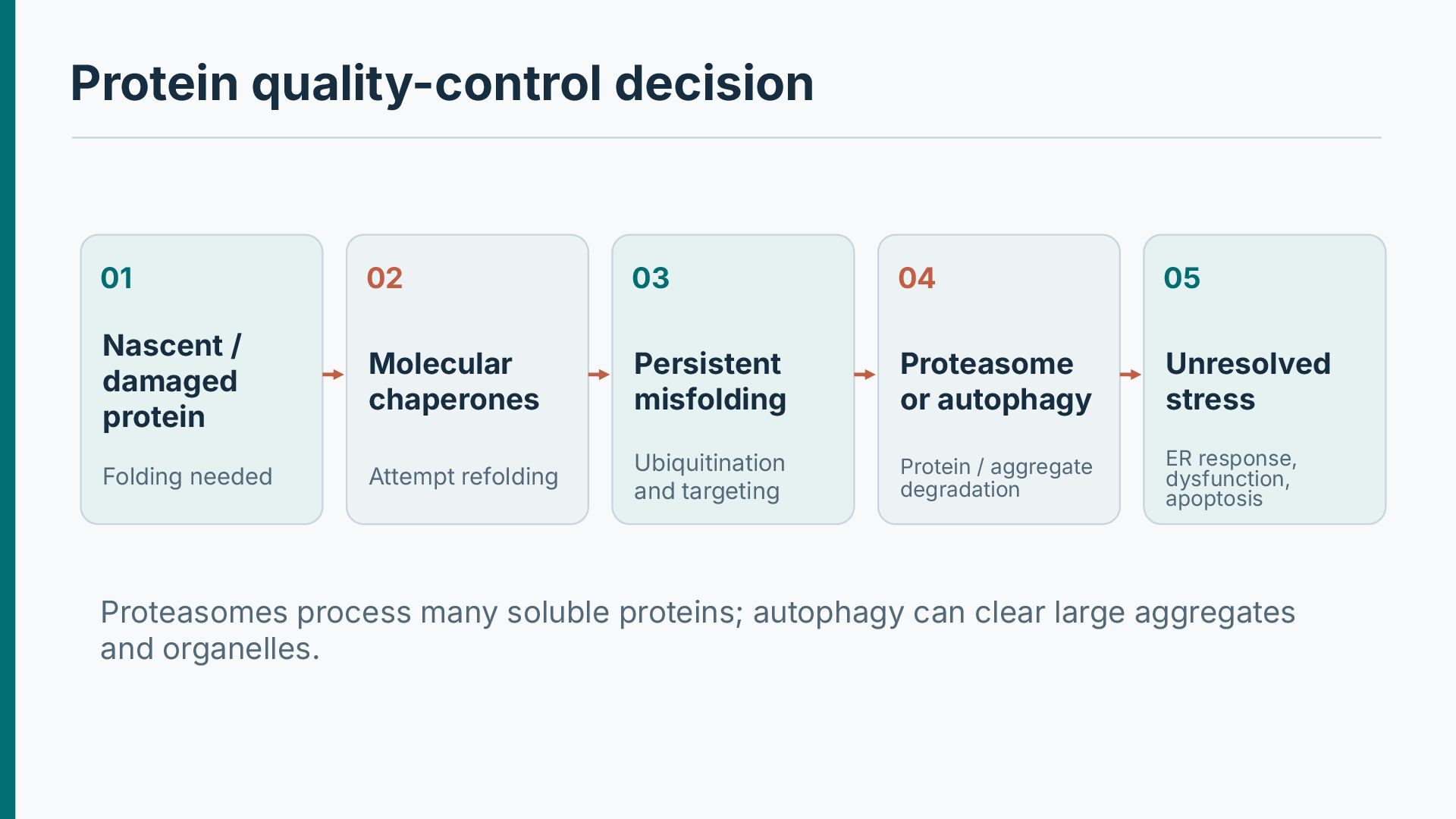
damaged protein Molecular chaperones Persistent misfolding Proteasome or autophagy Unresolved stress Folding needed Attempt refolding Ubiquitination and targeting Protein / aggregate degradation ER response, dysfunction, apoptosis Proteasomes process many soluble proteins; autophagy can clear large aggregates and organelles.
E2 and E3 activities build ubiquitin signals on protein substrates. The 26S proteasome degrades many ubiquitinated proteins. It removes damaged proteins and controls signaling and cell-cycle regulators. Overwhelmed proteostasis can contribute to degenerative pathology.
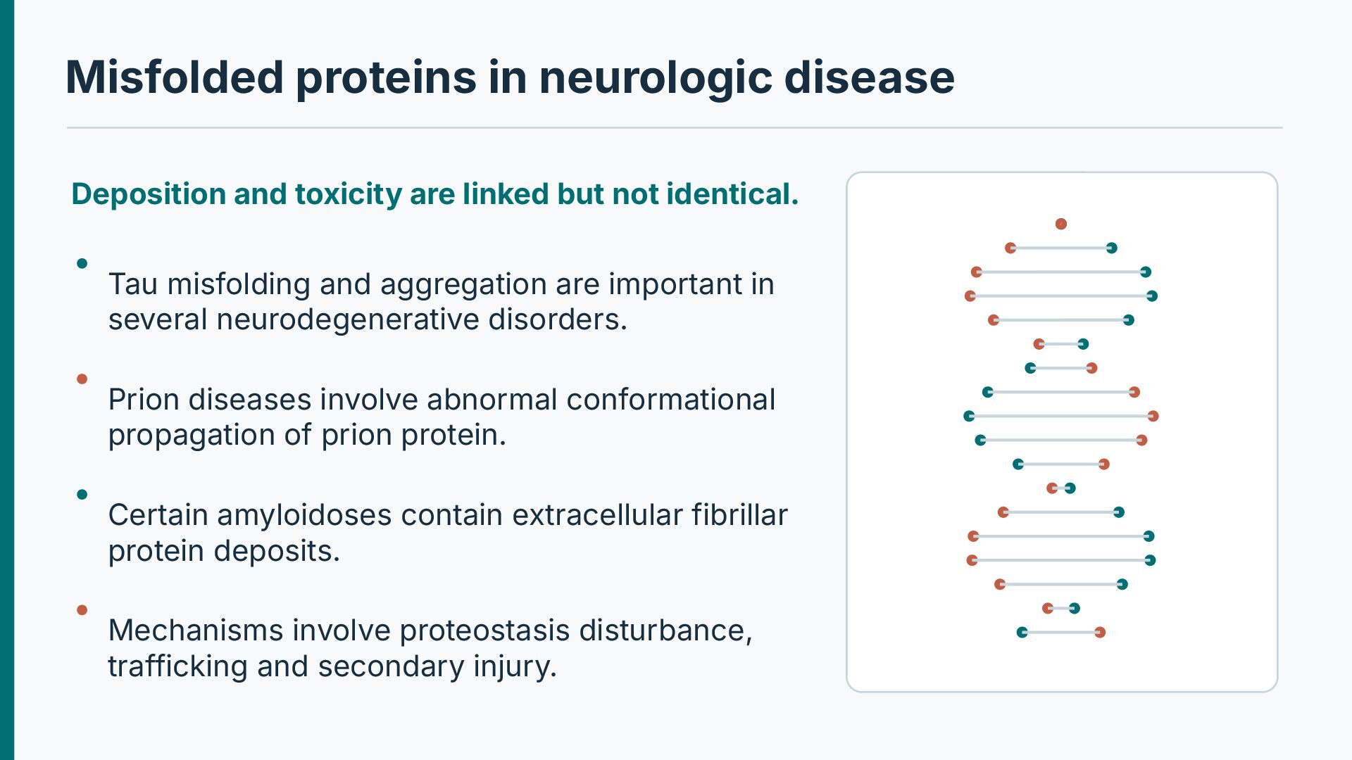
but not identical. Tau misfolding and aggregation are important in several neurodegenerative disorders. Prion diseases involve abnormal conformational propagation of prion protein. Certain amyloidoses contain extracellular fibrillar protein deposits. Mechanisms involve proteostasis disturbance, trafficking and secondary injury.
· Chapter: Cell Injury, Cell Death, and Adaptations For further study: correlate the molecular mechanisms with gross and microscopic pathology. Illustrative pathways are original schematics; selected histology images are retained from the supplied teaching material.


 abusuraihsakhri
 trycycle
 ianozsvald
 ykiyota
 rajeshrinet
 signer
 neutron63zf
 jarvist
 institutdelelevage
 hanbao
 ktatsuya
 akasan
 katarinadahlin
 scottboms
 nikkihalliwell
 axbom
 matthewcrist
 lynnandtonic
 inesmontani
 palkan
 techseoconnect
 speakerdeck
 jonoalderson
 marktimemedia