from the vascular compartment. It may occur externally, into tissue or within a body cavity. Small tissue hemorrhages have descriptive size-based names. A hematoma is a localized collection of extravasated blood.
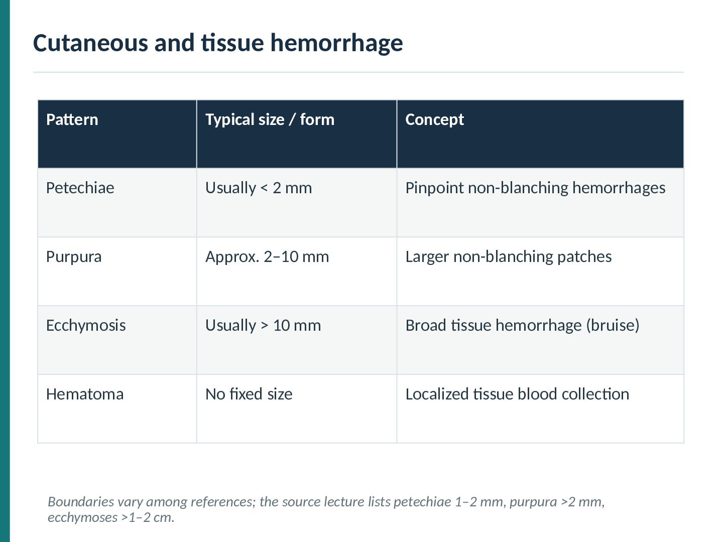
Petechiae Usually < 2 mm Pinpoint non-blanching hemorrhages Purpura Approx. 2–10 mm Larger non-blanching patches Ecchymosis Usually > 10 mm Broad tissue hemorrhage (bruise) Hematoma No fixed size Localized tissue blood collection Boundaries vary among references; the source lecture lists petechiae 1–2 mm, purpura >2 mm, ecchymoses >1–2 cm.
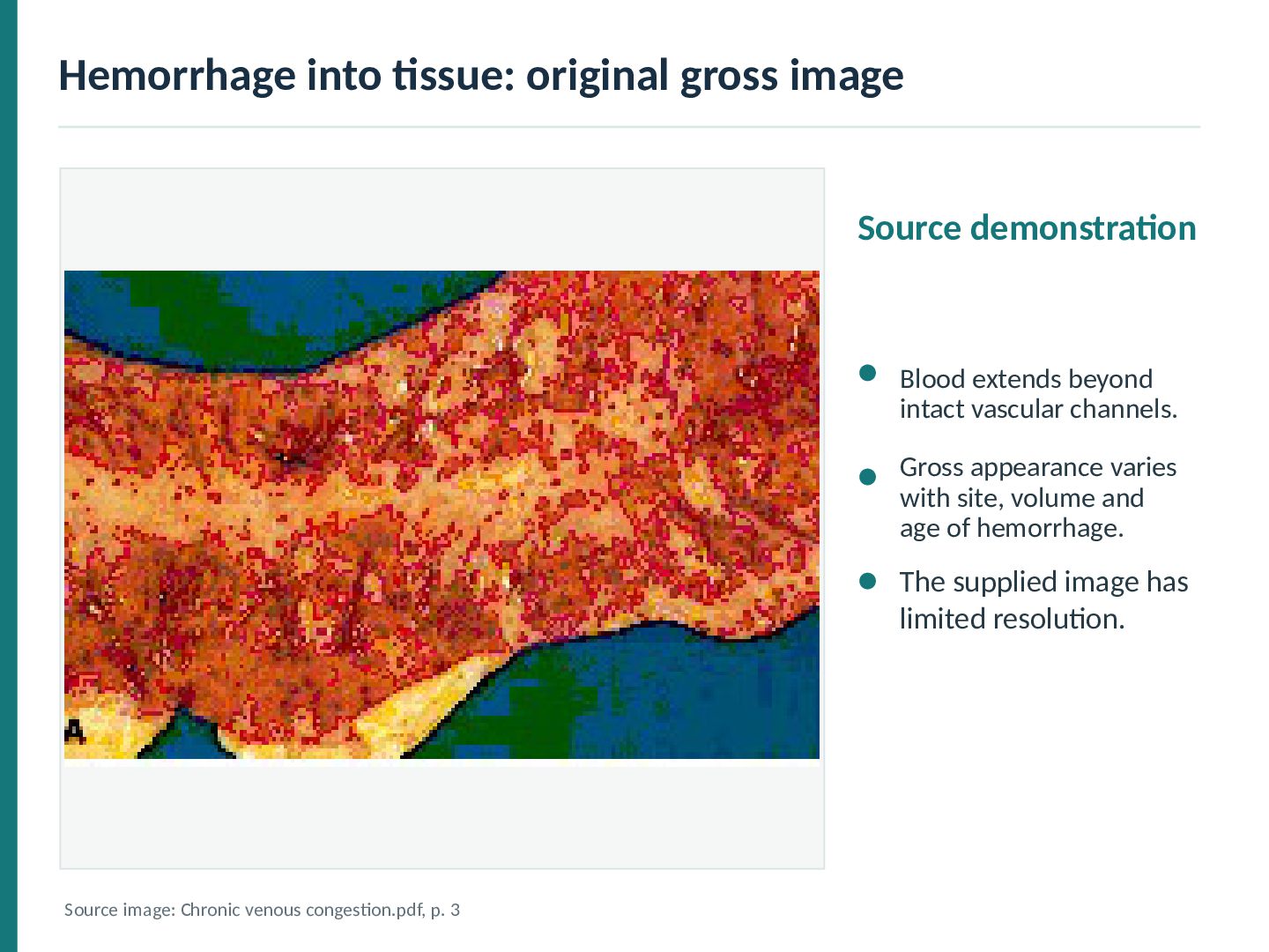
beyond intact vascular channels. Gross appearance varies with site, volume and age of hemorrhage. The supplied image has limited resolution. Source image: Chronic venous congestion.pdf, p. 3
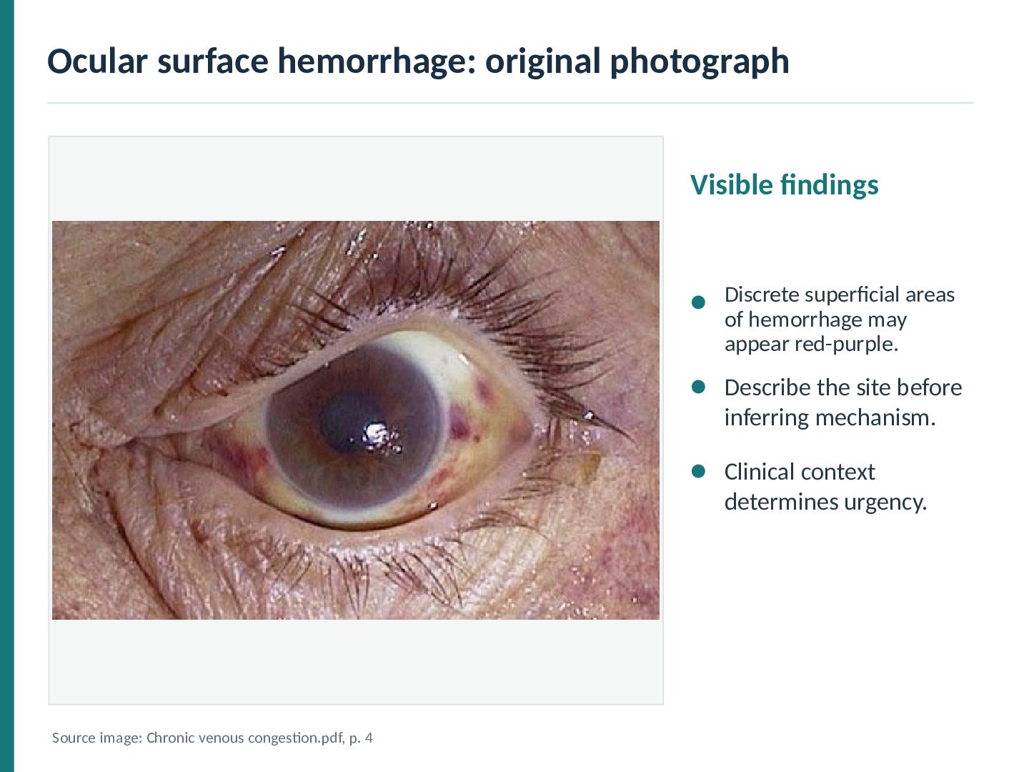
of hemorrhage may appear red-purple. Describe the site before inferring mechanism. Clinical context determines urgency. Source image: Chronic venous congestion.pdf, p. 4
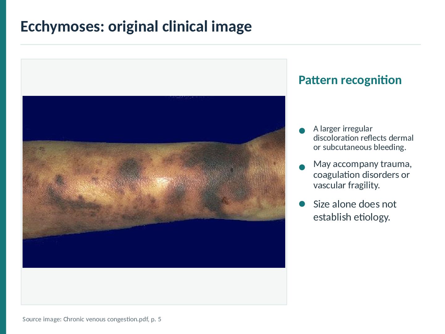
reflects dermal or subcutaneous bleeding. May accompany trauma, coagulation disorders or vascular fragility. Size alone does not establish etiology. Source image: Chronic venous congestion.pdf, p. 5
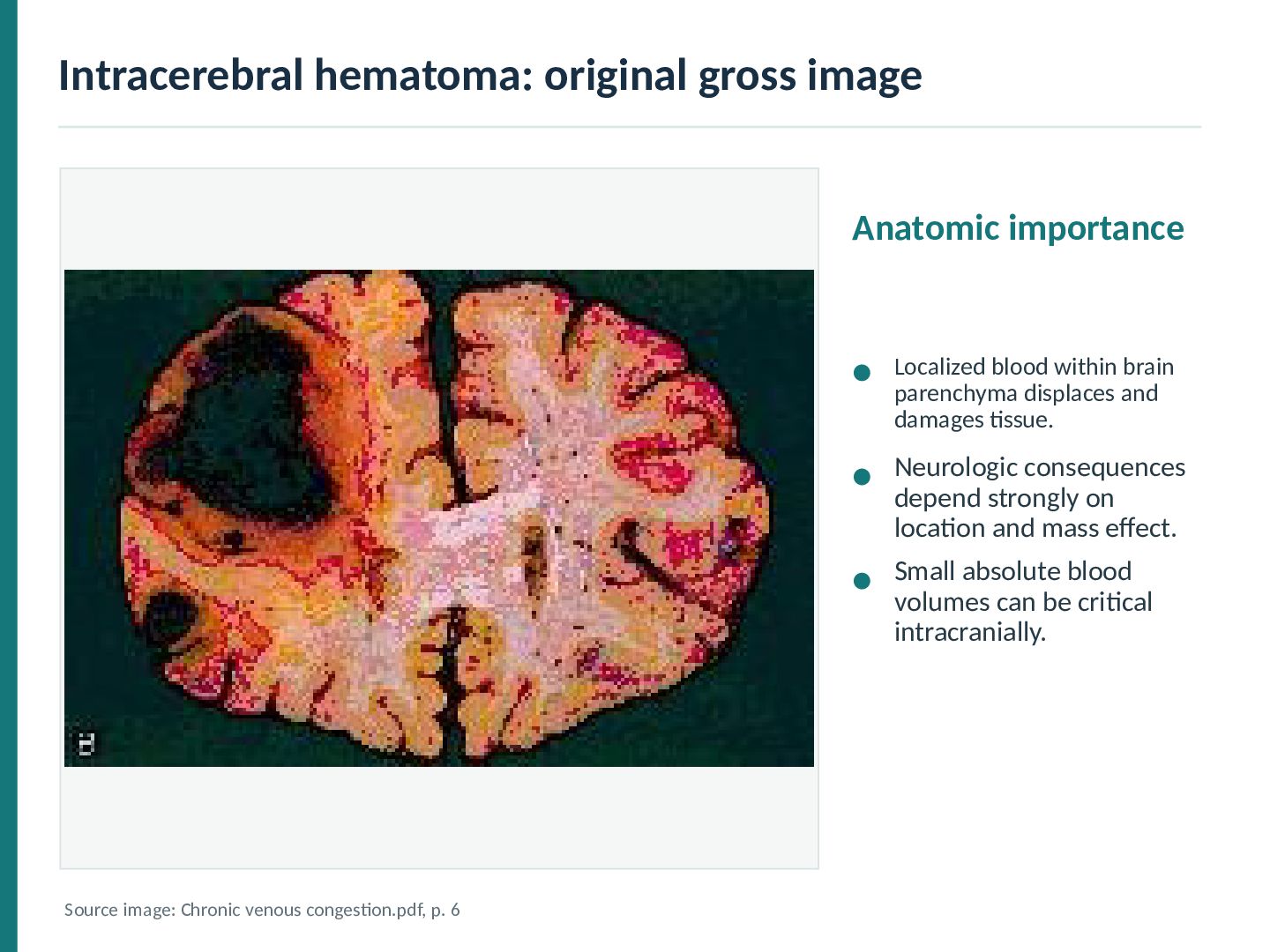
brain parenchyma displaces and damages tissue. Neurologic consequences depend strongly on location and mass effect. Small absolute blood volumes can be critical intracranially. Source image: Chronic venous congestion.pdf, p. 6
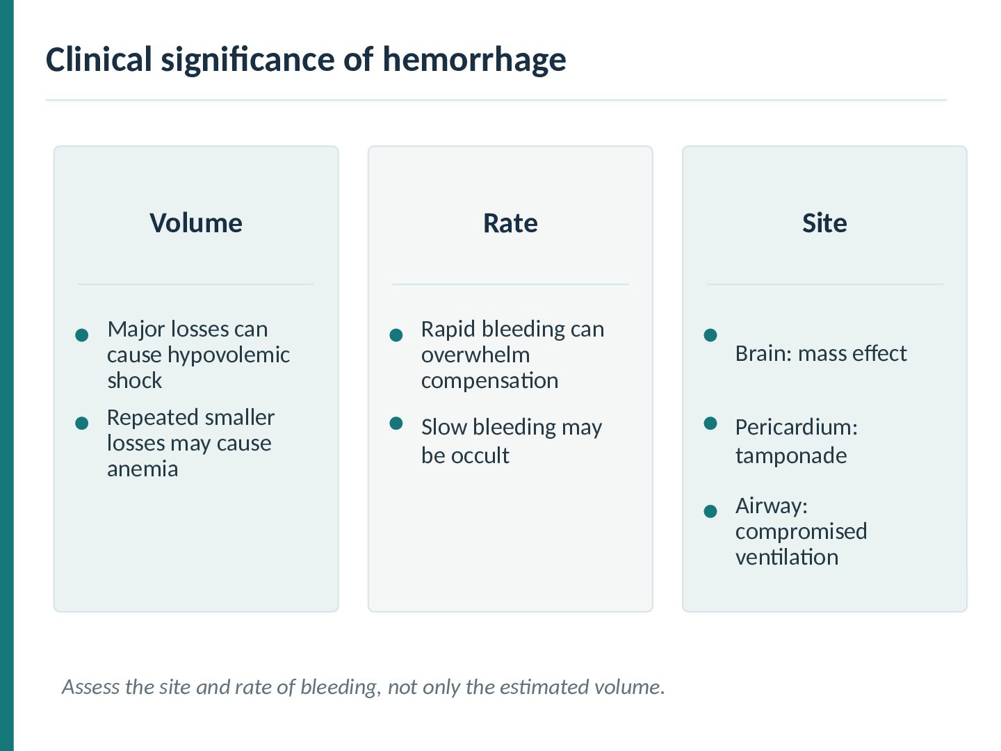
cause hypovolemic shock Rapid bleeding can overwhelm compensation Brain: mass effect Repeated smaller losses may cause anemia Slow bleeding may be occult Pericardium: tamponade Airway: compromised ventilation Assess the site and rate of bleeding, not only the estimated volume.
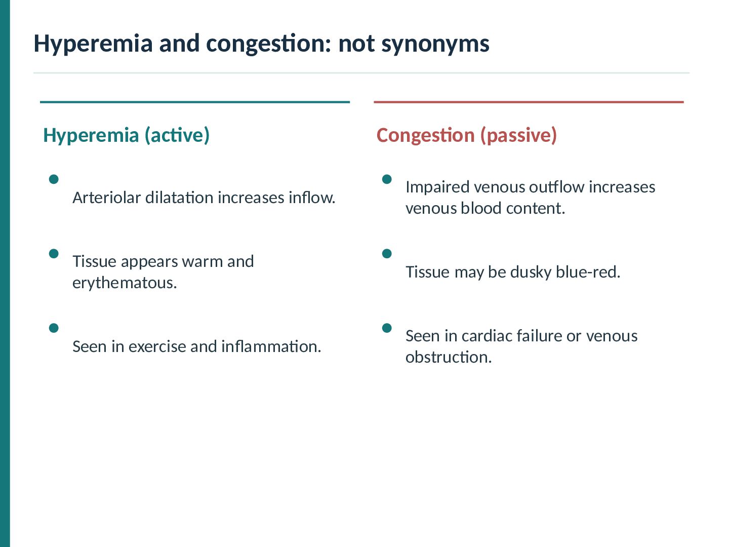
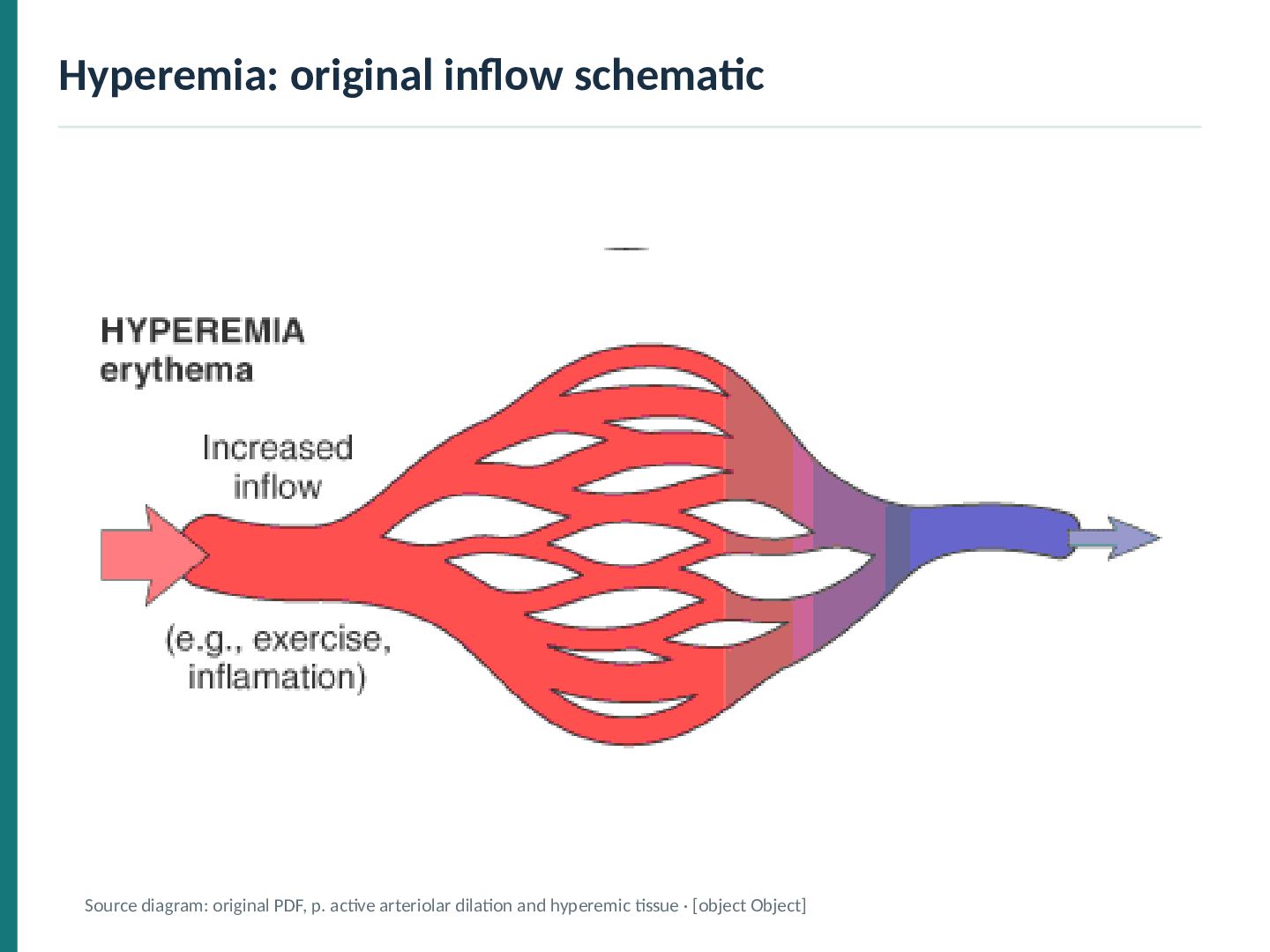
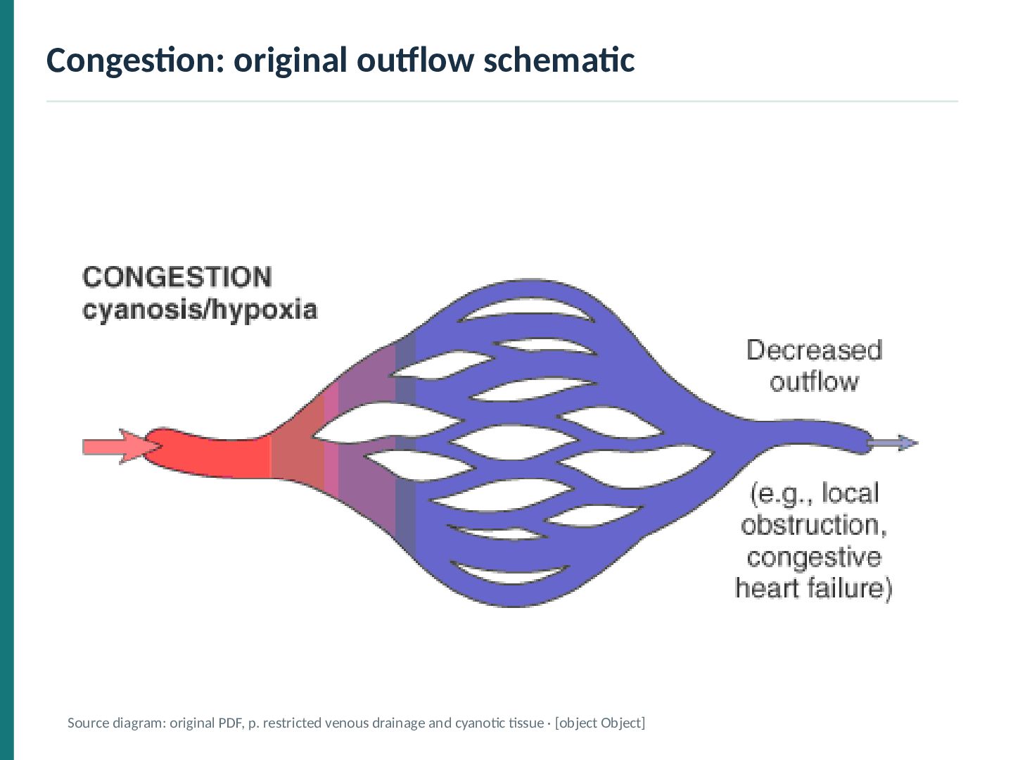
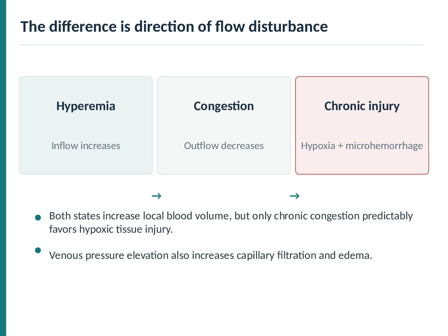
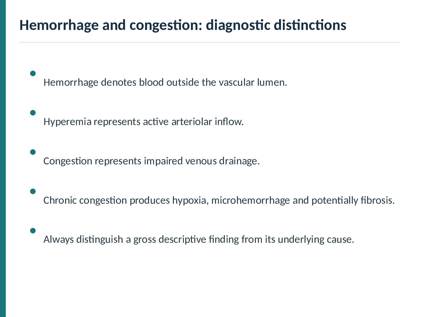
dilatation increases inflow. Impaired venous outflow increases venous blood content. Tissue appears warm and erythematous. Tissue may be dusky blue-red. Seen in exercise and inflammation. Seen in cardiac failure or venous obstruction.
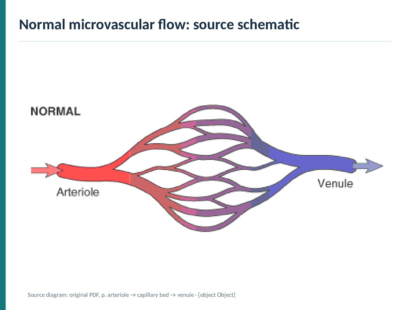
injury Inflow increases Outflow decreases Hypoxia + microhemorrhage → → Both states increase local blood volume, but only chronic congestion predictably favors hypoxic tissue injury. Venous pressure elevation also increases capillary filtration and edema.
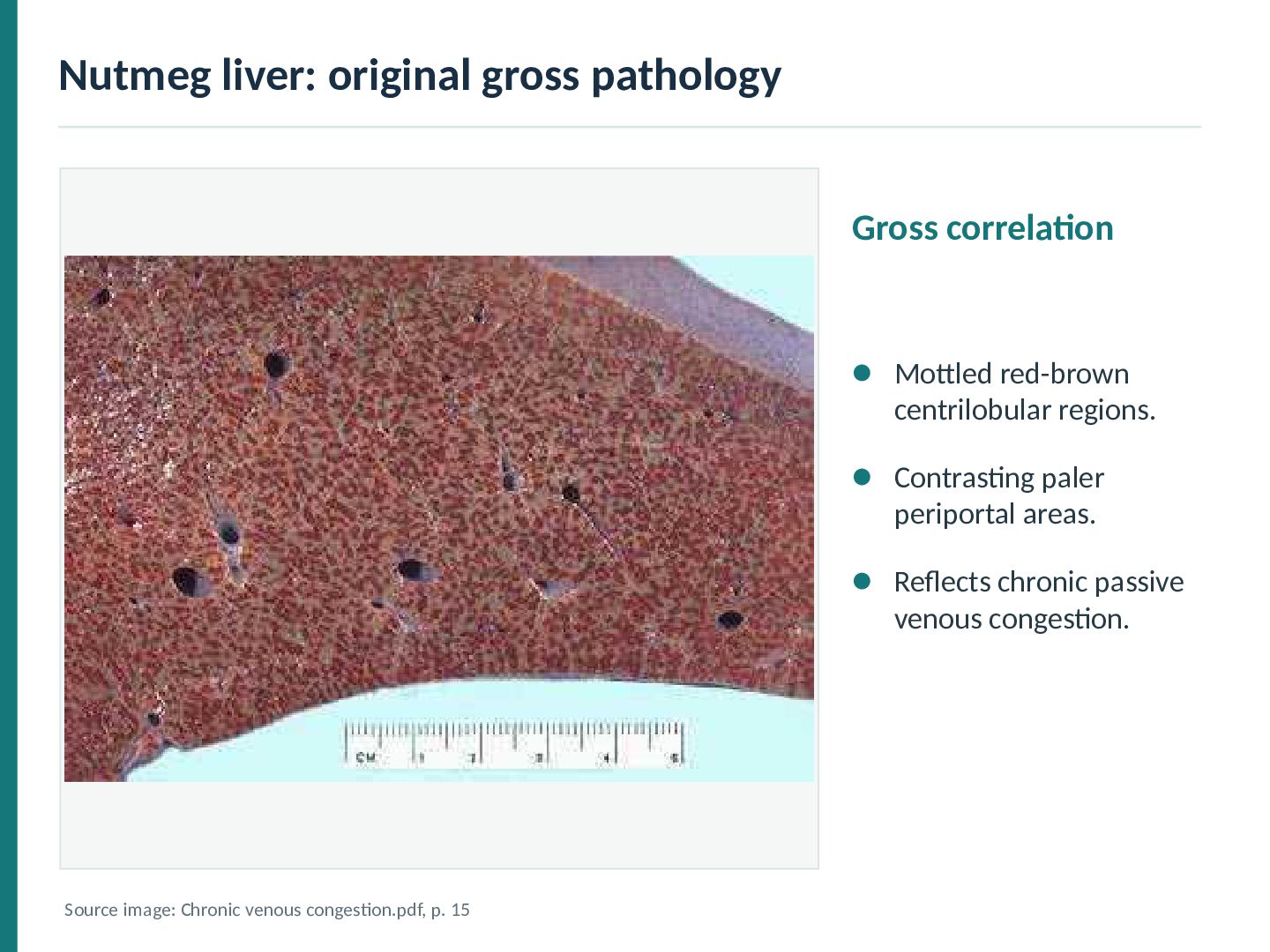
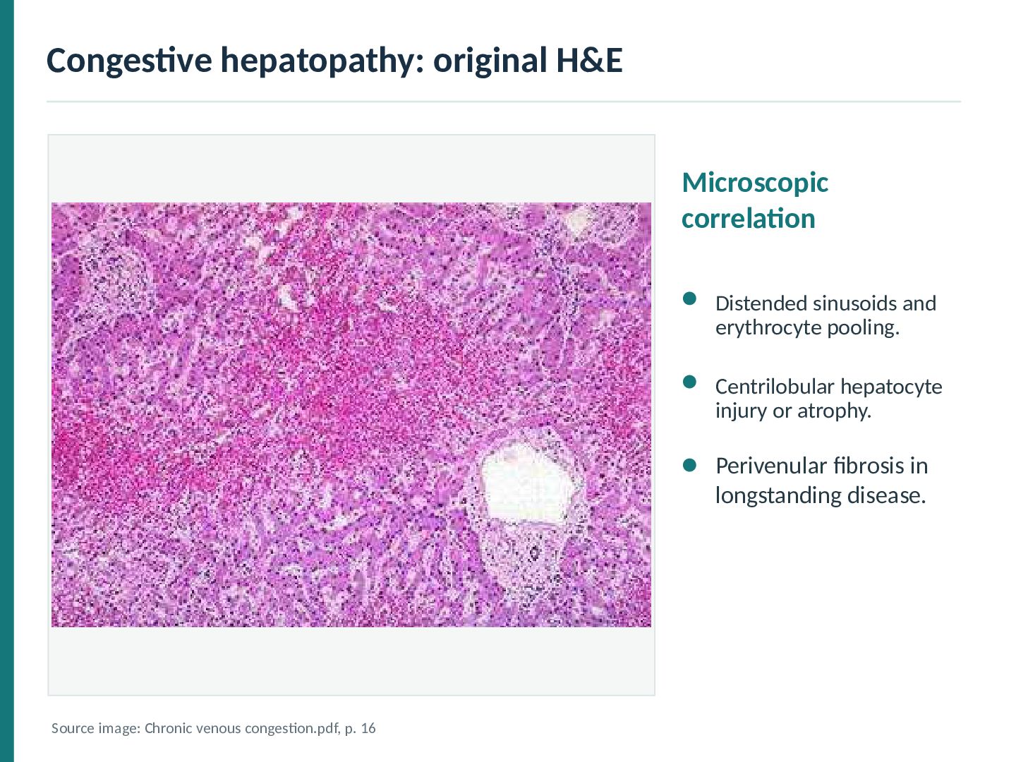
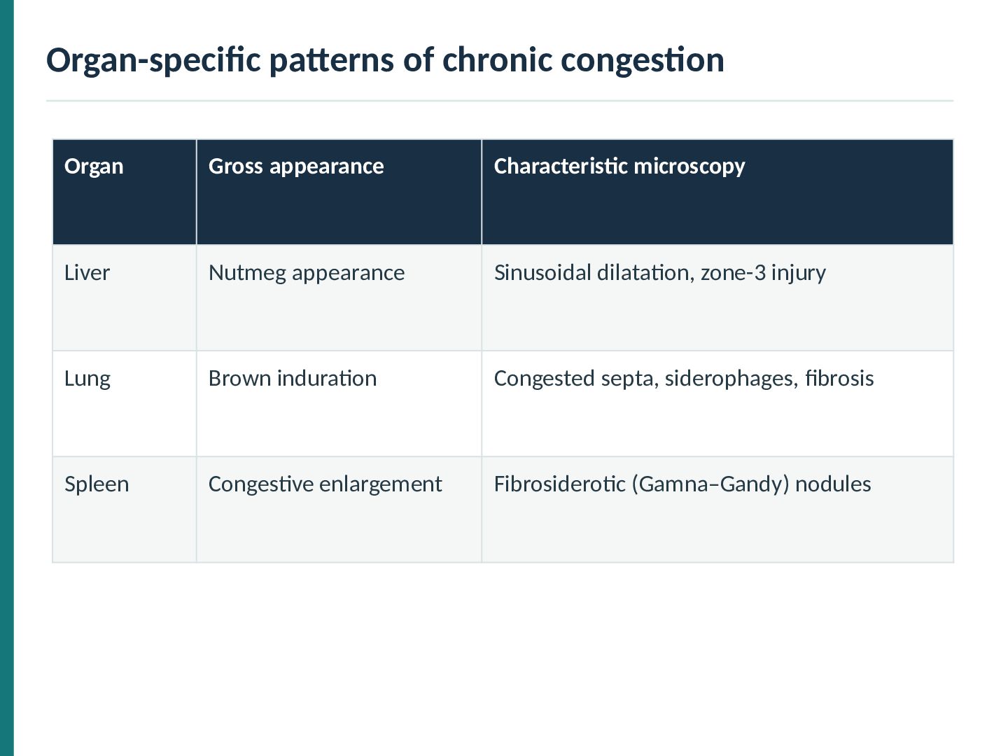
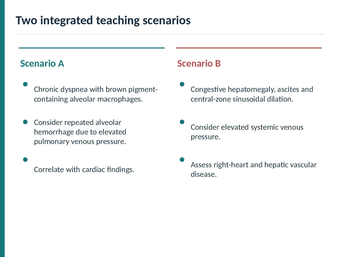
most prominent around central veins (zone 3). Dark-red congested centrilobular regions contrast with paler peripheral parenchyma. Sinusoidal dilation, hepatocyte atrophy and occasional hemorrhage are characteristic. Long-standing severe disease may produce centrilobular fibrosis.
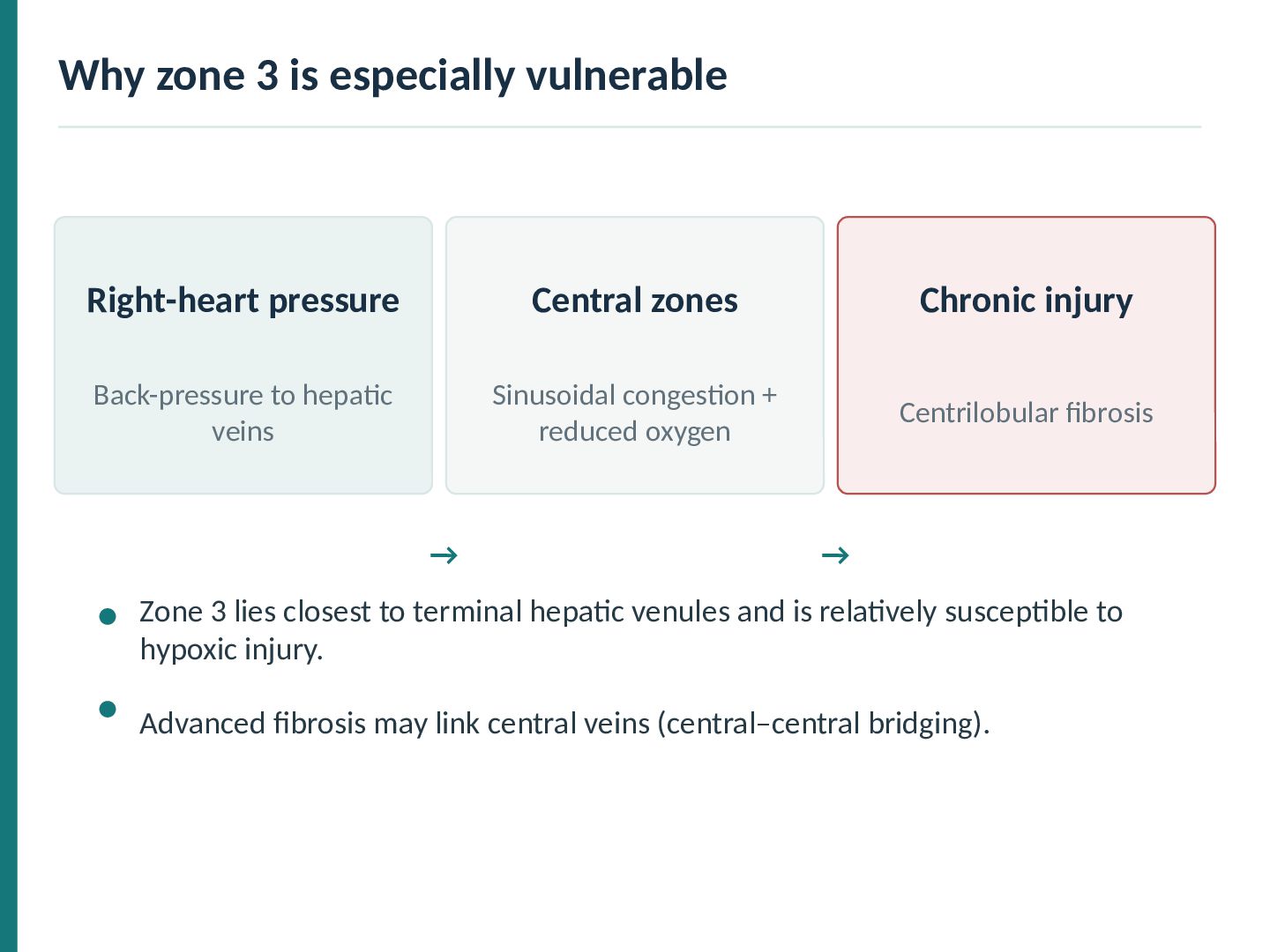
Chronic injury Back-pressure to hepatic veins Sinusoidal congestion + reduced oxygen Centrilobular fibrosis → → Zone 3 lies closest to terminal hepatic venules and is relatively susceptible to hypoxic injury. Advanced fibrosis may link central veins (central–central bridging).
a traditional term for advanced fibrosis with chronic rightheart disease. Centrilobular and central–central bridging patterns may dominate. True architectural cirrhosis is not present in every case of congestive hepatopathy. Morphology should be interpreted alongside cardiac and vascular findings.
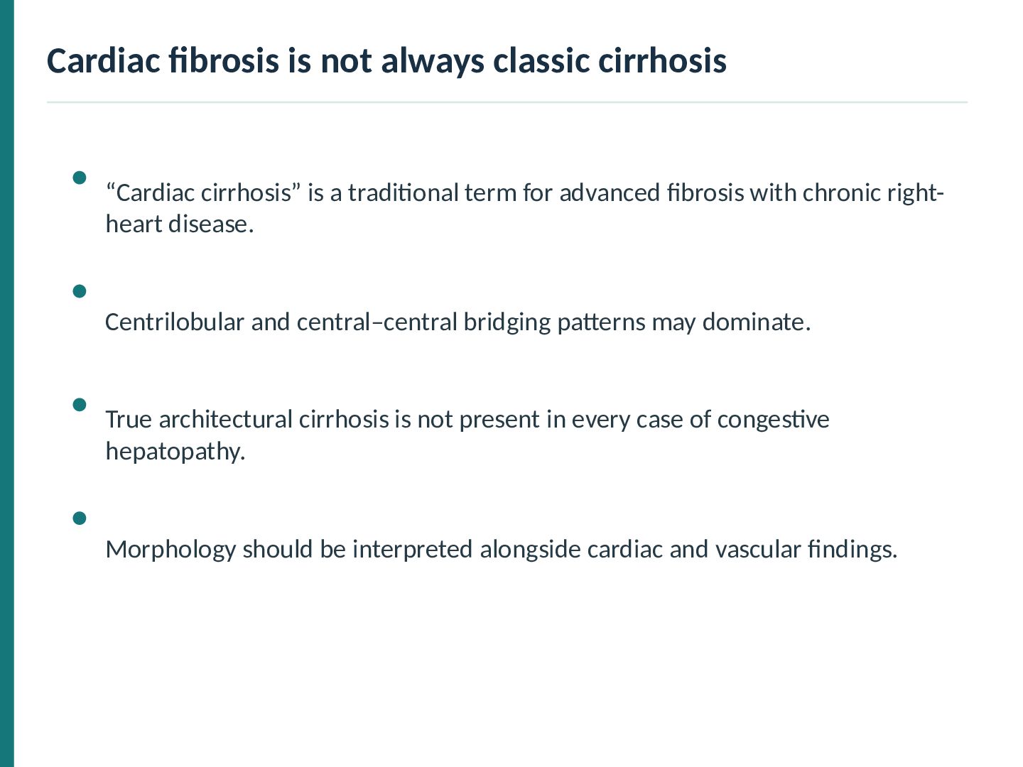
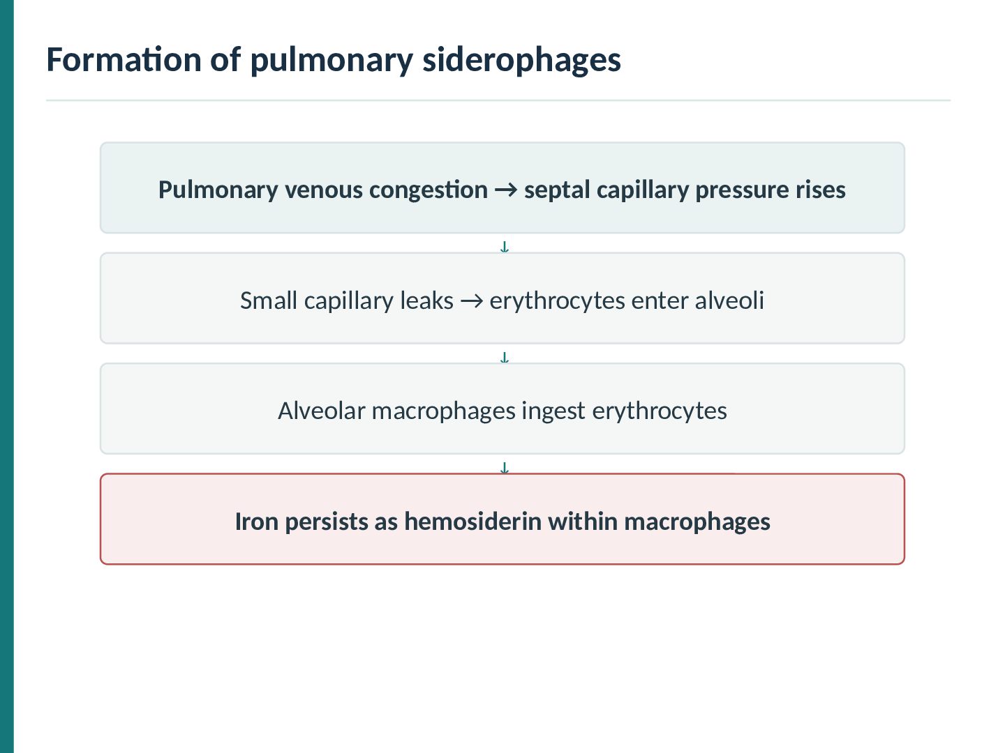
reddish-brown parenchyma in prolonged pulmonary venous congestion. Chronic pressure elevation may produce hemosiderin deposition and fibrosis. Classic term: brown induration. Source image: Chronic venous congestion.pdf, p. 17
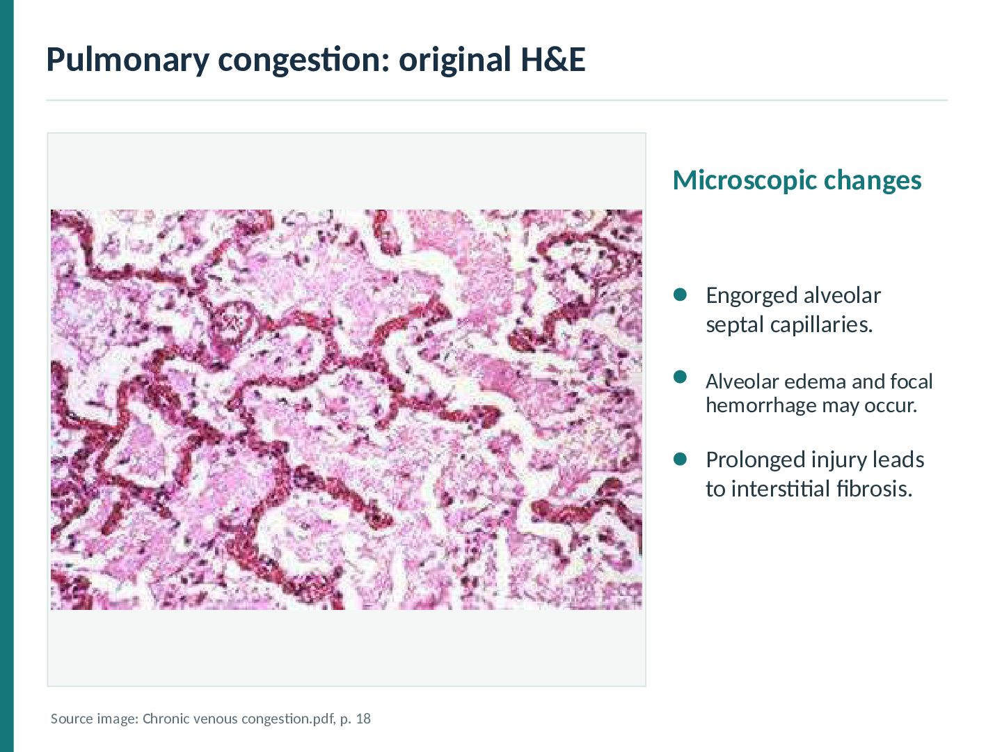
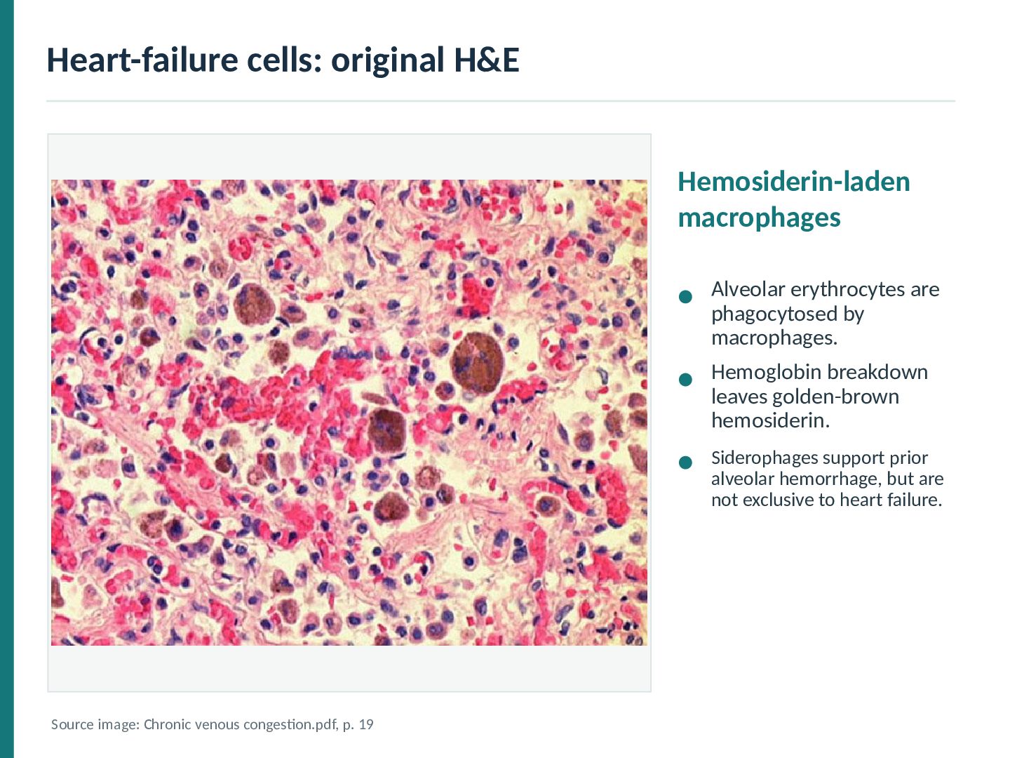
by macrophages. Hemoglobin breakdown leaves golden-brown hemosiderin. Siderophages support prior alveolar hemorrhage, but are not exclusive to heart failure. Source image: Chronic venous congestion.pdf, p. 19
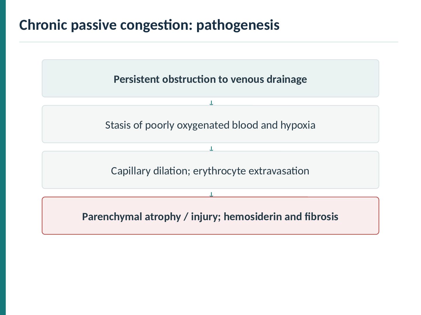
pressure rises ↓ Small capillary leaks → erythrocytes enter alveoli ↓ Alveolar macrophages ingest erythrocytes ↓ Iron persists as hemosiderin within macrophages
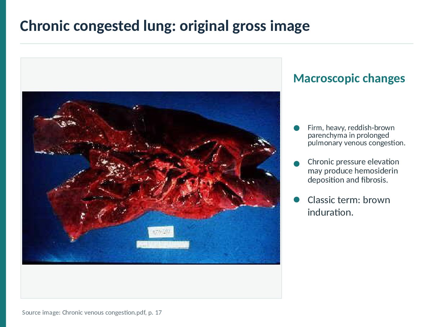
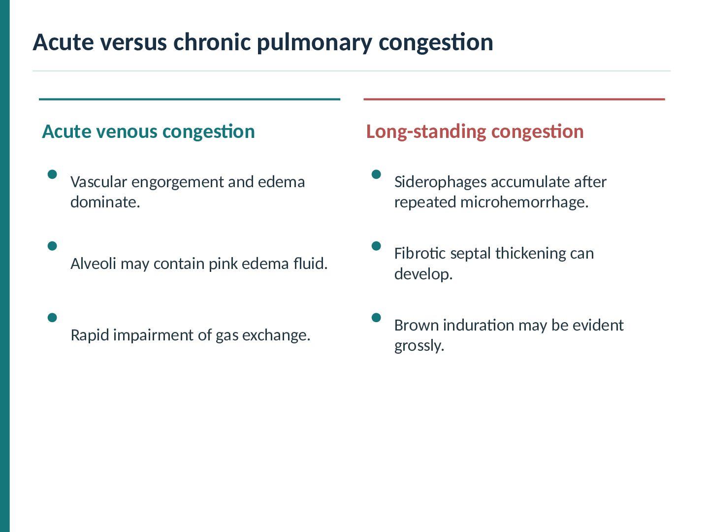
Vascular engorgement and edema dominate. Siderophages accumulate after repeated microhemorrhage. Alveoli may contain pink edema fluid. Fibrotic septal thickening can develop. Rapid impairment of gas exchange. Brown induration may be evident grossly.
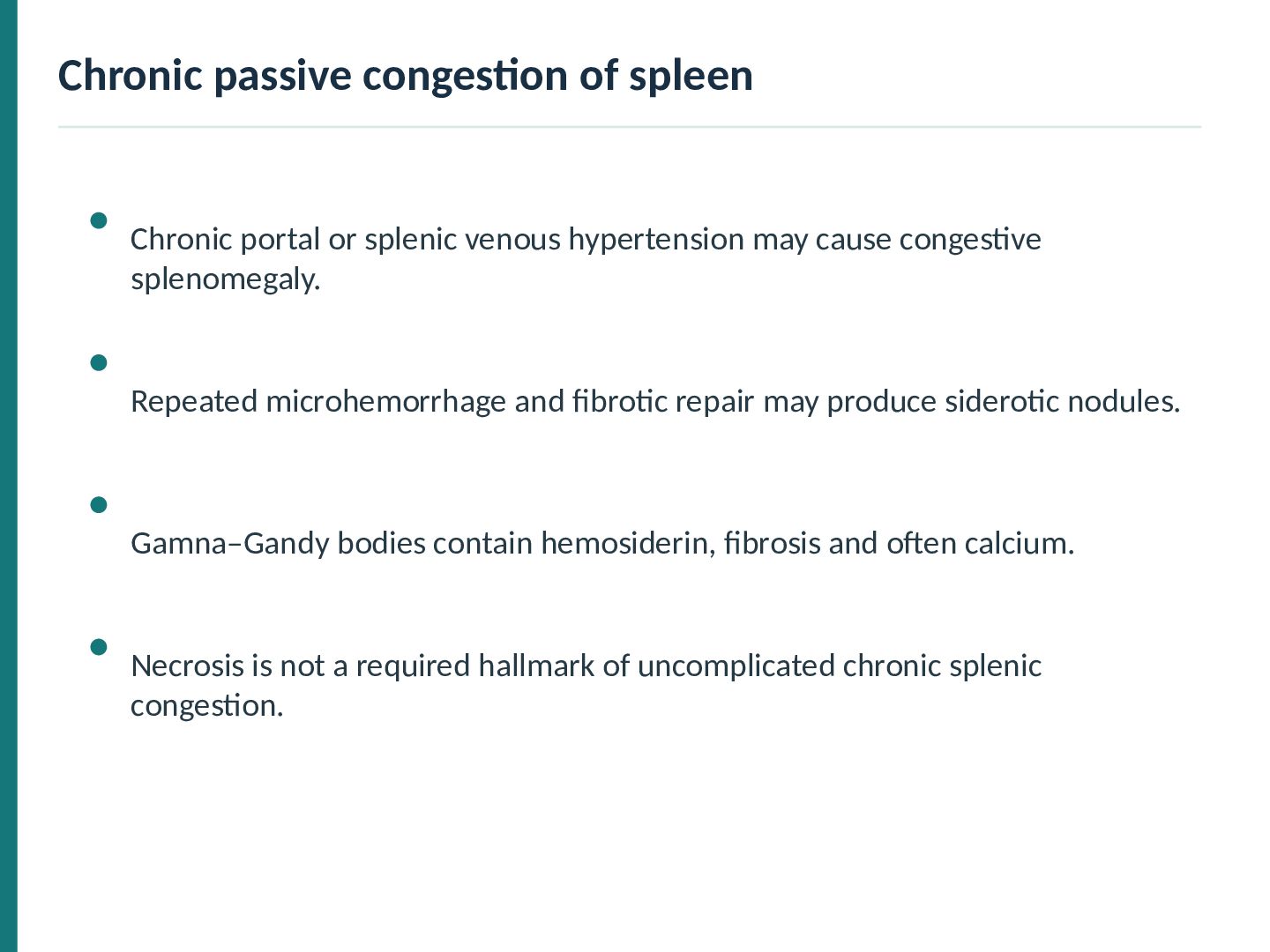
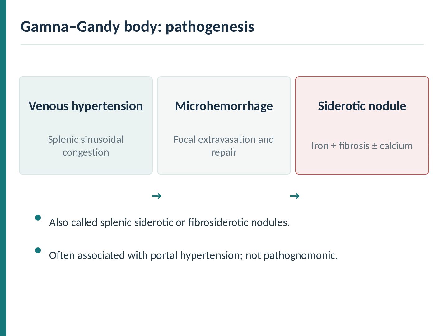
hypertension may cause congestive splenomegaly. Repeated microhemorrhage and fibrotic repair may produce siderotic nodules. Gamna–Gandy bodies contain hemosiderin, fibrosis and often calcium. Necrosis is not a required hallmark of uncomplicated chronic splenic congestion.
congestion Focal extravasation and repair Iron + fibrosis ± calcium → → Also called splenic siderotic or fibrosiderotic nodules. Often associated with portal hypertension; not pathognomonic.
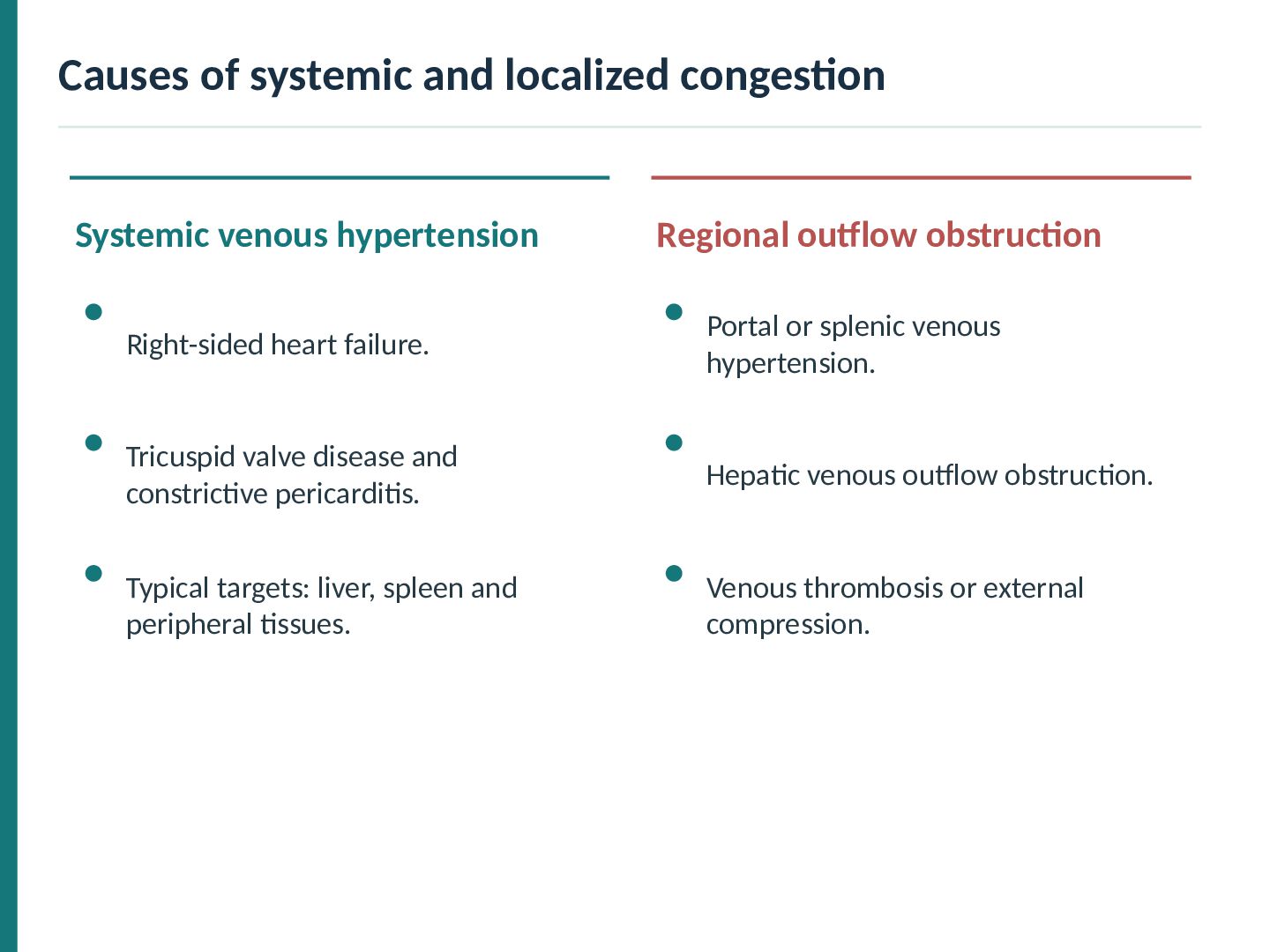
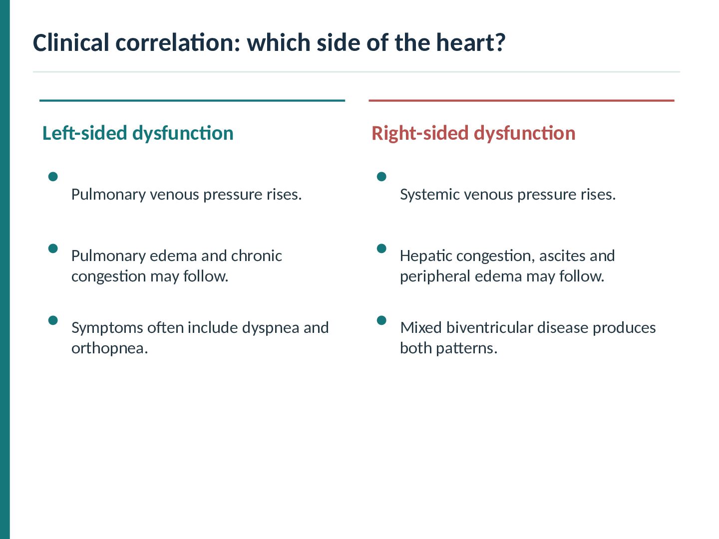
dysfunction Pulmonary venous pressure rises. Systemic venous pressure rises. Pulmonary edema and chronic congestion may follow. Hepatic congestion, ascites and peripheral edema may follow. Symptoms often include dyspnea and orthopnea. Mixed biventricular disease produces both patterns.
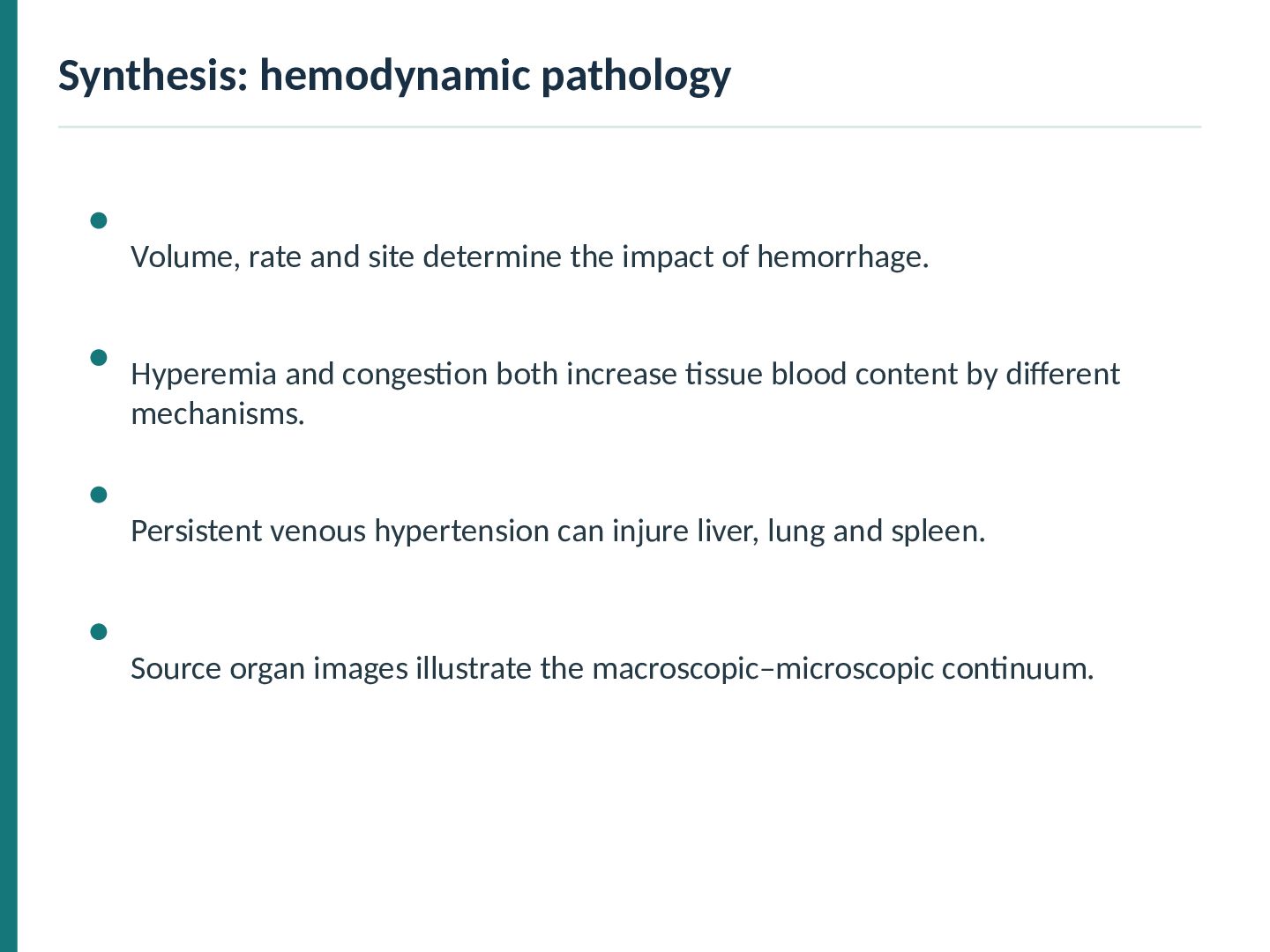
of hemorrhage. Hyperemia and congestion both increase tissue blood content by different mechanisms. Persistent venous hypertension can injure liver, lung and spleen. Source organ images illustrate the macroscopic–microscopic continuum.


 abusuraihsakhri
 jarvist
 institutdelelevage
 arthur1
 rmaruy
 sshimizu2006
 okana2ki
 trycycle
 tagtag
 konakalab
 akasan
 lychee1223
 jacobtomlinson
 sabderemane
 reverentgeek
 aleyda
 michielstock
 techseoconnect
 palkan
 coloredviolet
 inesmontani
 sugarenia
 baasie