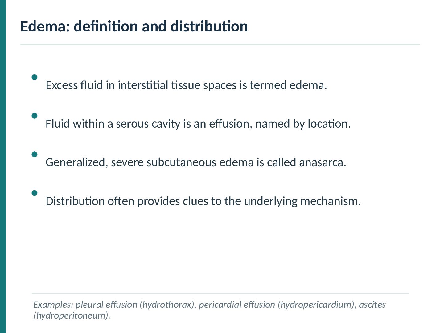
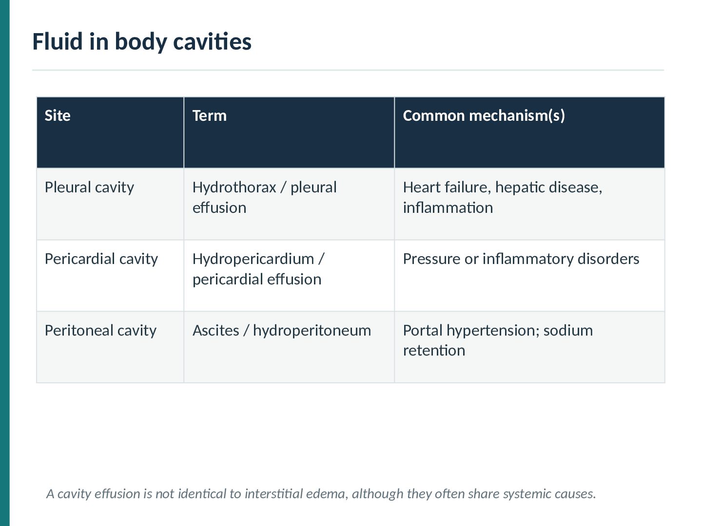
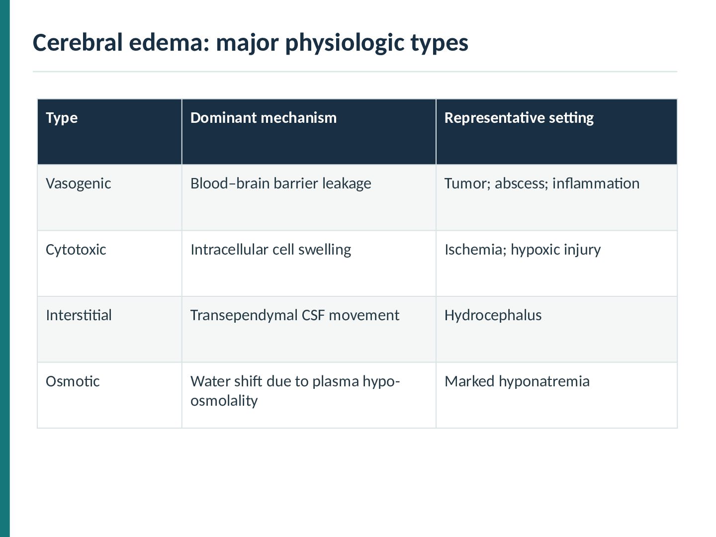
is termed edema. Fluid within a serous cavity is an effusion, named by location. Generalized, severe subcutaneous edema is called anasarca. Distribution often provides clues to the underlying mechanism. Examples: pleural effusion (hydrothorax), pericardial effusion (hydropericardium), ascites (hydroperitoneum).
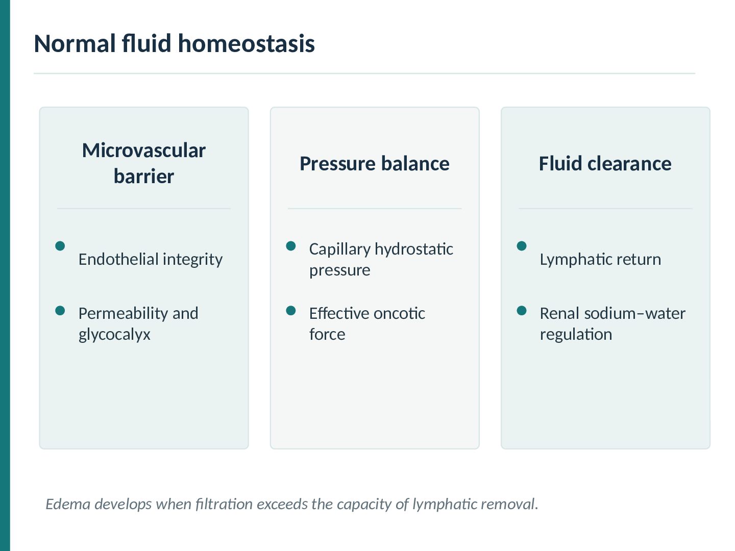
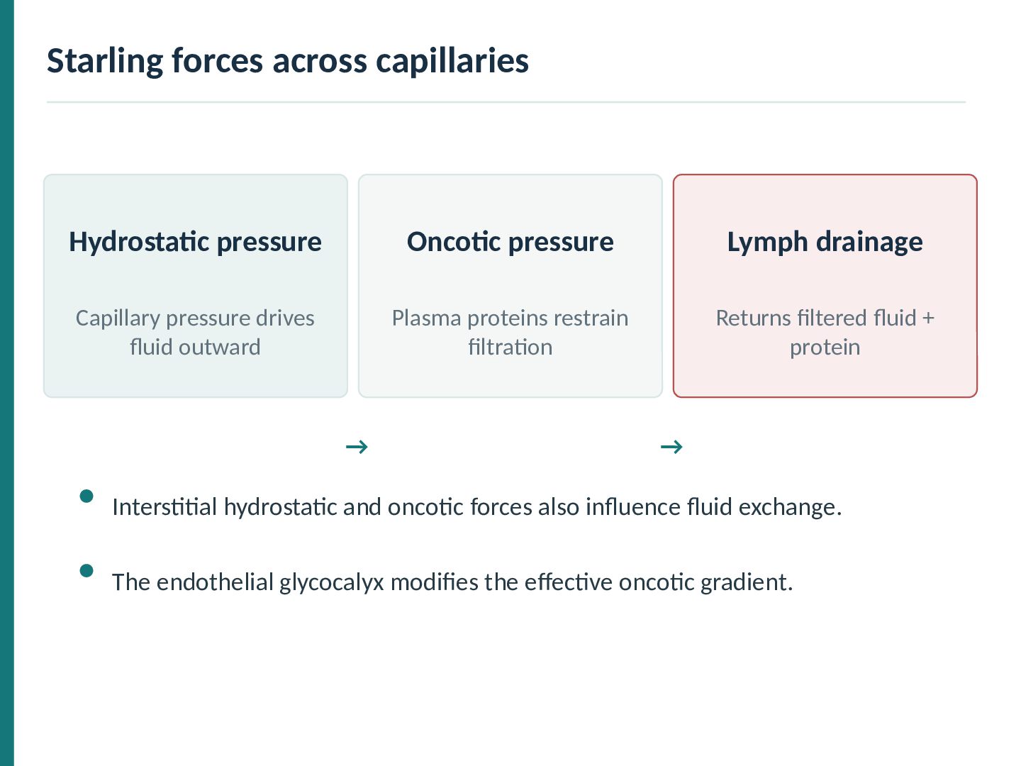
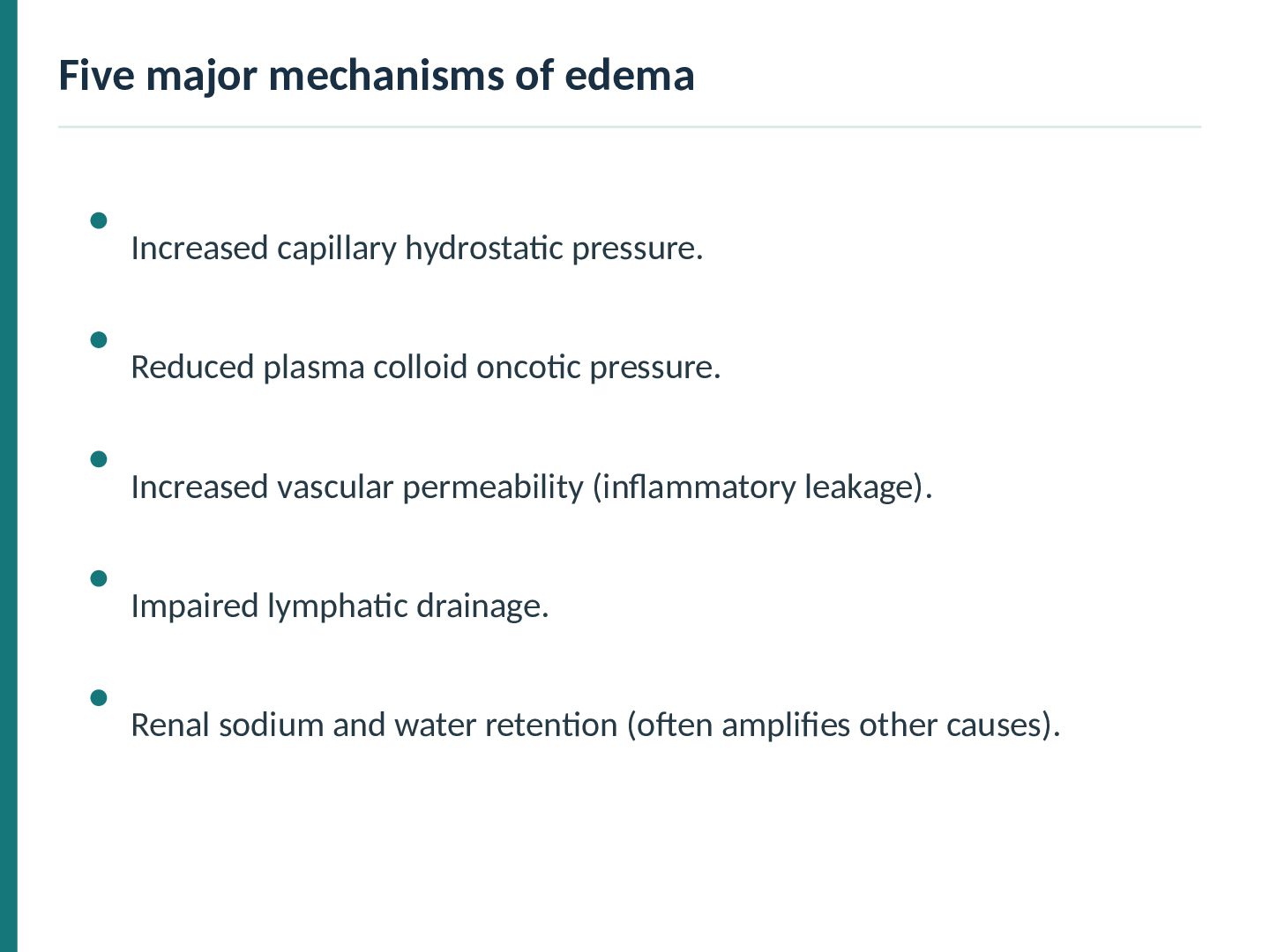
integrity Capillary hydrostatic pressure Lymphatic return Permeability and glycocalyx Effective oncotic force Renal sodium–water regulation Edema develops when filtration exceeds the capacity of lymphatic removal.
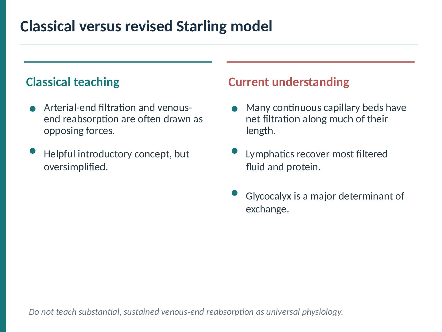
filtration and venousend reabsorption are often drawn as opposing forces. Many continuous capillary beds have net filtration along much of their length. Helpful introductory concept, but oversimplified. Lymphatics recover most filtered fluid and protein. Glycocalyx is a major determinant of exchange. Do not teach substantial, sustained venous-end reabsorption as universal physiology.
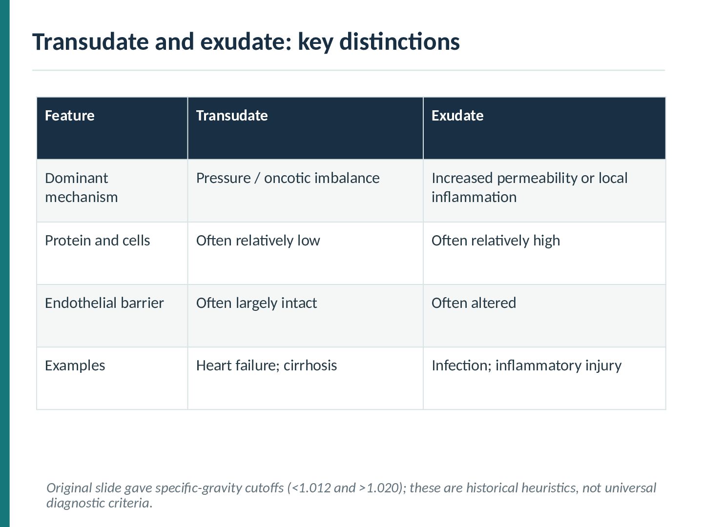
Pressure / oncotic imbalance Increased permeability or local inflammation Protein and cells Often relatively low Often relatively high Endothelial barrier Often largely intact Often altered Examples Heart failure; cirrhosis Infection; inflammatory injury Original slide gave specific-gravity cutoffs (<1.012 and >1.020); these are historical heuristics, not universal diagnostic criteria.
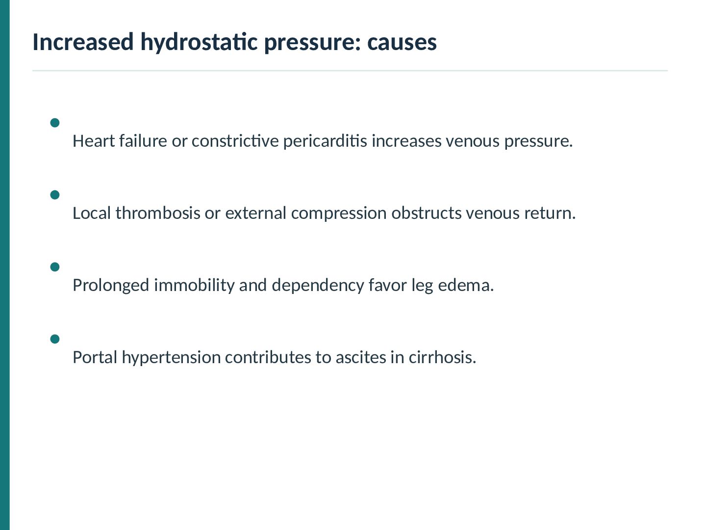
venous pressure. Local thrombosis or external compression obstructs venous return. Prolonged immobility and dependency favor leg edema. Portal hypertension contributes to ascites in cirrhosis.
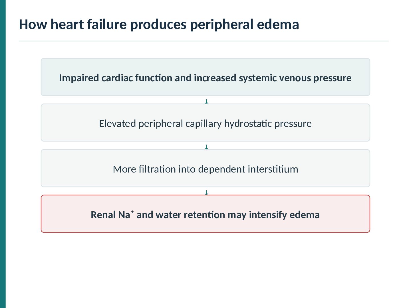
increased systemic venous pressure ↓ Elevated peripheral capillary hydrostatic pressure ↓ More filtration into dependent interstitium ↓ Renal Na⁺ and water retention may intensify edema
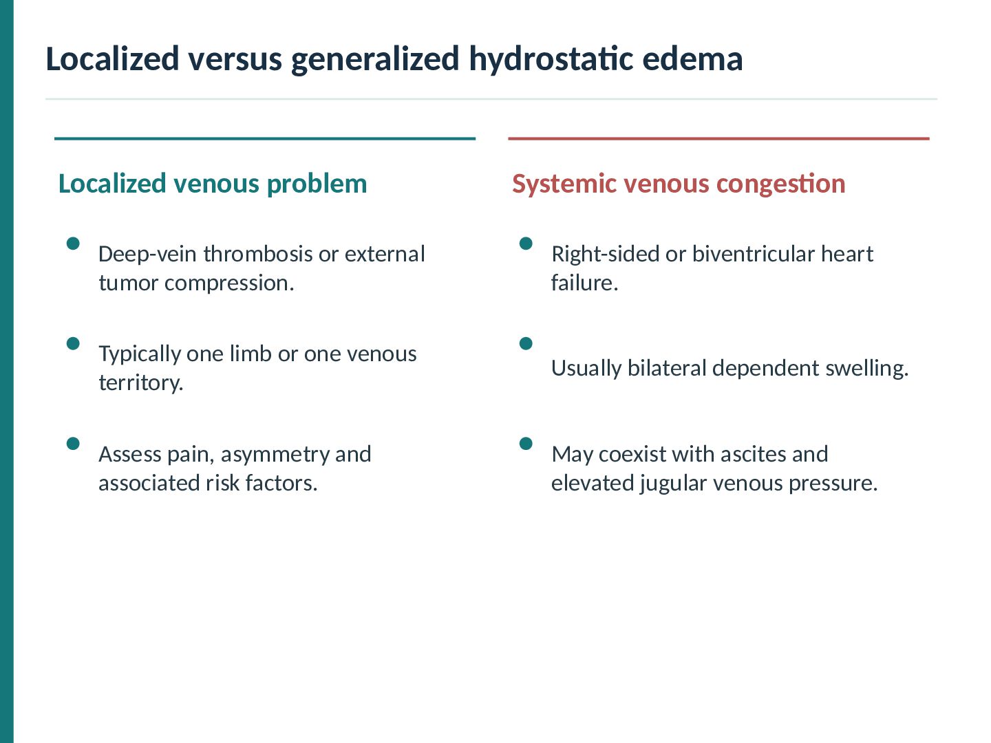
congestion Deep-vein thrombosis or external tumor compression. Right-sided or biventricular heart failure. Typically one limb or one venous territory. Usually bilateral dependent swelling. Assess pain, asymmetry and associated risk factors. May coexist with ascites and elevated jugular venous pressure.
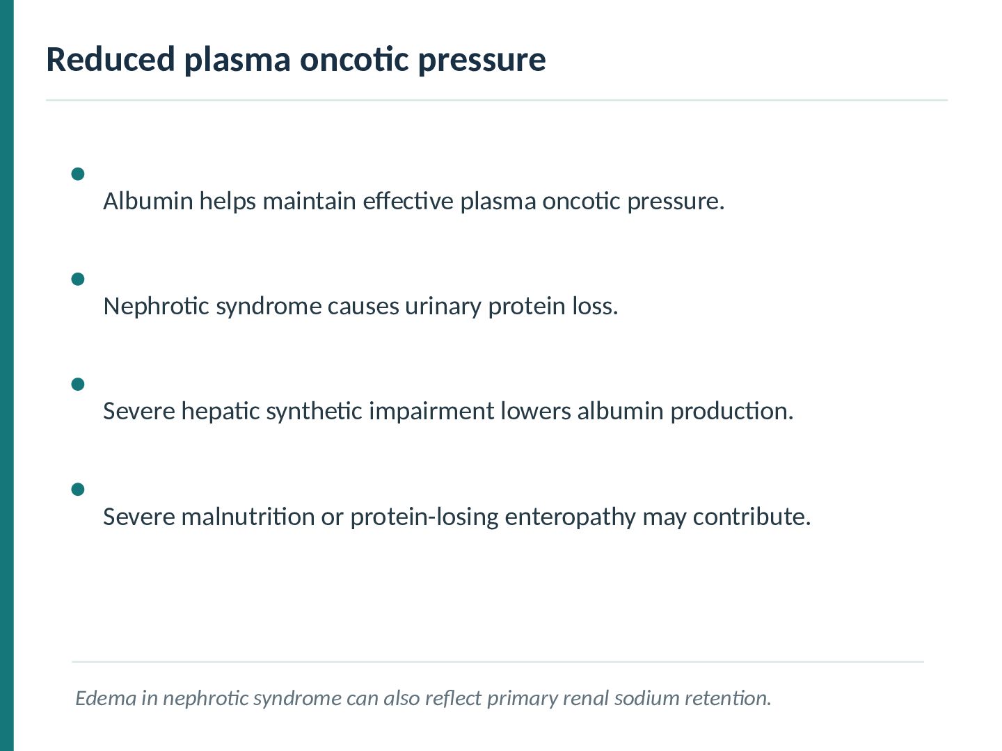
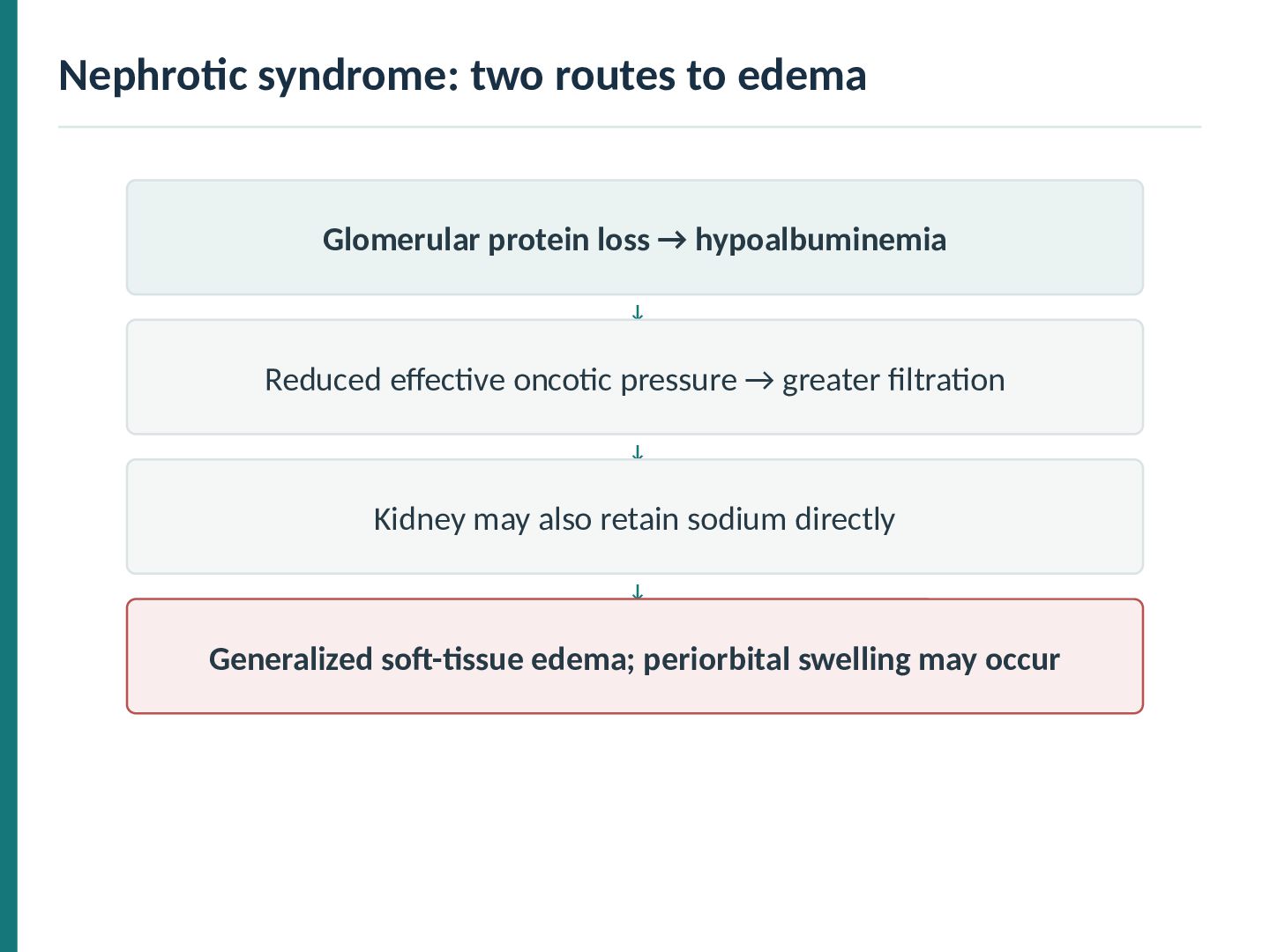
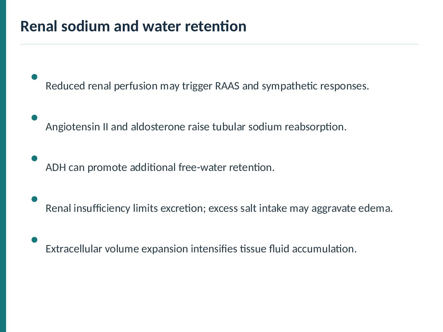
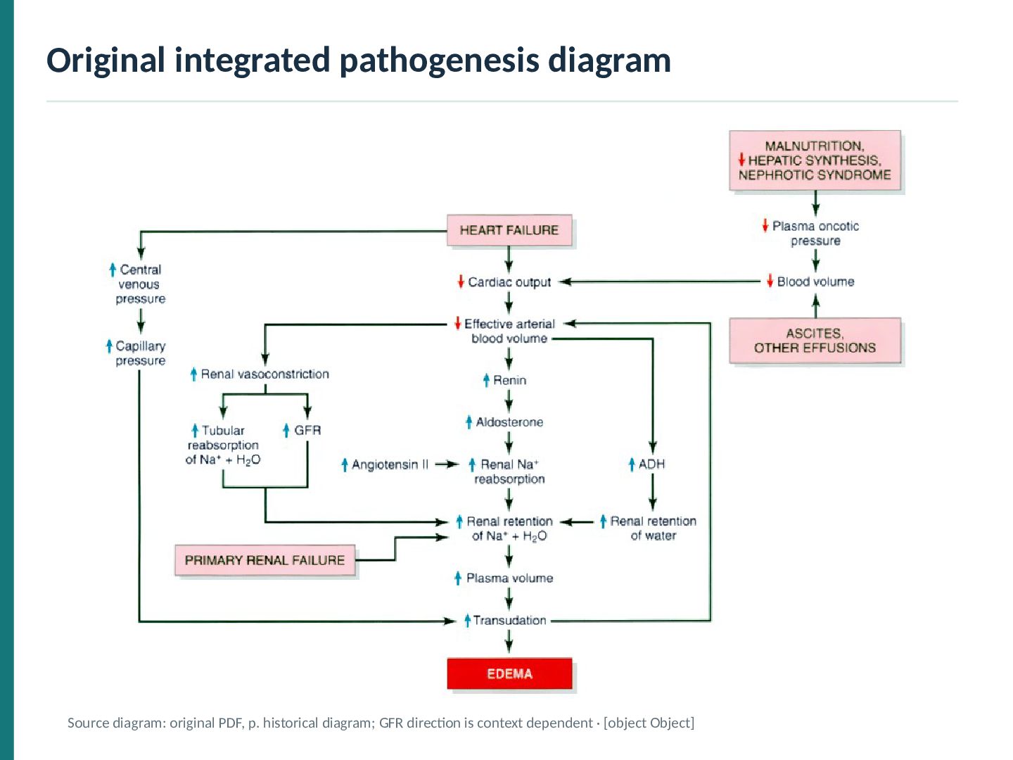
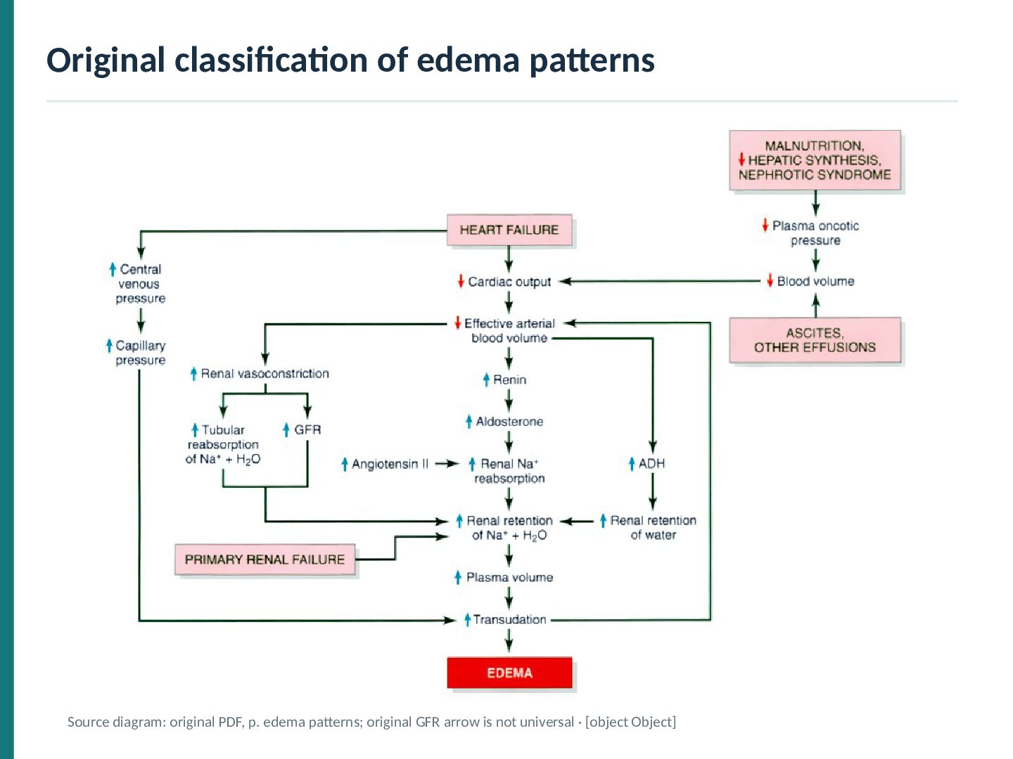
pressure. Nephrotic syndrome causes urinary protein loss. Severe hepatic synthetic impairment lowers albumin production. Severe malnutrition or protein-losing enteropathy may contribute. Edema in nephrotic syndrome can also reflect primary renal sodium retention.
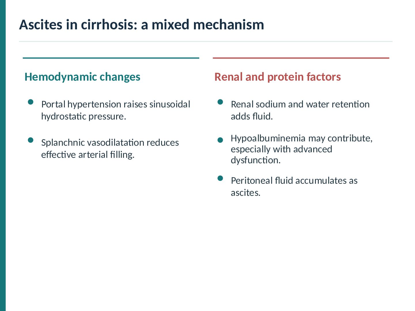
protein factors Portal hypertension raises sinusoidal hydrostatic pressure. Renal sodium and water retention adds fluid. Splanchnic vasodilatation reduces effective arterial filling. Hypoalbuminemia may contribute, especially with advanced dysfunction. Peritoneal fluid accumulates as ascites.
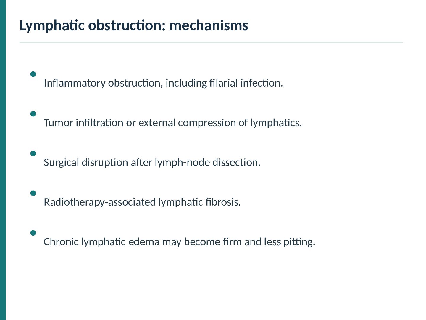
or external compression of lymphatics. Surgical disruption after lymph-node dissection. Radiotherapy-associated lymphatic fibrosis. Chronic lymphatic edema may become firm and less pitting.
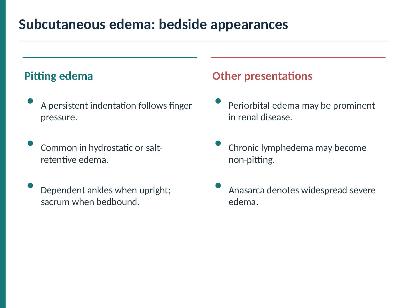
indentation follows finger pressure. Periorbital edema may be prominent in renal disease. Common in hydrostatic or saltretentive edema. Chronic lymphedema may become non-pitting. Dependent ankles when upright; sacrum when bedbound. Anasarca denotes widespread severe edema.
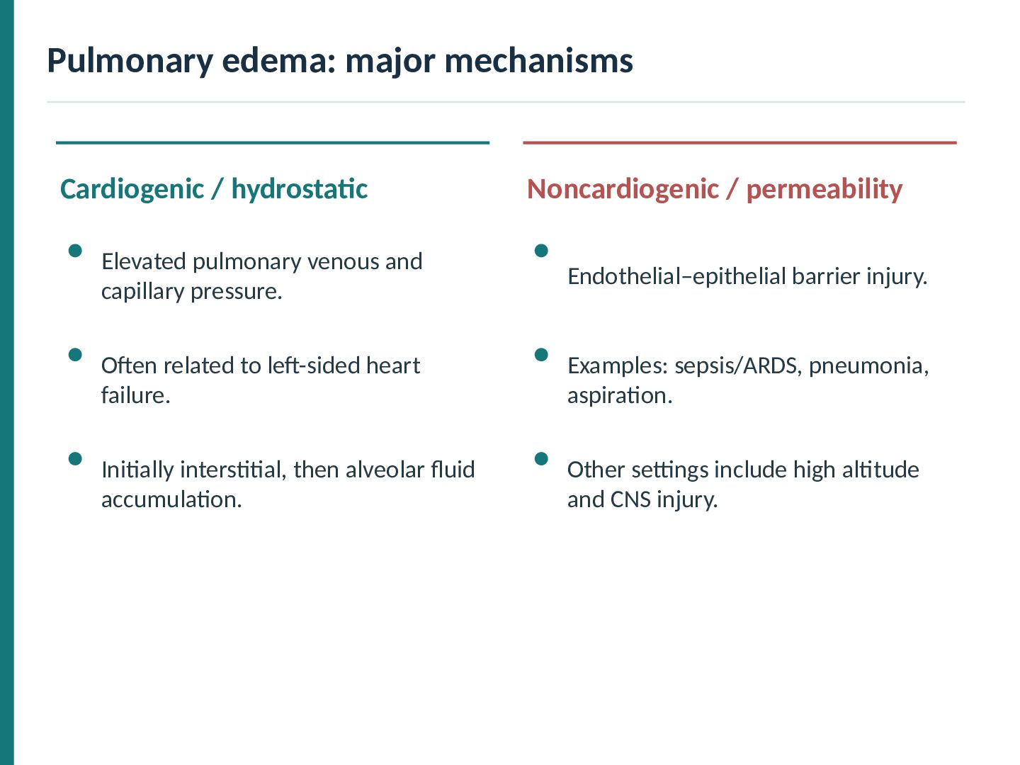
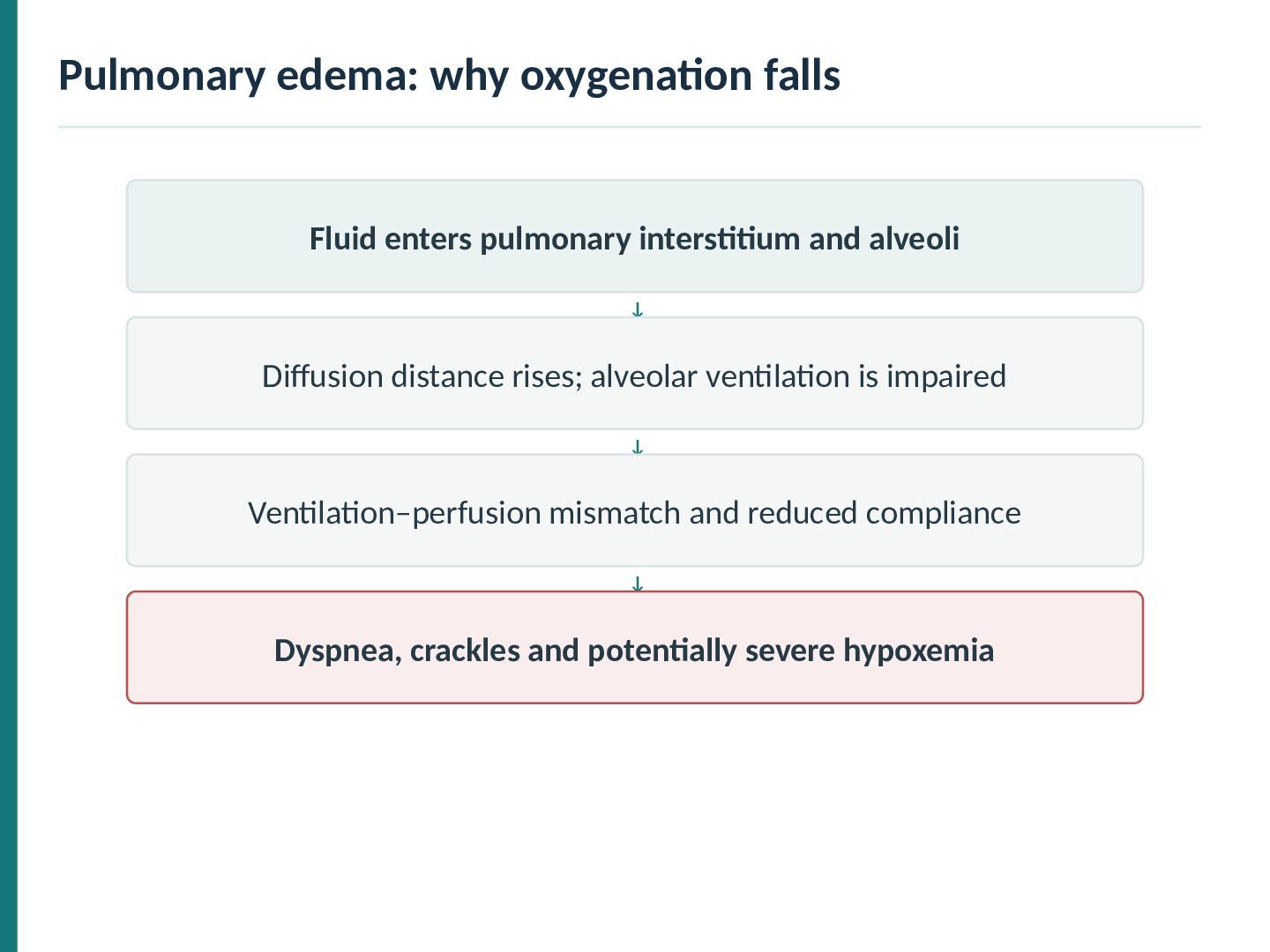
Elevated pulmonary venous and capillary pressure. Endothelial–epithelial barrier injury. Often related to left-sided heart failure. Examples: sepsis/ARDS, pneumonia, aspiration. Initially interstitial, then alveolar fluid accumulation. Other settings include high altitude and CNS injury.
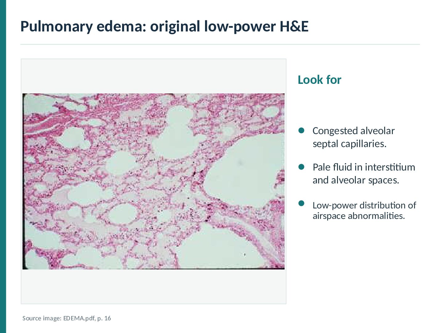
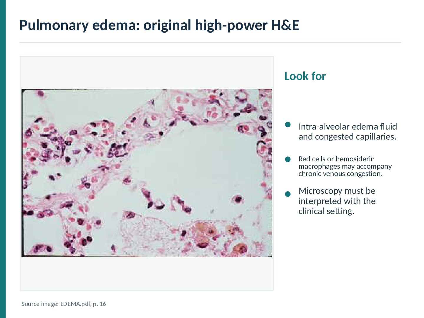
and congested capillaries. Red cells or hemosiderin macrophages may accompany chronic venous congestion. Microscopy must be interpreted with the clinical setting. Source image: EDEMA.pdf, p. 16
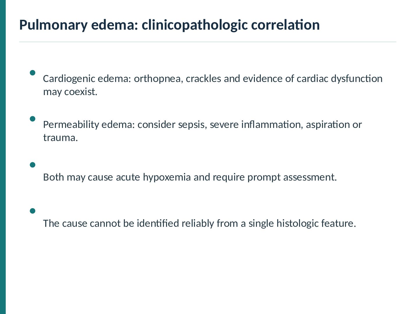
of cardiac dysfunction may coexist. Permeability edema: consider sepsis, severe inflammation, aspiration or trauma. Both may cause acute hypoxemia and require prompt assessment. The cause cannot be identified reliably from a single histologic feature.
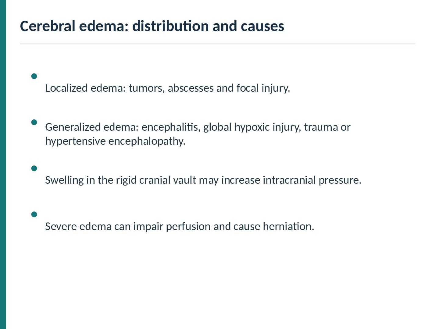
focal injury. Generalized edema: encephalitis, global hypoxic injury, trauma or hypertensive encephalopathy. Swelling in the rigid cranial vault may increase intracranial pressure. Severe edema can impair perfusion and cause herniation.
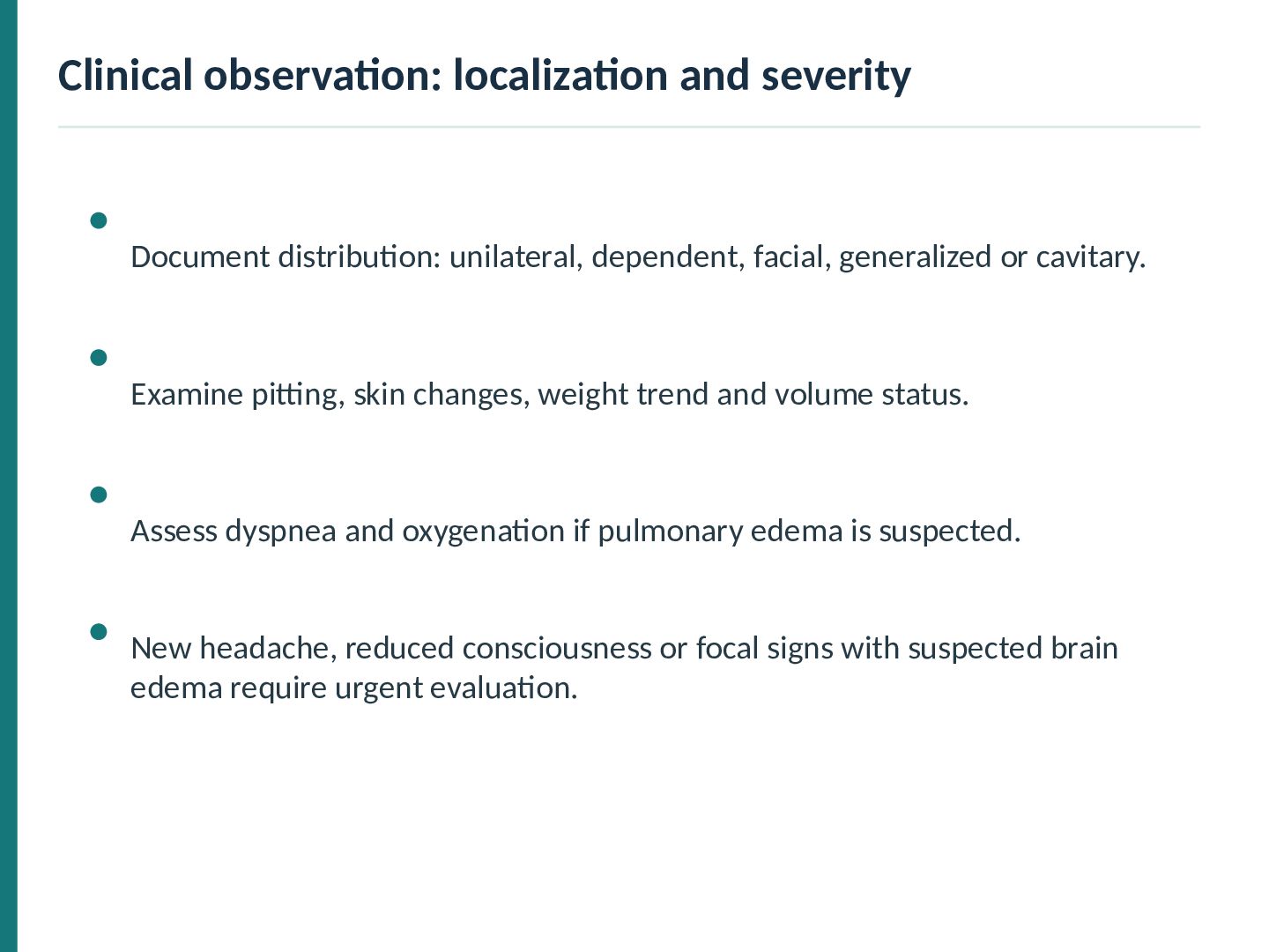
generalized or cavitary. Examine pitting, skin changes, weight trend and volume status. Assess dyspnea and oxygenation if pulmonary edema is suspected. New headache, reduced consciousness or focal signs with suspected brain edema require urgent evaluation.
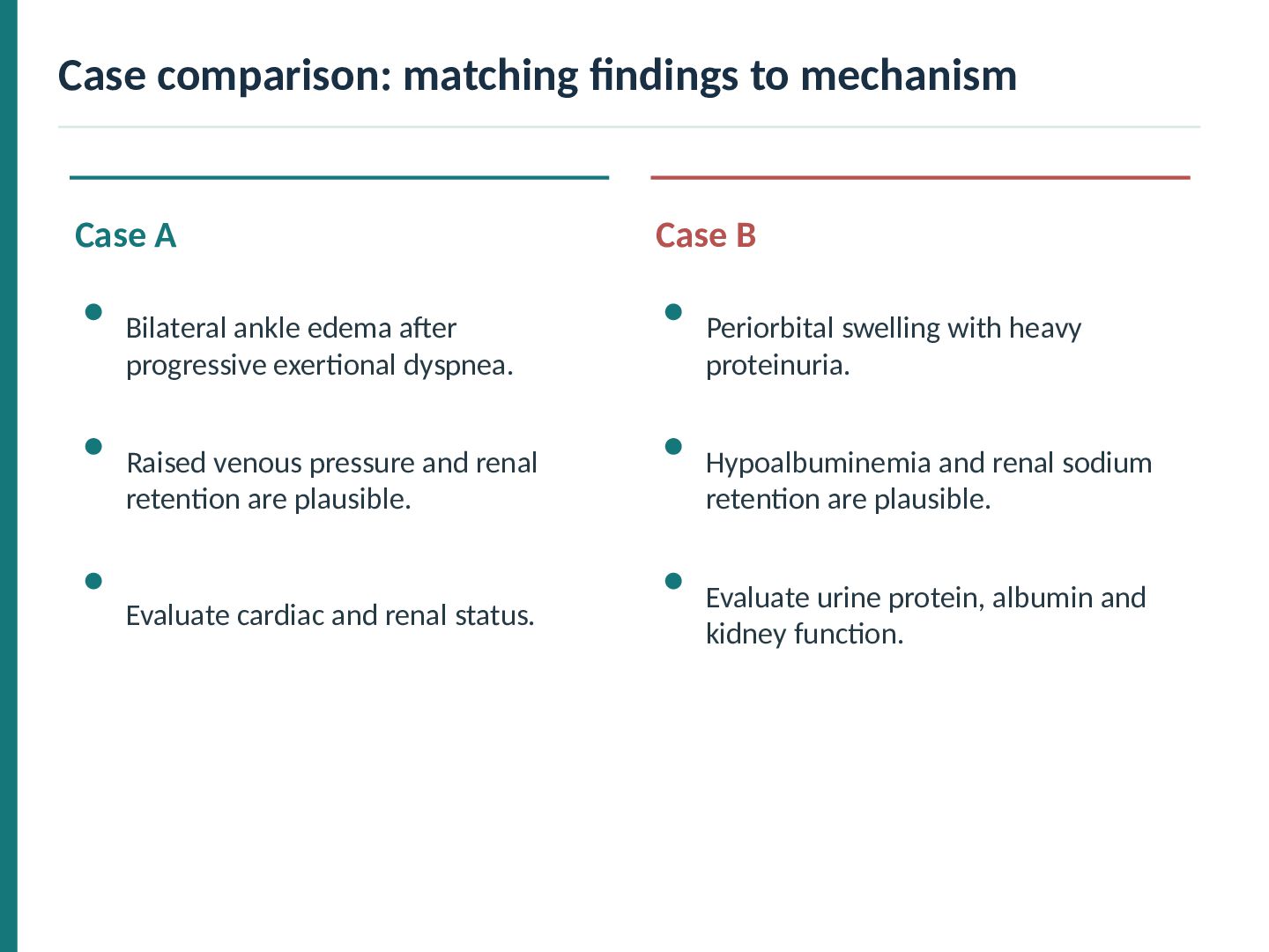
Bilateral ankle edema after progressive exertional dyspnea. Periorbital swelling with heavy proteinuria. Raised venous pressure and renal retention are plausible. Hypoalbuminemia and renal sodium retention are plausible. Evaluate cardiac and renal status. Evaluate urine protein, albumin and kidney function.
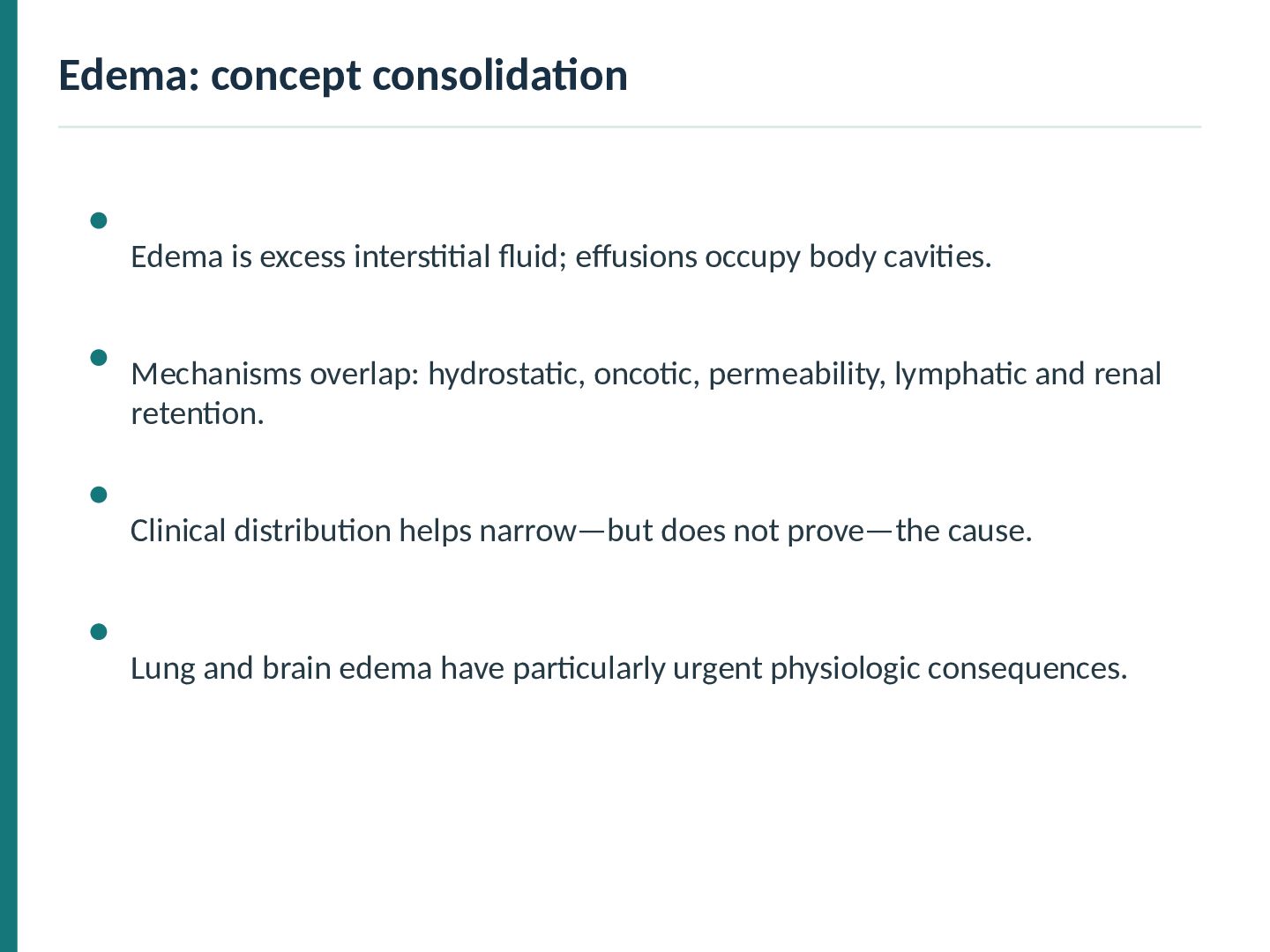
body cavities. Mechanisms overlap: hydrostatic, oncotic, permeability, lymphatic and renal retention. Clinical distribution helps narrow—but does not prove—the cause. Lung and brain edema have particularly urgent physiologic consequences.


 abusuraihsakhri
 sshimizu2006
 institutdelelevage
 trycycle
 jagupath
 okana2ki
 lychee1223
 konakalab
 mickey_kubo
 takusamar
 tagtag
 naospon
 watany
 bkeepers
 aleyda
 pauljervisheath
 katarinadahlin
 kimpetersen
 rkendrick25
 62gerente
 jakevdp
 rakko
 reverentgeek
 jacobtomlinson