clearance. Material may be an endogenous normal constituent, an abnormal product or an exogenous substance. It may be synthesized within the affected cell or delivered from elsewhere. Deposits may be temporary or persistent, innocuous or toxic. Localization may be cytoplasmic, lysosomal or, less often, nuclear.
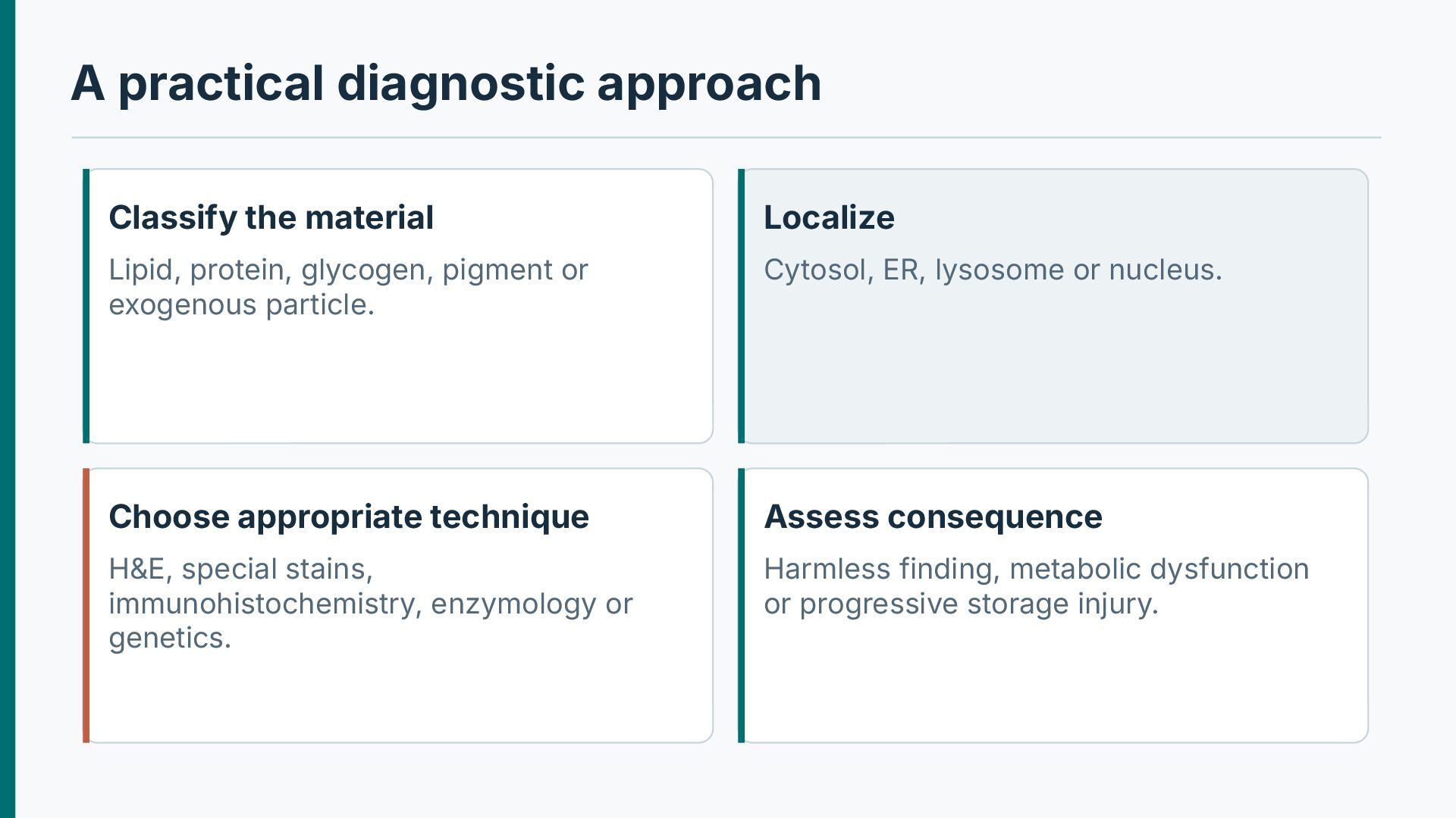
identifies the accumulated substance. Ask whether the material is intracellular or extracellular. Assess size, color, staining characteristics and organ distribution. Correlate with systemic metabolic state, medication use and genetic history. Confirm the biochemical identity with a fit-forpurpose assay where necessary.
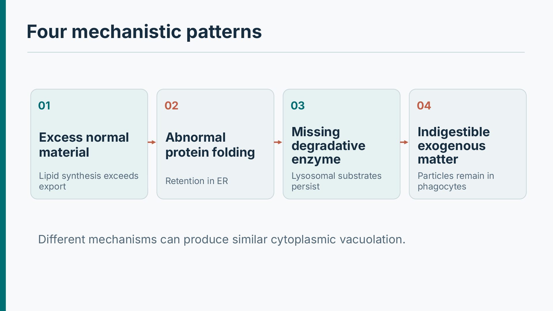
Abnormal protein folding Missing degradative enzyme Indigestible exogenous matter Lipid synthesis exceeds export Retention in ER Lysosomal substrates persist Particles remain in phagocytes Different mechanisms can produce similar cytoplasmic vacuolation.
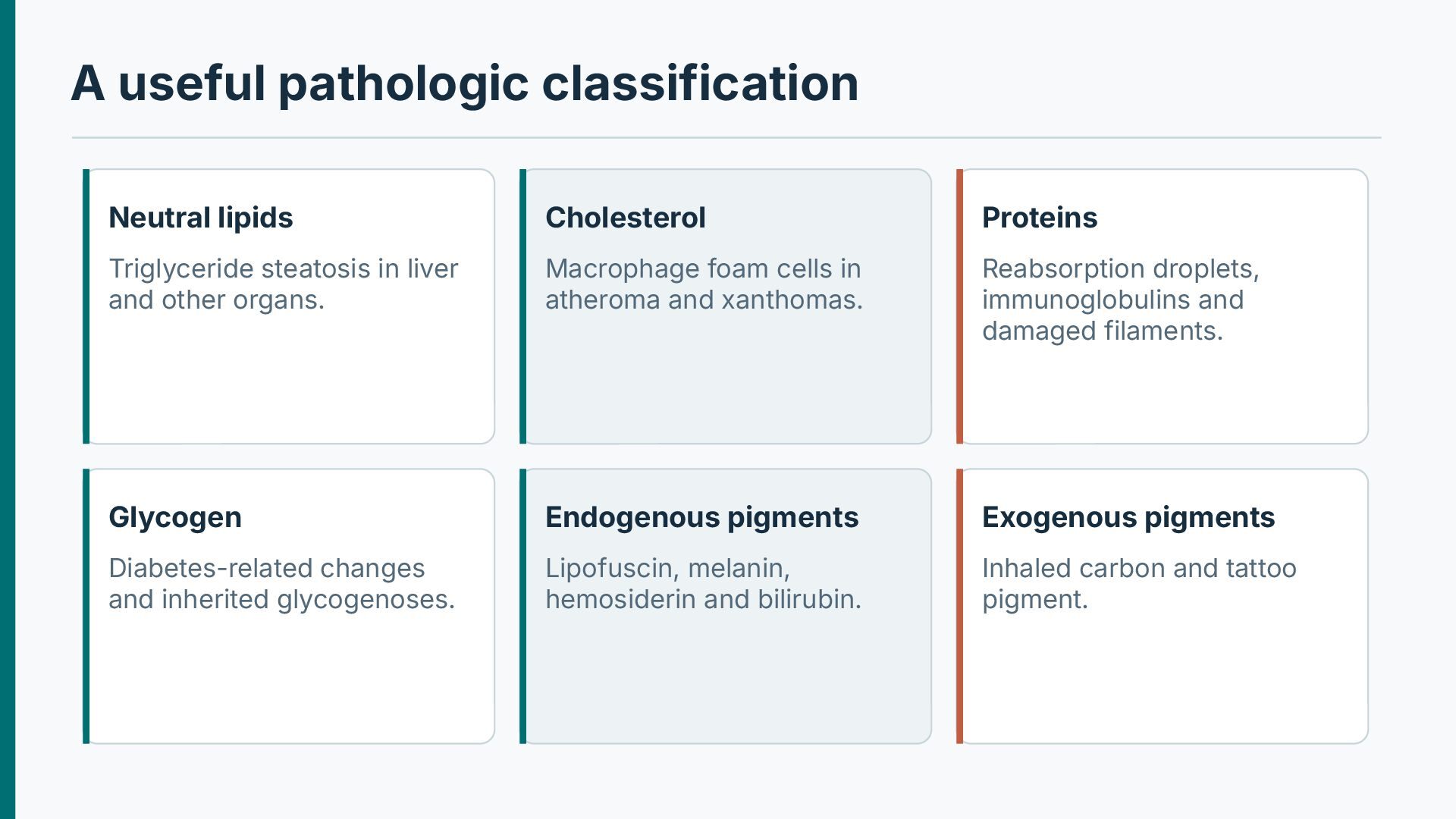
in liver and other organs. Macrophage foam cells in atheroma and xanthomas. Reabsorption droplets, immunoglobulins and damaged filaments. Glycogen Endogenous pigments Exogenous pigments Diabetes-related changes and inherited glycogenoses. Lipofuscin, melanin, hemosiderin and bilirubin. Inhaled carbon and tattoo pigment.
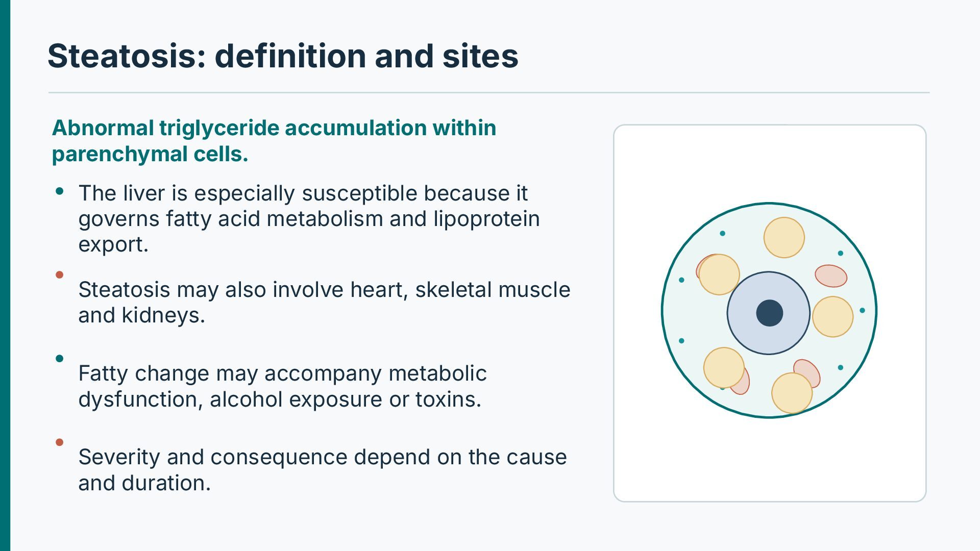
The liver is especially susceptible because it governs fatty acid metabolism and lipoprotein export. Steatosis may also involve heart, skeletal muscle and kidneys. Fatty change may accompany metabolic dysfunction, alcohol exposure or toxins. Severity and consequence depend on the cause and duration.
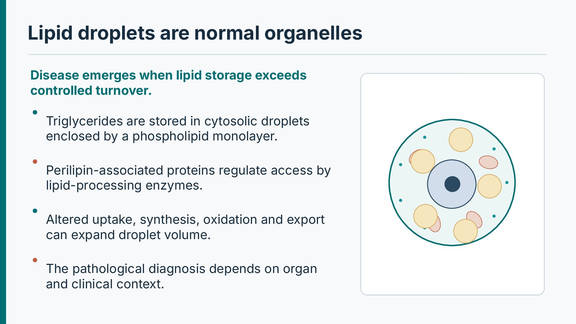
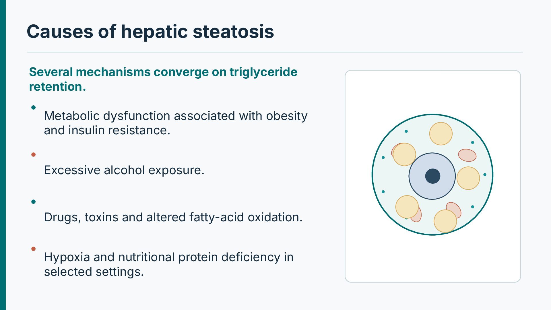
exceeds controlled turnover. Triglycerides are stored in cytosolic droplets enclosed by a phospholipid monolayer. Perilipin-associated proteins regulate access by lipid-processing enzymes. Altered uptake, synthesis, oxidation and export can expand droplet volume. The pathological diagnosis depends on organ and clinical context.
Metabolic dysfunction associated with obesity and insulin resistance. Excessive alcohol exposure. Drugs, toxins and altered fatty-acid oxidation. Hypoxia and nutritional protein deficiency in selected settings.
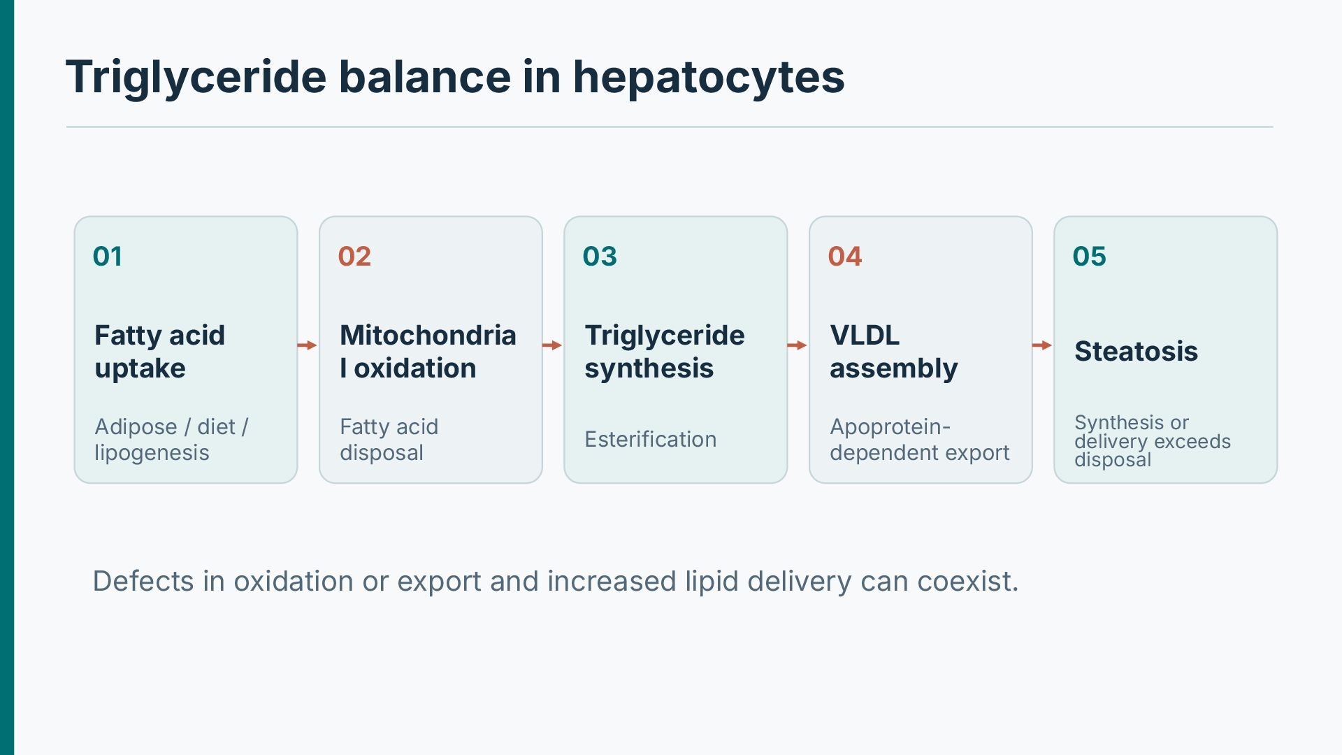
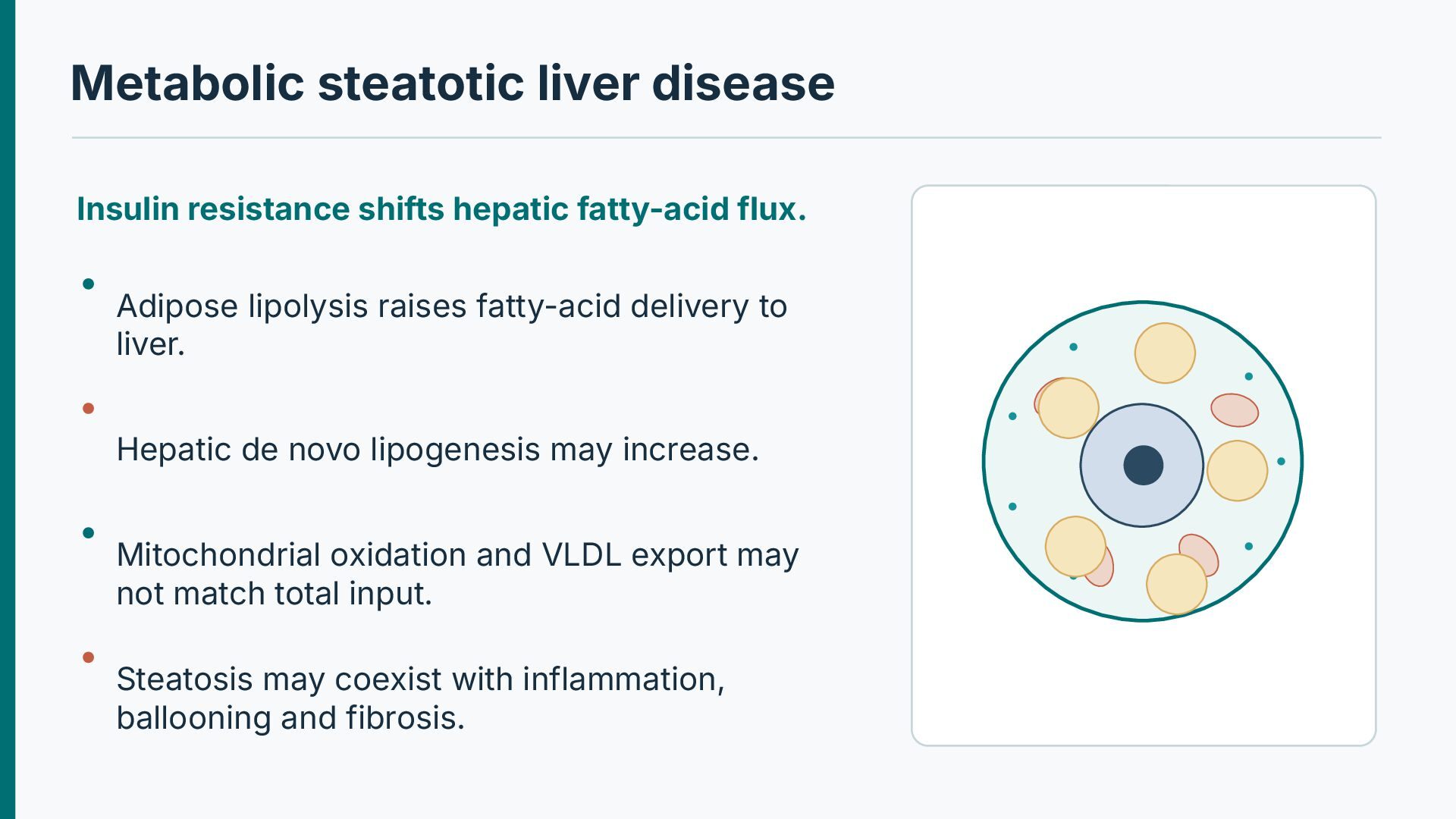
Adipose lipolysis raises fatty-acid delivery to liver. Hepatic de novo lipogenesis may increase. Mitochondrial oxidation and VLDL export may not match total input. Steatosis may coexist with inflammation, ballooning and fibrosis.
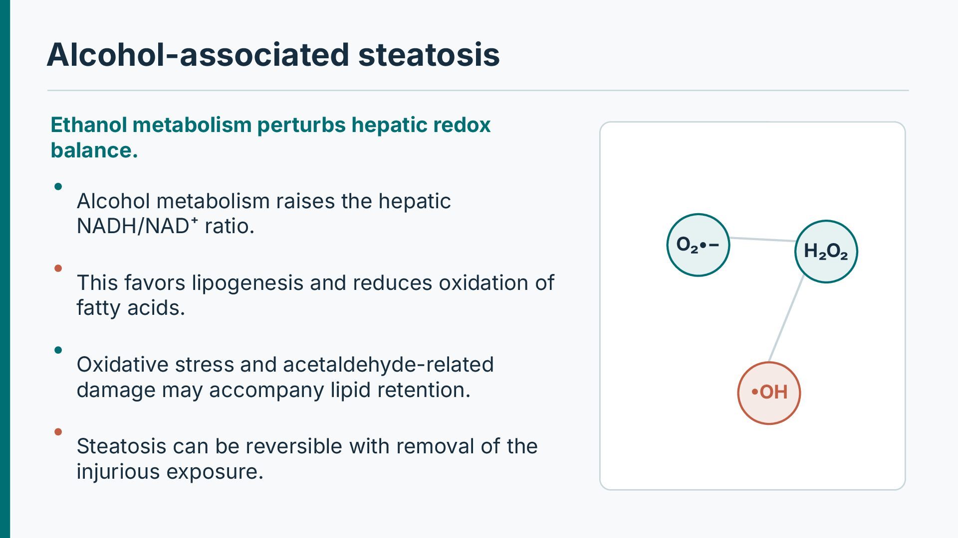
raises the hepatic NADH/NAD⁺ ratio. O₂•− H₂O₂ This favors lipogenesis and reduces oxidation of fatty acids. Oxidative stress and acetaldehyde-related damage may accompany lipid retention. Steatosis can be reversible with removal of the injurious exposure. •OH
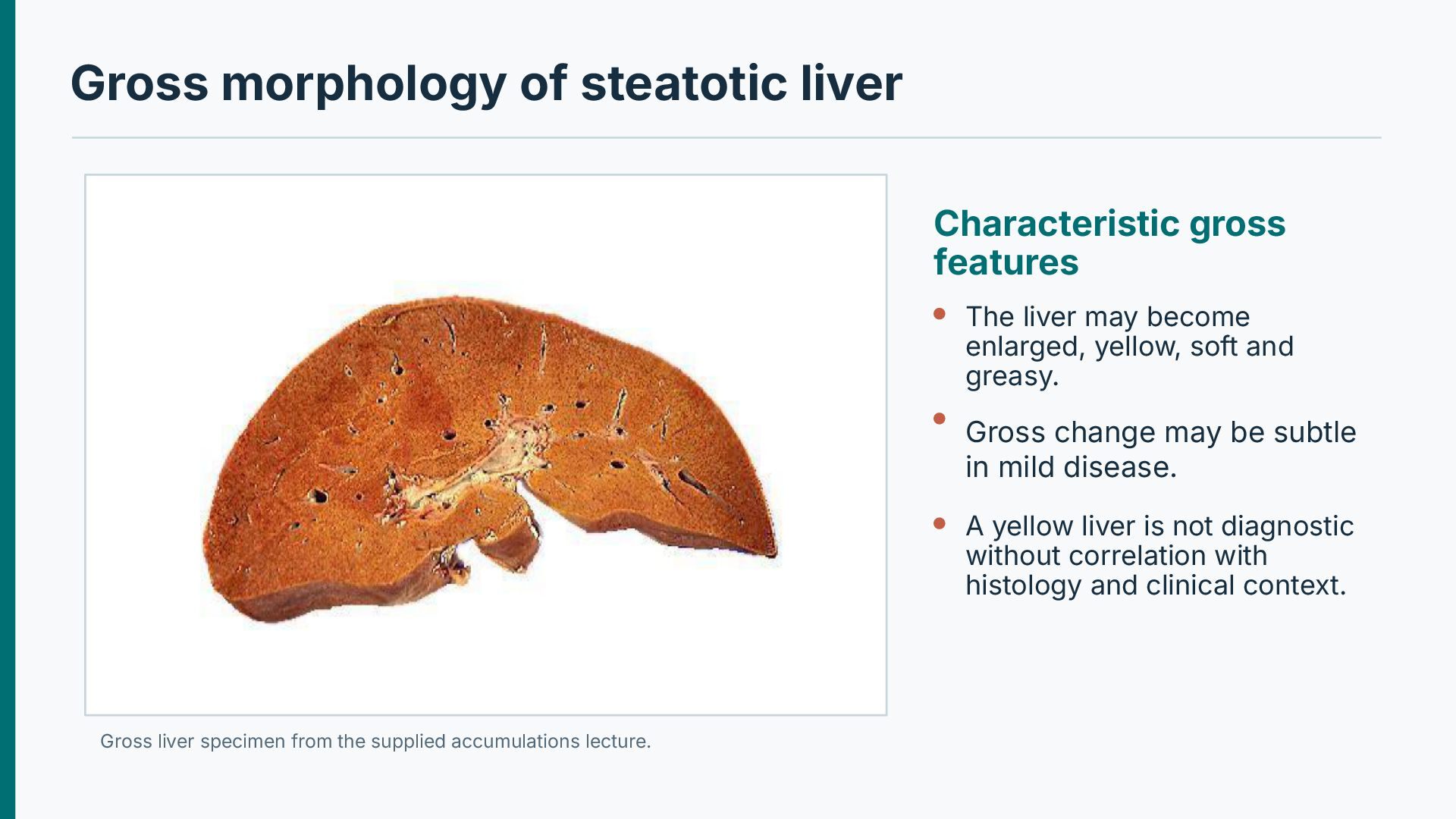
may become enlarged, yellow, soft and greasy. Gross change may be subtle in mild disease. A yellow liver is not diagnostic without correlation with histology and clinical context. Gross liver specimen from the supplied accumulations lecture.
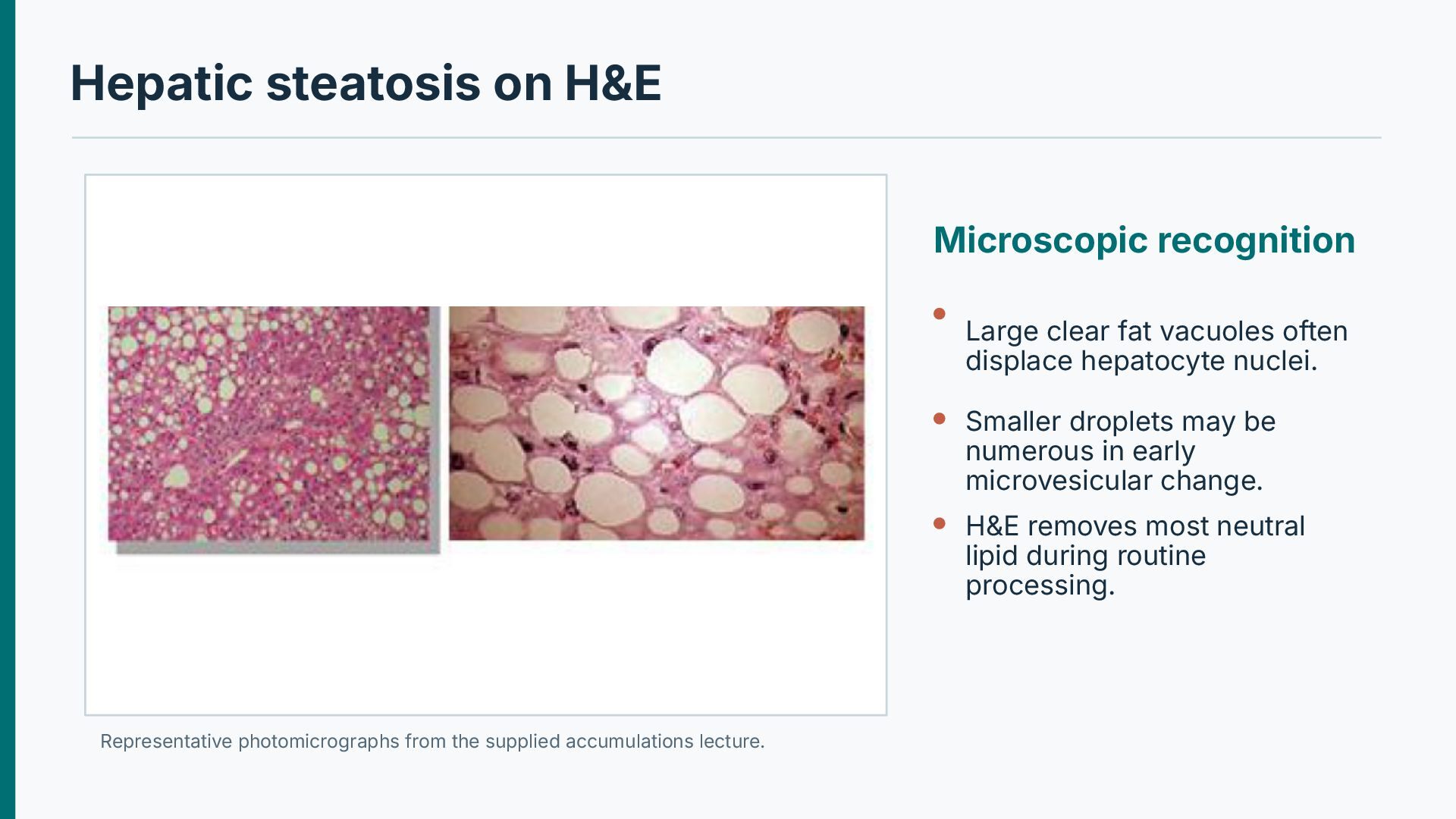
often displace hepatocyte nuclei. Smaller droplets may be numerous in early microvesicular change. H&E removes most neutral lipid during routine processing. Representative photomicrographs from the supplied accumulations lecture.
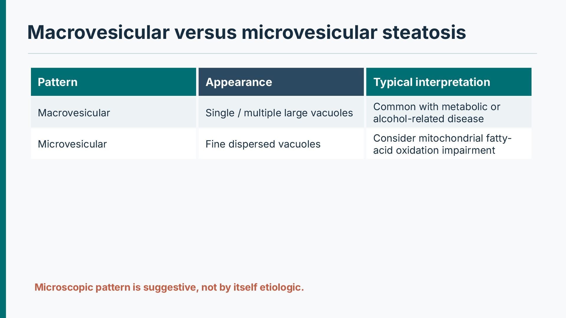
/ multiple large vacuoles Common with metabolic or alcohol-related disease Microvesicular Fine dispersed vacuoles Consider mitochondrial fattyacid oxidation impairment Microscopic pattern is suggestive, not by itself etiologic.
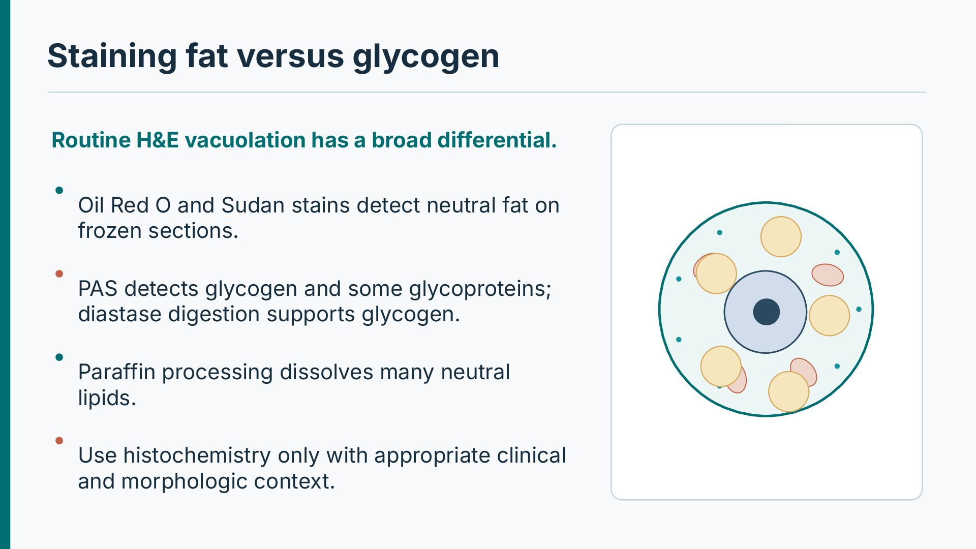
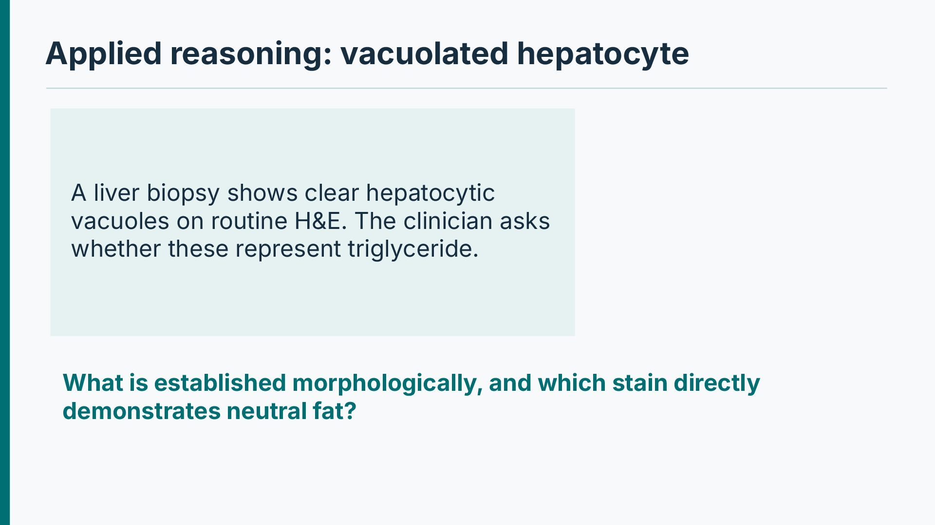
differential. Oil Red O and Sudan stains detect neutral fat on frozen sections. PAS detects glycogen and some glycoproteins; diastase digestion supports glycogen. Paraffin processing dissolves many neutral lipids. Use histochemistry only with appropriate clinical and morphologic context.
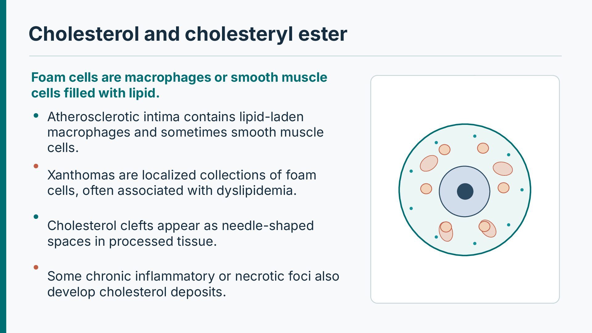
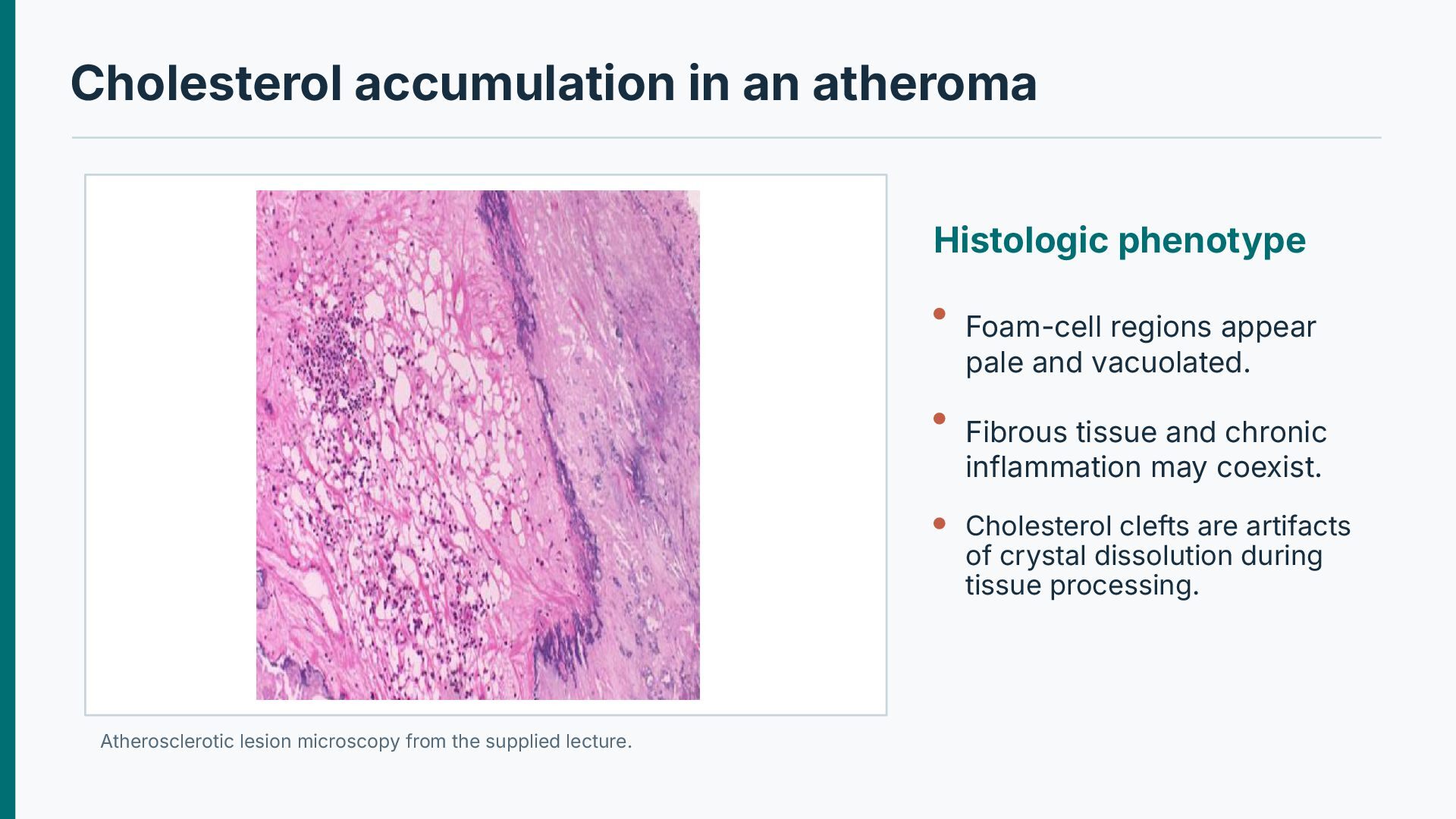
muscle cells filled with lipid. Atherosclerotic intima contains lipid-laden macrophages and sometimes smooth muscle cells. Xanthomas are localized collections of foam cells, often associated with dyslipidemia. Cholesterol clefts appear as needle-shaped spaces in processed tissue. Some chronic inflammatory or necrotic foci also develop cholesterol deposits.
pale and vacuolated. Fibrous tissue and chronic inflammation may coexist. Cholesterol clefts are artifacts of crystal dissolution during tissue processing. Atherosclerotic lesion microscopy from the supplied lecture.
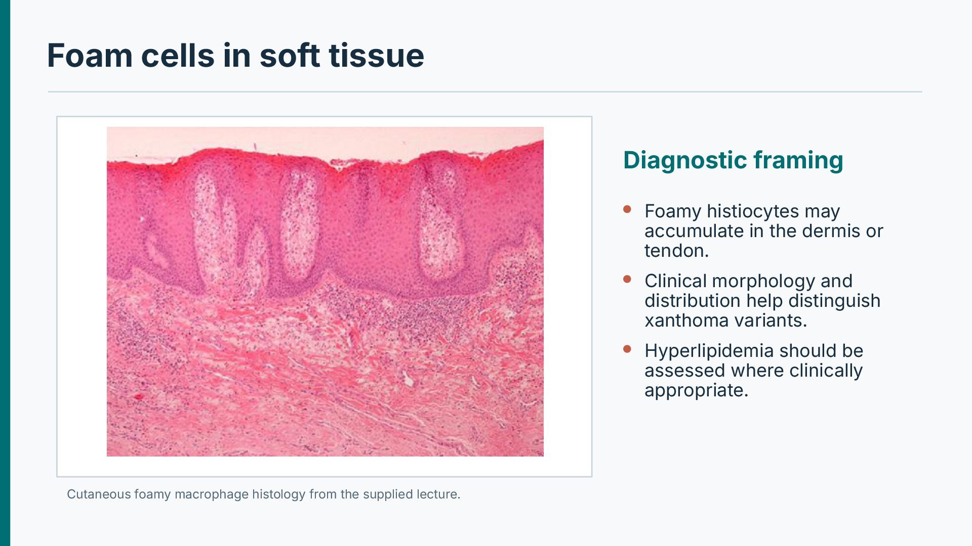
accumulate in the dermis or tendon. Clinical morphology and distribution help distinguish xanthoma variants. Hyperlipidemia should be assessed where clinically appropriate. Cutaneous foamy macrophage histology from the supplied lecture.
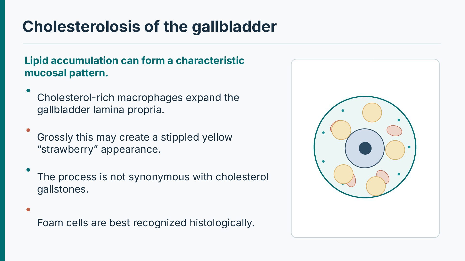
mucosal pattern. Cholesterol-rich macrophages expand the gallbladder lamina propria. Grossly this may create a stippled yellow “strawberry” appearance. The process is not synonymous with cholesterol gallstones. Foam cells are best recognized histologically.
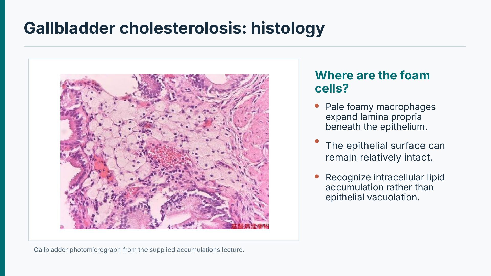
macrophages expand lamina propria beneath the epithelium. The epithelial surface can remain relatively intact. Recognize intracellular lipid accumulation rather than epithelial vacuolation. Gallbladder photomicrograph from the supplied accumulations lecture.
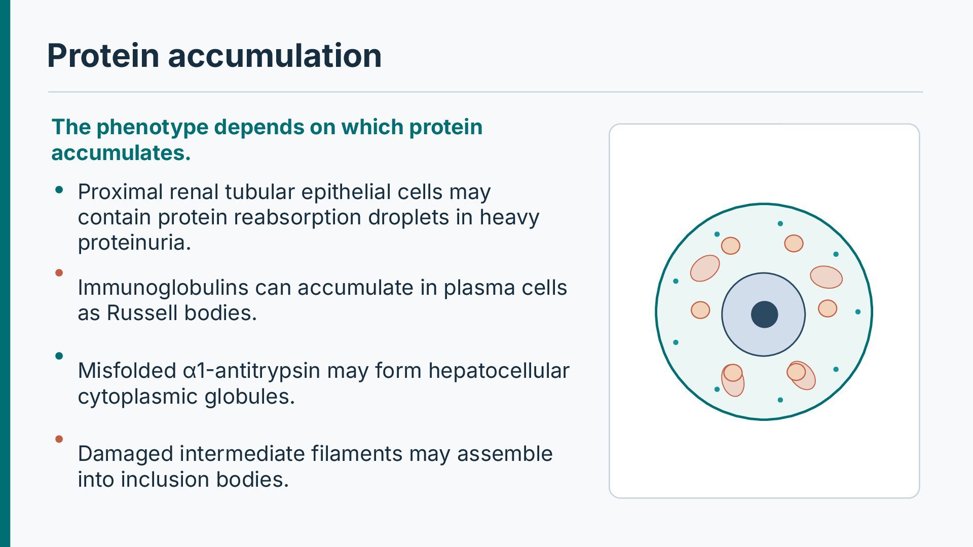
renal tubular epithelial cells may contain protein reabsorption droplets in heavy proteinuria. Immunoglobulins can accumulate in plasma cells as Russell bodies. Misfolded α1-antitrypsin may form hepatocellular cytoplasmic globules. Damaged intermediate filaments may assemble into inclusion bodies.
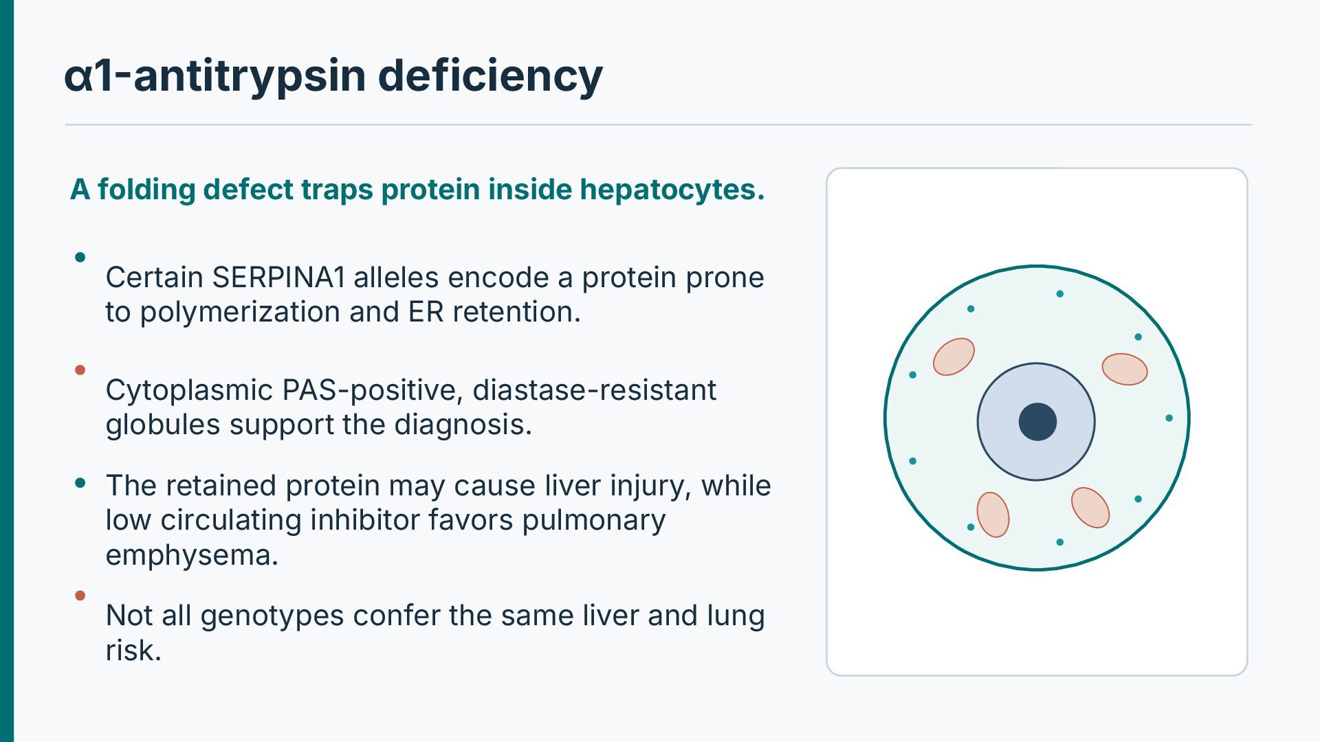
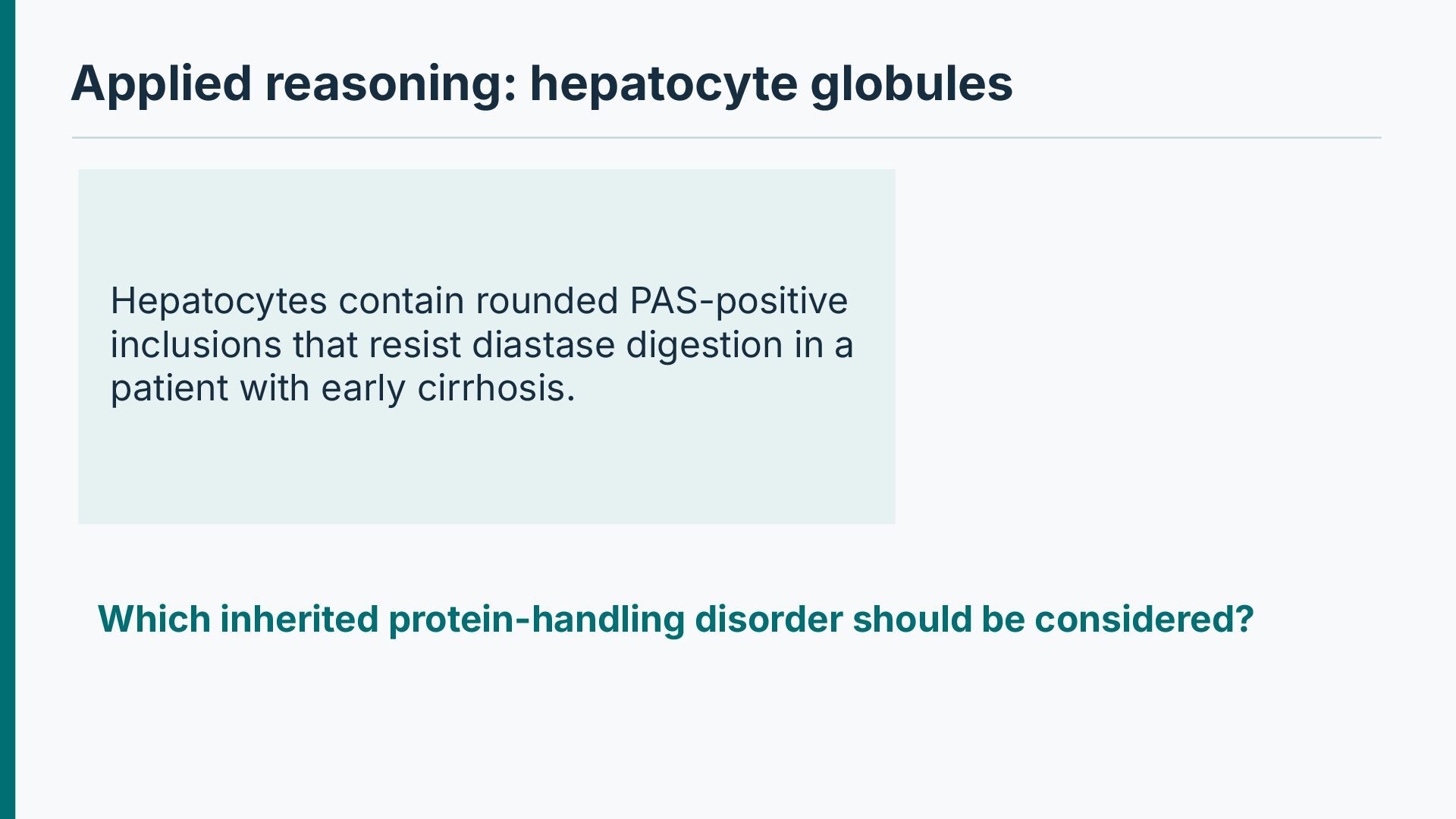
SERPINA1 alleles encode a protein prone to polymerization and ER retention. Cytoplasmic PAS-positive, diastase-resistant globules support the diagnosis. The retained protein may cause liver injury, while low circulating inhibitor favors pulmonary emphysema. Not all genotypes confer the same liver and lung risk.
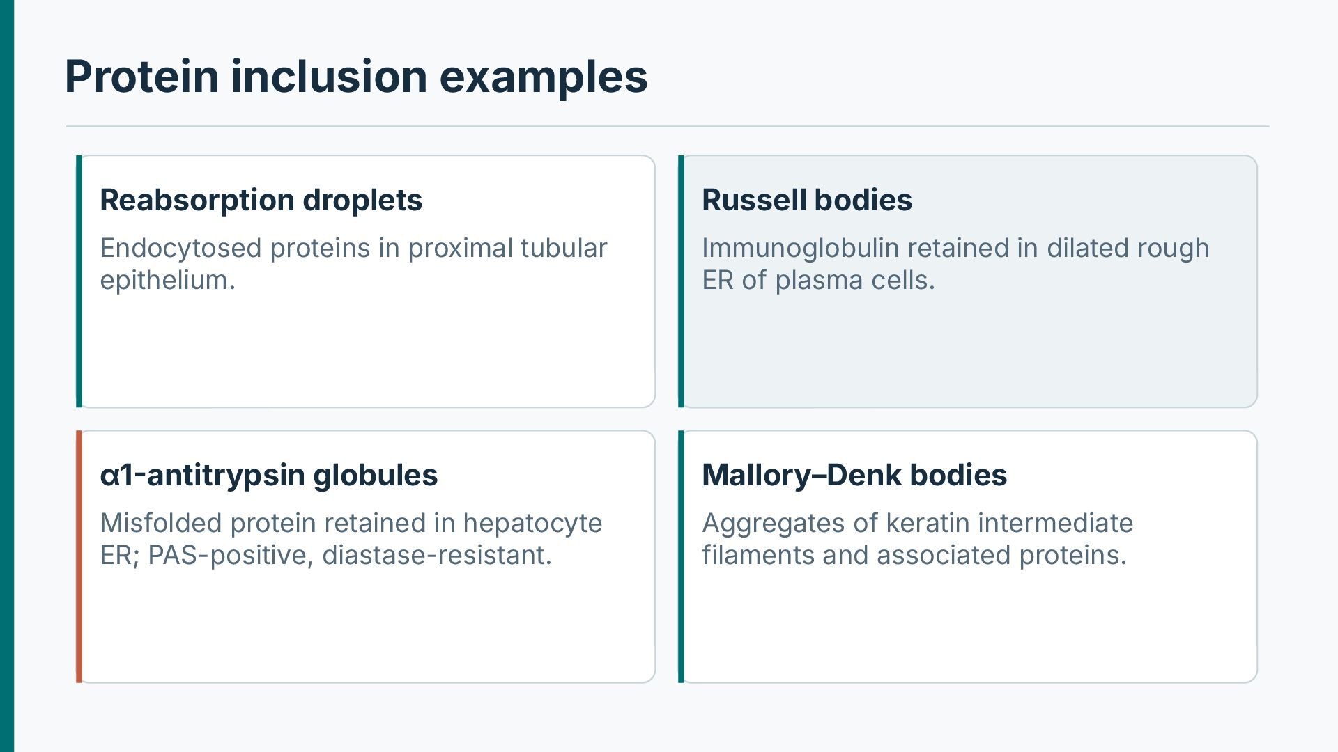
proximal tubular epithelium. Immunoglobulin retained in dilated rough ER of plasma cells. α1-antitrypsin globules Mallory–Denk bodies Misfolded protein retained in hepatocyte ER; PAS-positive, diastase-resistant. Aggregates of keratin intermediate filaments and associated proteins.
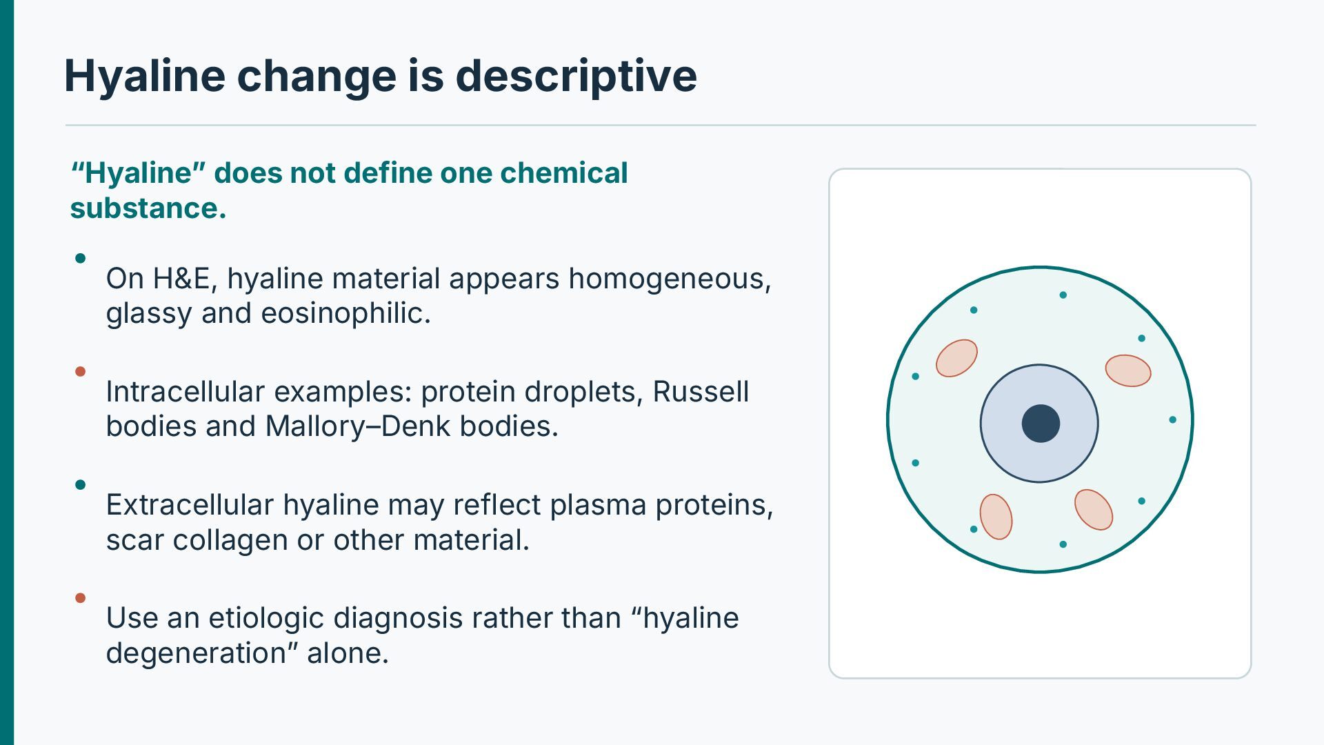
substance. On H&E, hyaline material appears homogeneous, glassy and eosinophilic. Intracellular examples: protein droplets, Russell bodies and Mallory–Denk bodies. Extracellular hyaline may reflect plasma proteins, scar collagen or other material. Use an etiologic diagnosis rather than “hyaline degeneration” alone.
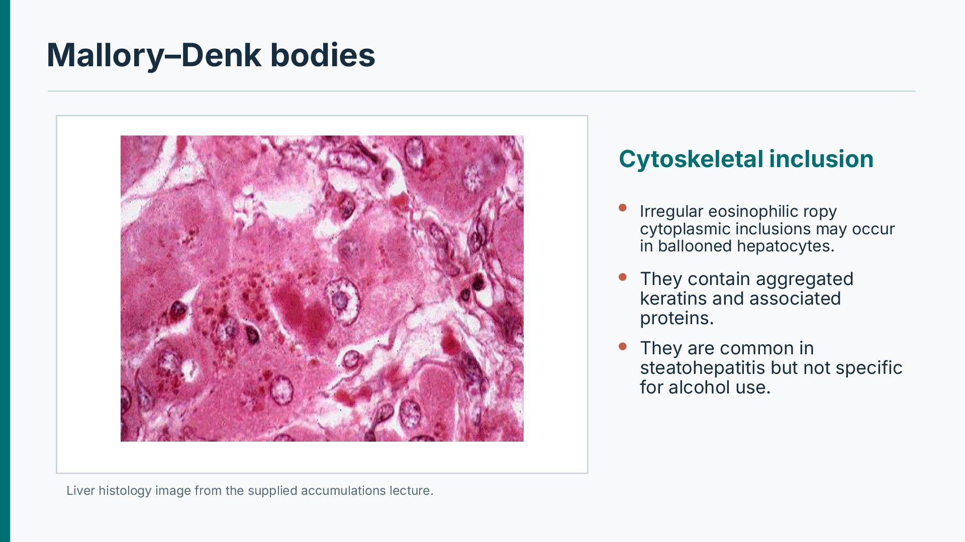
occur in ballooned hepatocytes. They contain aggregated keratins and associated proteins. They are common in steatohepatitis but not specific for alcohol use. Liver histology image from the supplied accumulations lecture.
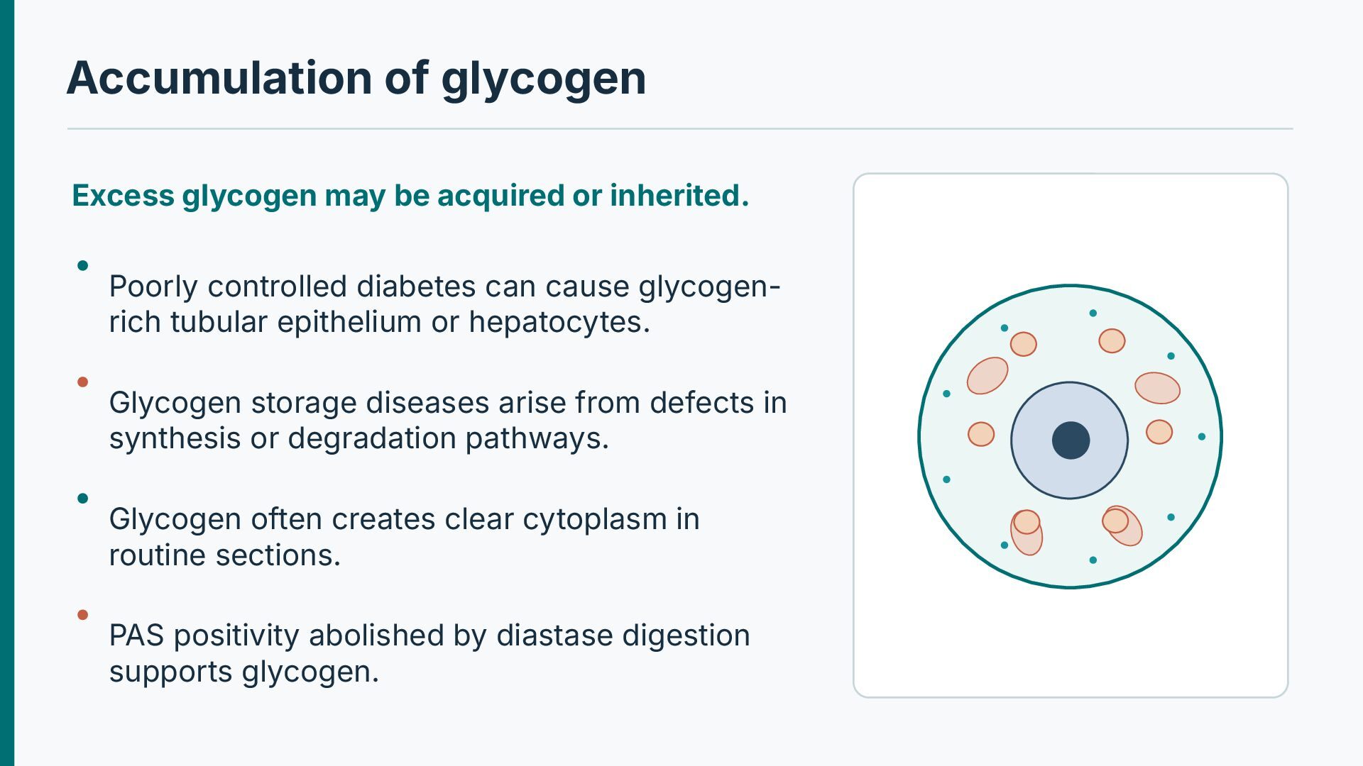
Poorly controlled diabetes can cause glycogenrich tubular epithelium or hepatocytes. Glycogen storage diseases arise from defects in synthesis or degradation pathways. Glycogen often creates clear cytoplasm in routine sections. PAS positivity abolished by diastase digestion supports glycogen.
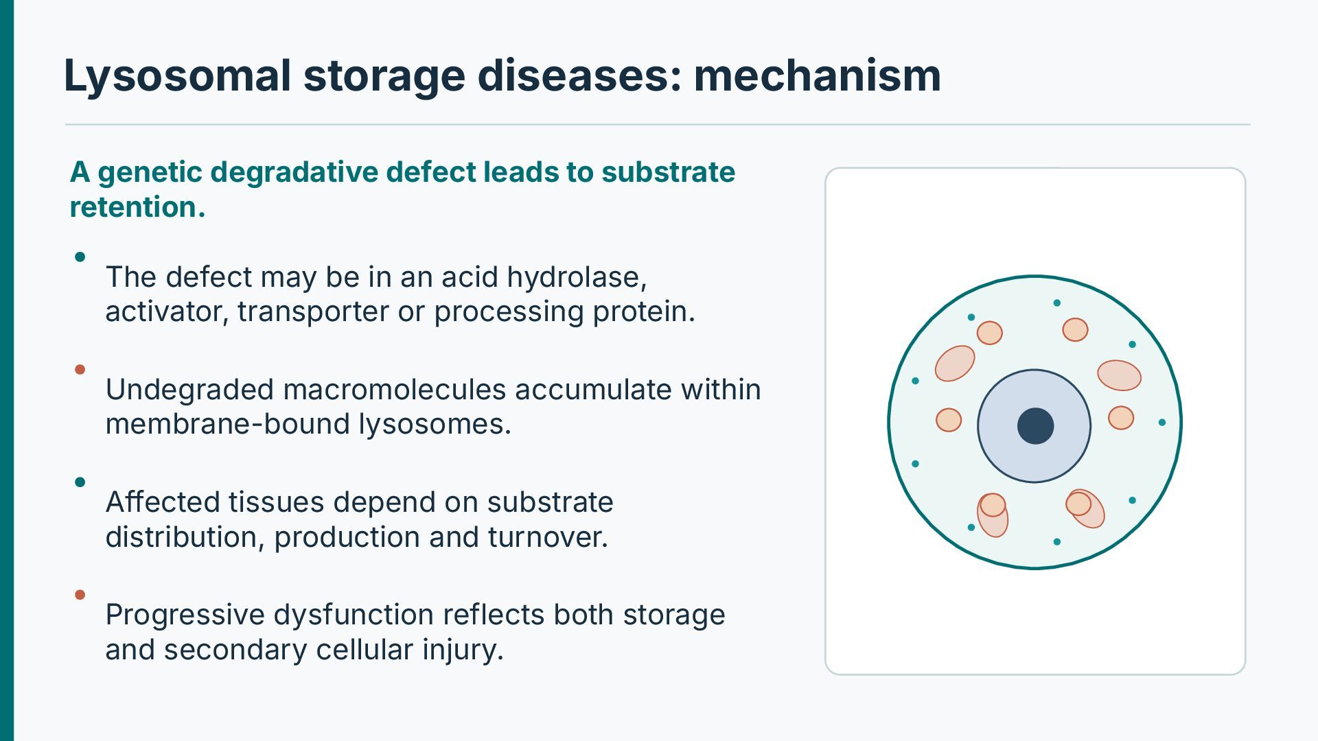
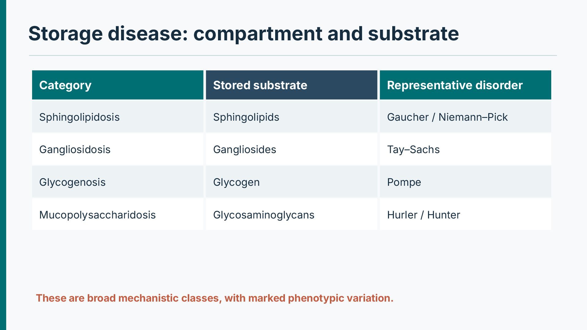
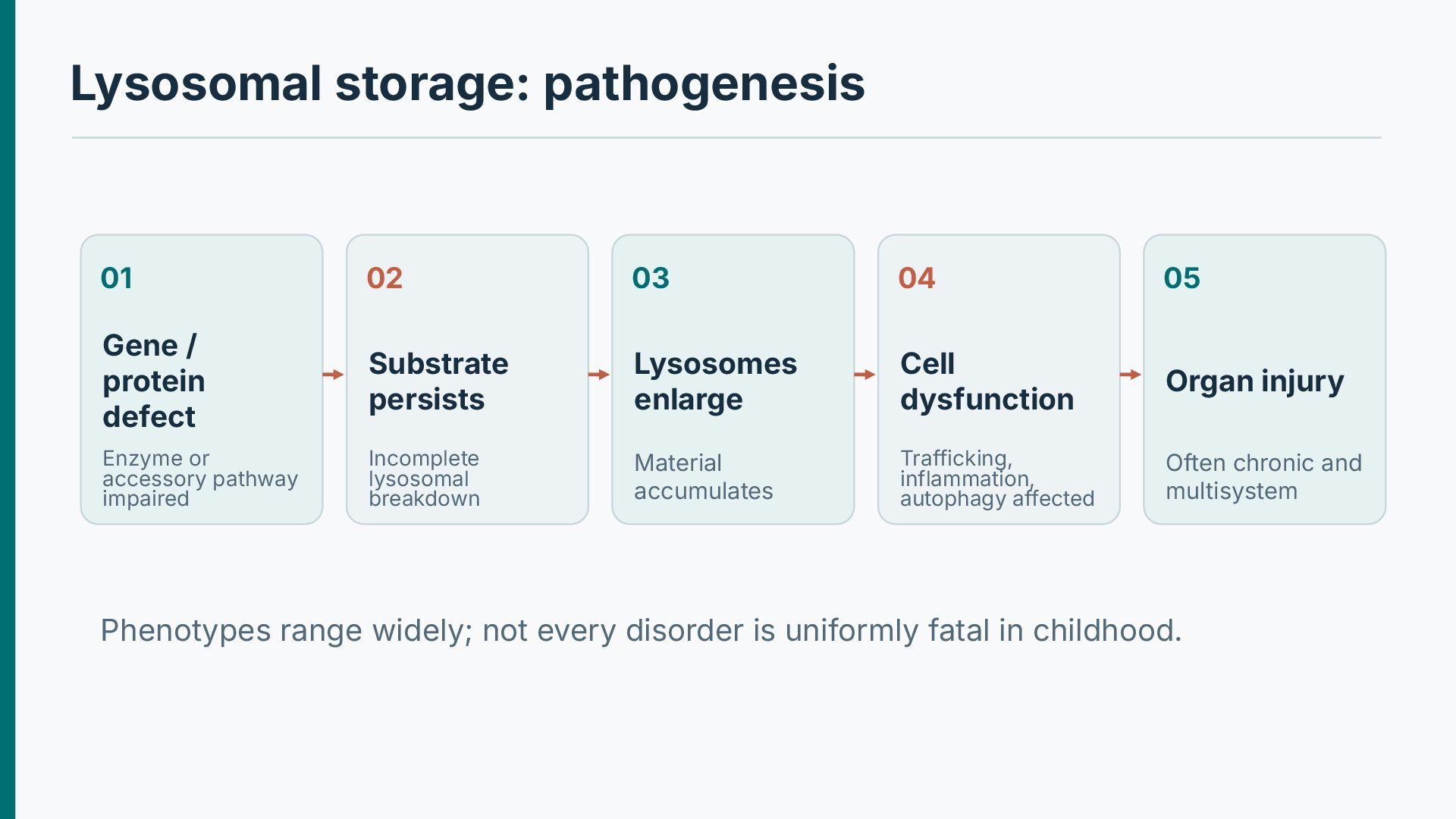
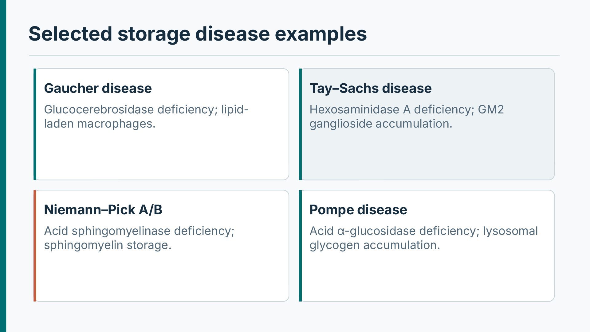
substrate retention. The defect may be in an acid hydrolase, activator, transporter or processing protein. Undegraded macromolecules accumulate within membrane-bound lysosomes. Affected tissues depend on substrate distribution, production and turnover. Progressive dysfunction reflects both storage and secondary cellular injury.
protein defect Substrate persists Lysosomes enlarge Cell dysfunction Organ injury Enzyme or accessory pathway impaired Incomplete lysosomal breakdown Material accumulates Trafficking, inflammation, autophagy affected Often chronic and multisystem Phenotypes range widely; not every disorder is uniformly fatal in childhood.
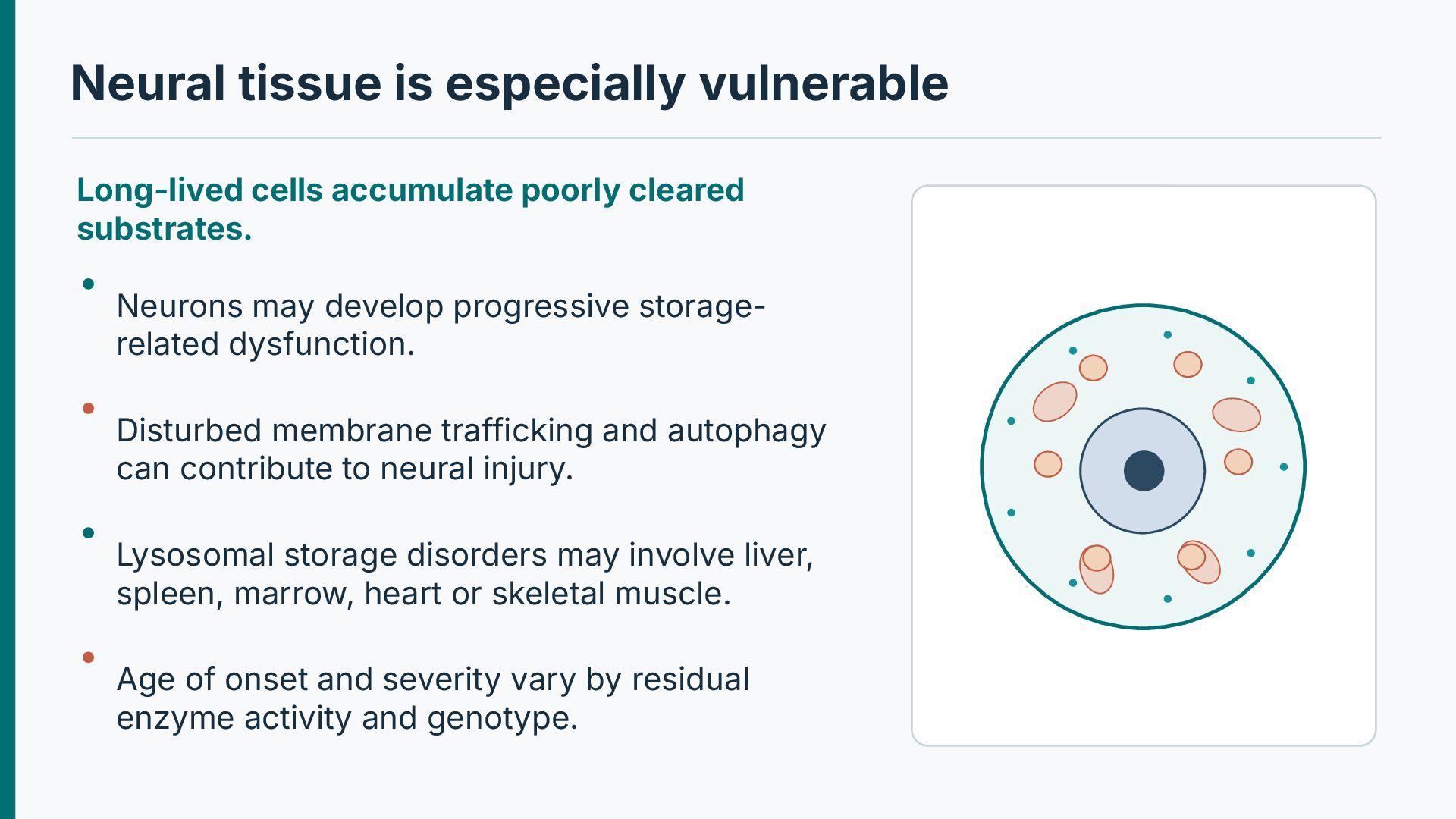
substrates. Neurons may develop progressive storagerelated dysfunction. Disturbed membrane trafficking and autophagy can contribute to neural injury. Lysosomal storage disorders may involve liver, spleen, marrow, heart or skeletal muscle. Age of onset and severity vary by residual enzyme activity and genotype.
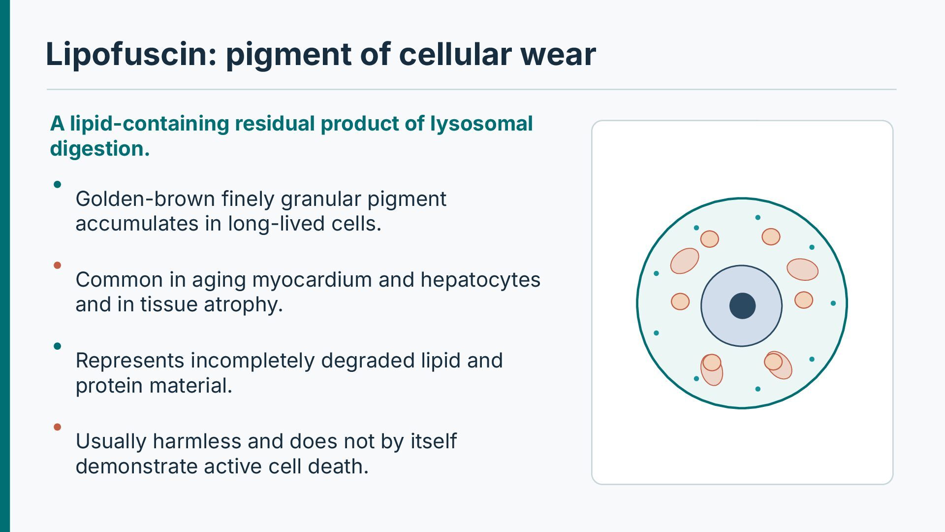
lysosomal digestion. Golden-brown finely granular pigment accumulates in long-lived cells. Common in aging myocardium and hepatocytes and in tissue atrophy. Represents incompletely degraded lipid and protein material. Usually harmless and does not by itself demonstrate active cell death.
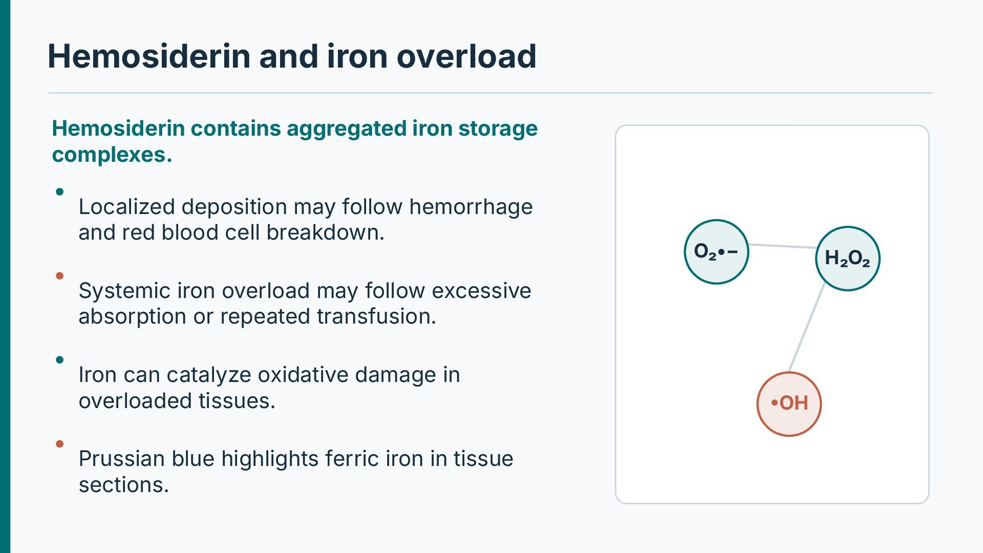
Localized deposition may follow hemorrhage and red blood cell breakdown. O₂•− H₂O₂ Systemic iron overload may follow excessive absorption or repeated transfusion. Iron can catalyze oxidative damage in overloaded tissues. Prussian blue highlights ferric iron in tissue sections. •OH
and context. Melanin is produced in melanocytes and transferred to adjacent epithelial cells. Bilirubin pigment may be evident in cholestatic hepatocytes or bile canaliculi. Inhaled carbon remains within pulmonary macrophages and lymph nodes. Tattoo pigments can persist in dermal macrophages.
by macrophages. Inhaled carbon accumulates in lung macrophages and draining nodes. Anthracotic pigment is commonly black and usually reflects environmental exposure. Tattoo pigment remains in dermal macrophages and fibroblastic tissue. Distribution and exposure history prevent confusion with endogenous pigment.
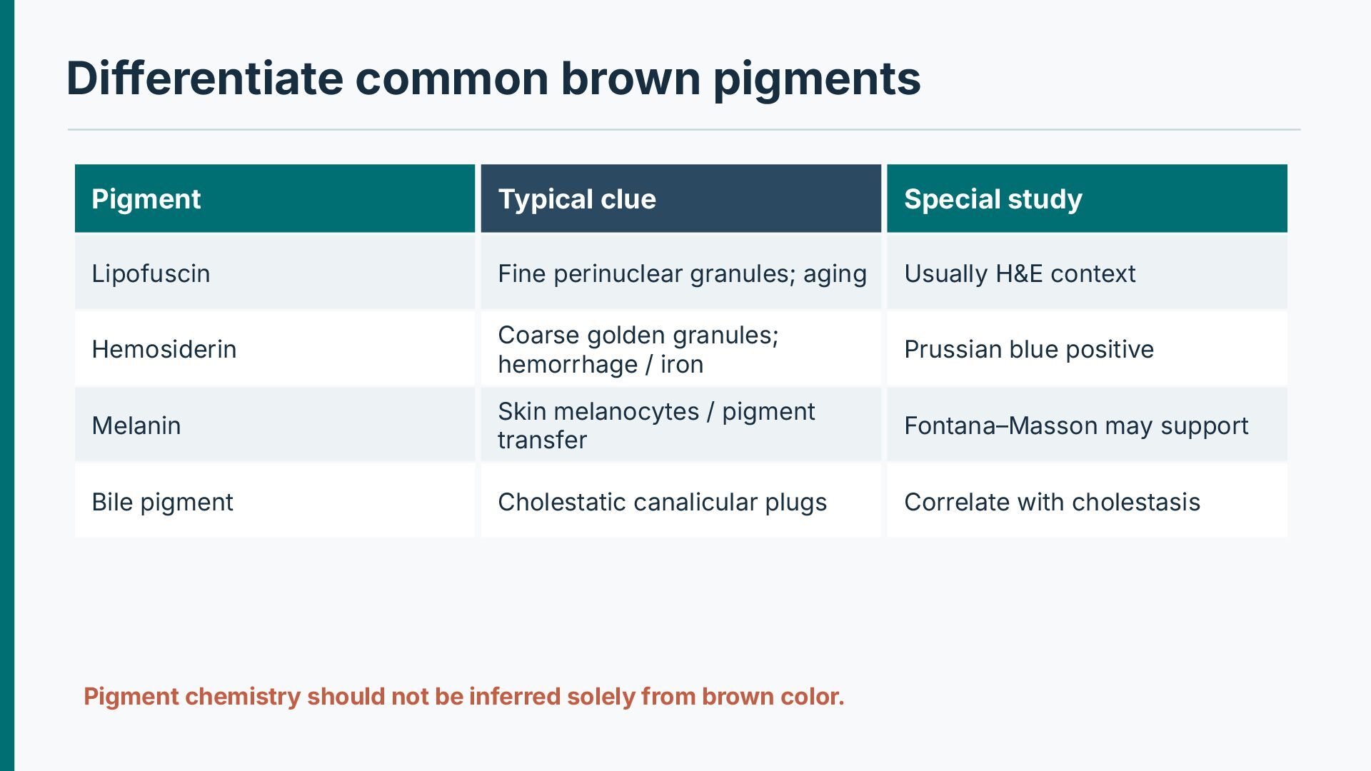
Fine perinuclear granules; aging Usually H&E context Hemosiderin Coarse golden granules; hemorrhage / iron Prussian blue positive Melanin Skin melanocytes / pigment transfer Fontana–Masson may support Bile pigment Cholestatic canalicular plugs Correlate with cholestasis Pigment chemistry should not be inferred solely from brown color.
vacuoles on routine H&E. The clinician asks whether these represent triglyceride. What is established morphologically, and which stain directly demonstrates neutral fat?
glycogen, pigment or exogenous particle. Cytosol, ER, lysosome or nucleus. Choose appropriate technique Assess consequence H&E, special stains, immunohistochemistry, enzymology or genetics. Harmless finding, metabolic dysfunction or progressive storage injury.
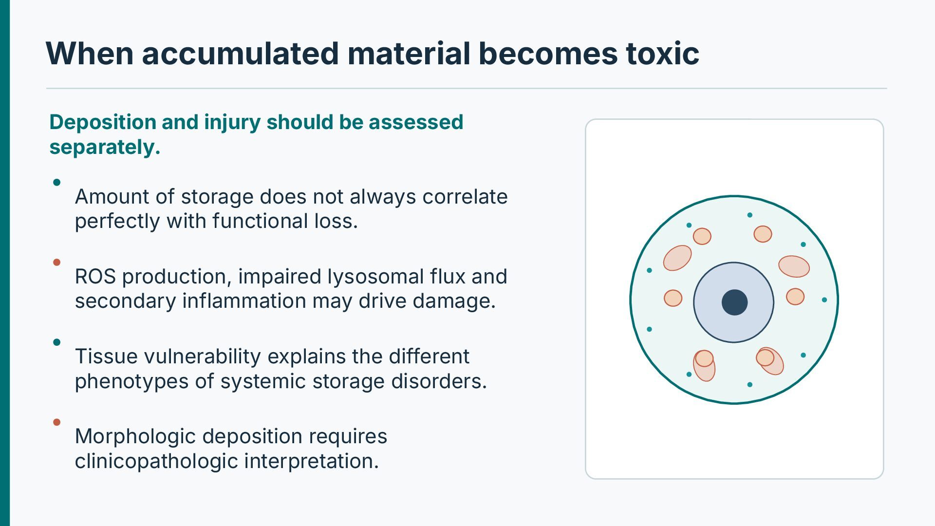
assessed separately. Amount of storage does not always correlate perfectly with functional loss. ROS production, impaired lysosomal flux and secondary inflammation may drive damage. Tissue vulnerability explains the different phenotypes of systemic storage disorders. Morphologic deposition requires clinicopathologic interpretation.
· Chapter: Cell Injury, Cell Death, and Adaptations For further study: correlate the molecular mechanisms with gross and microscopic pathology. Illustrative pathways are original schematics; selected histology images are retained from the supplied teaching material.


 abusuraihsakhri
 institutdelelevage
 trycycle
 aikinohara
 lychee1223
 syuchimu
 shumpei777
 masakiokuda
 estie
 arthur1
 akademiya2063
 aleyda
 thoeni
 katarinadahlin
 ivargrimstad
 tenderlove
 zakiyama
 addyosmani
 keathley
 tapps
 bluesmoon