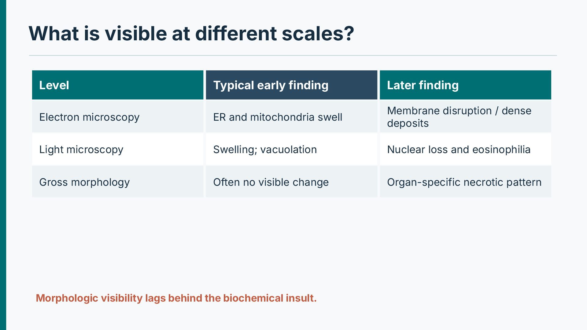
Later finding Electron microscopy ER and mitochondria swell Membrane disruption / dense deposits Light microscopy Swelling; vacuolation Nuclear loss and eosinophilia Gross morphology Often no visible change Organ-specific necrotic pattern Morphologic visibility lags behind the biochemical insult.
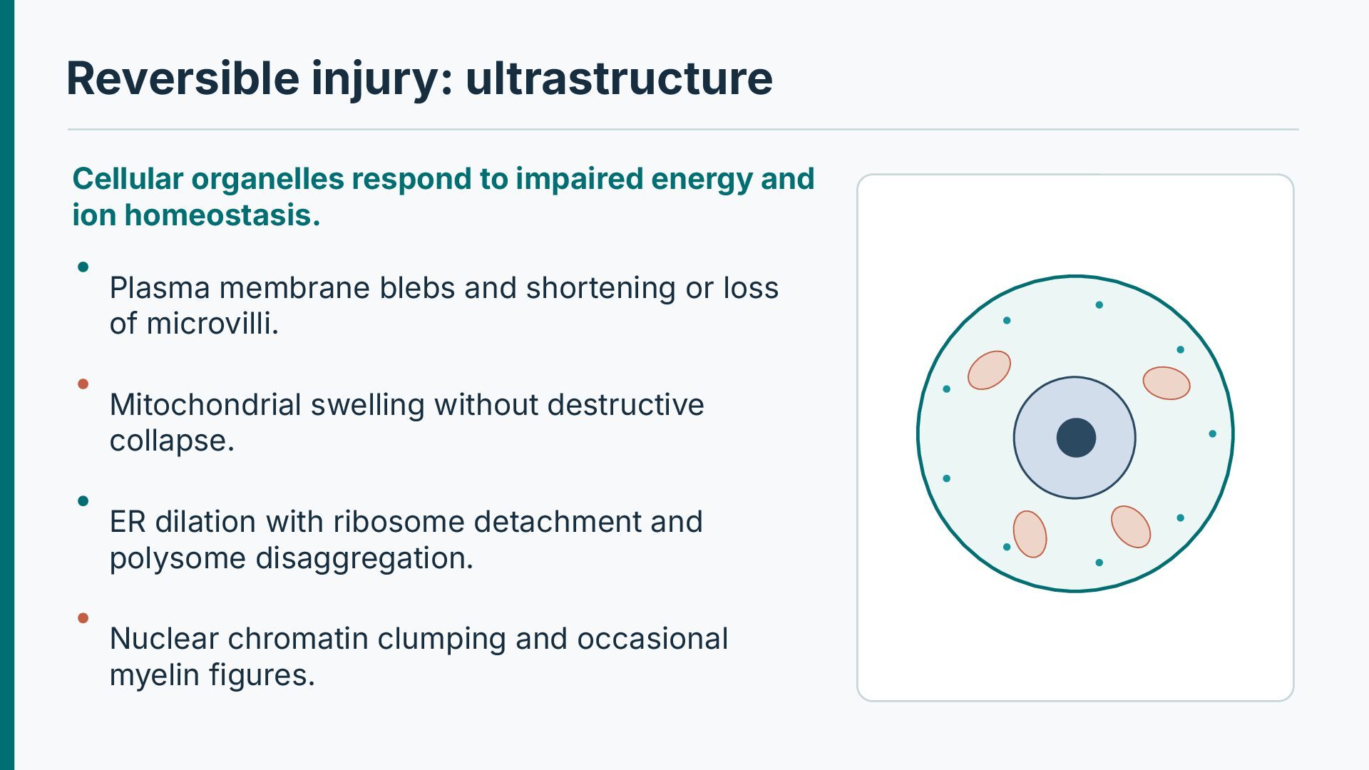
ion homeostasis. Plasma membrane blebs and shortening or loss of microvilli. Mitochondrial swelling without destructive collapse. ER dilation with ribosome detachment and polysome disaggregation. Nuclear chromatin clumping and occasional myelin figures.
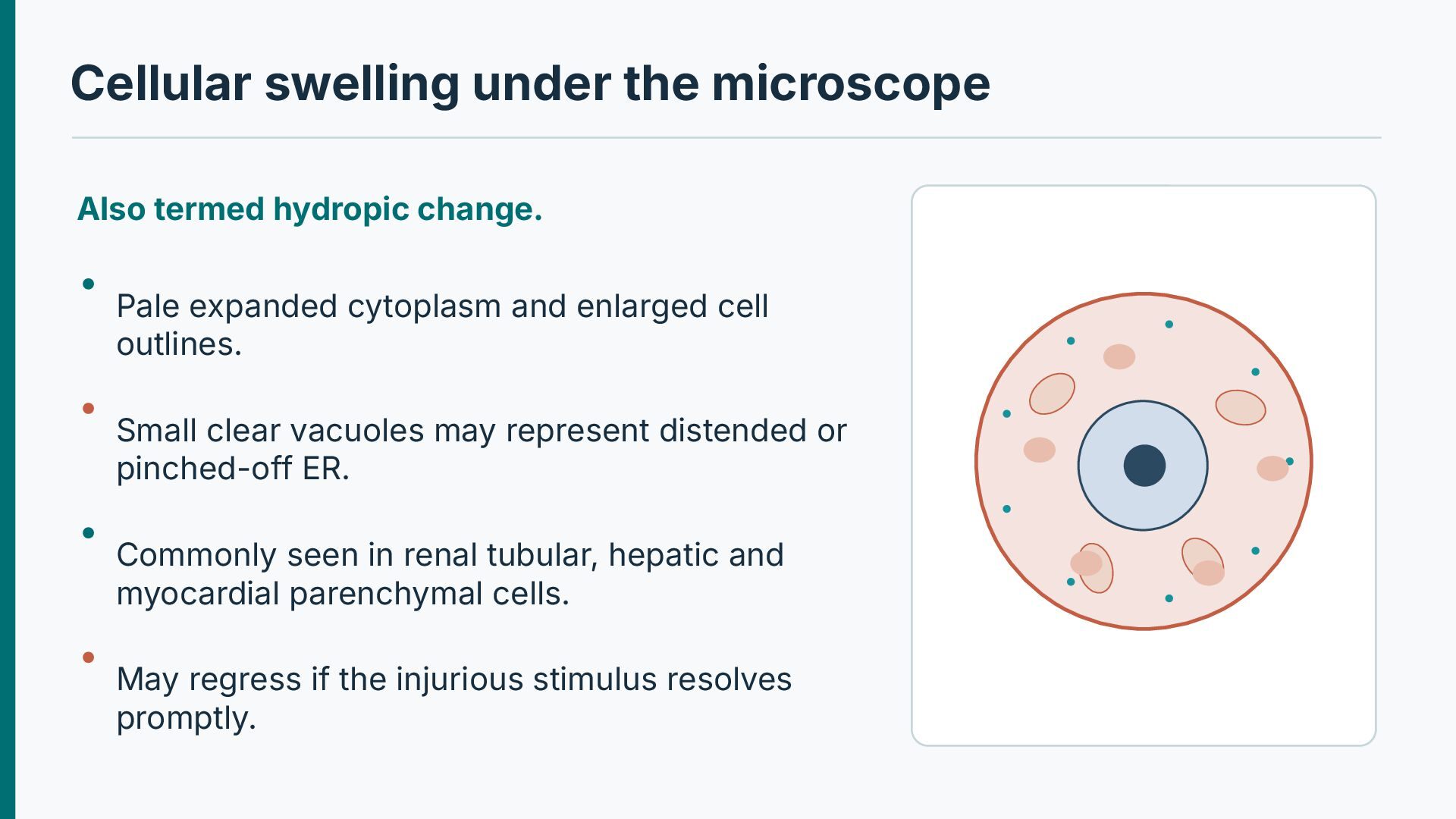
expanded cytoplasm and enlarged cell outlines. Small clear vacuoles may represent distended or pinched-off ER. Commonly seen in renal tubular, hepatic and myocardial parenchymal cells. May regress if the injurious stimulus resolves promptly.
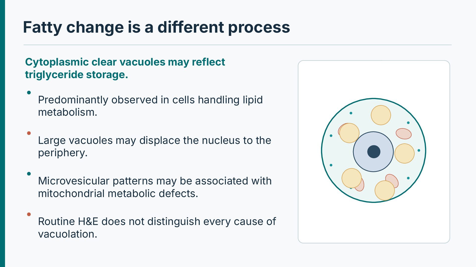
reflect triglyceride storage. Predominantly observed in cells handling lipid metabolism. Large vacuoles may displace the nucleus to the periphery. Microvesicular patterns may be associated with mitochondrial metabolic defects. Routine H&E does not distinguish every cause of vacuolation.
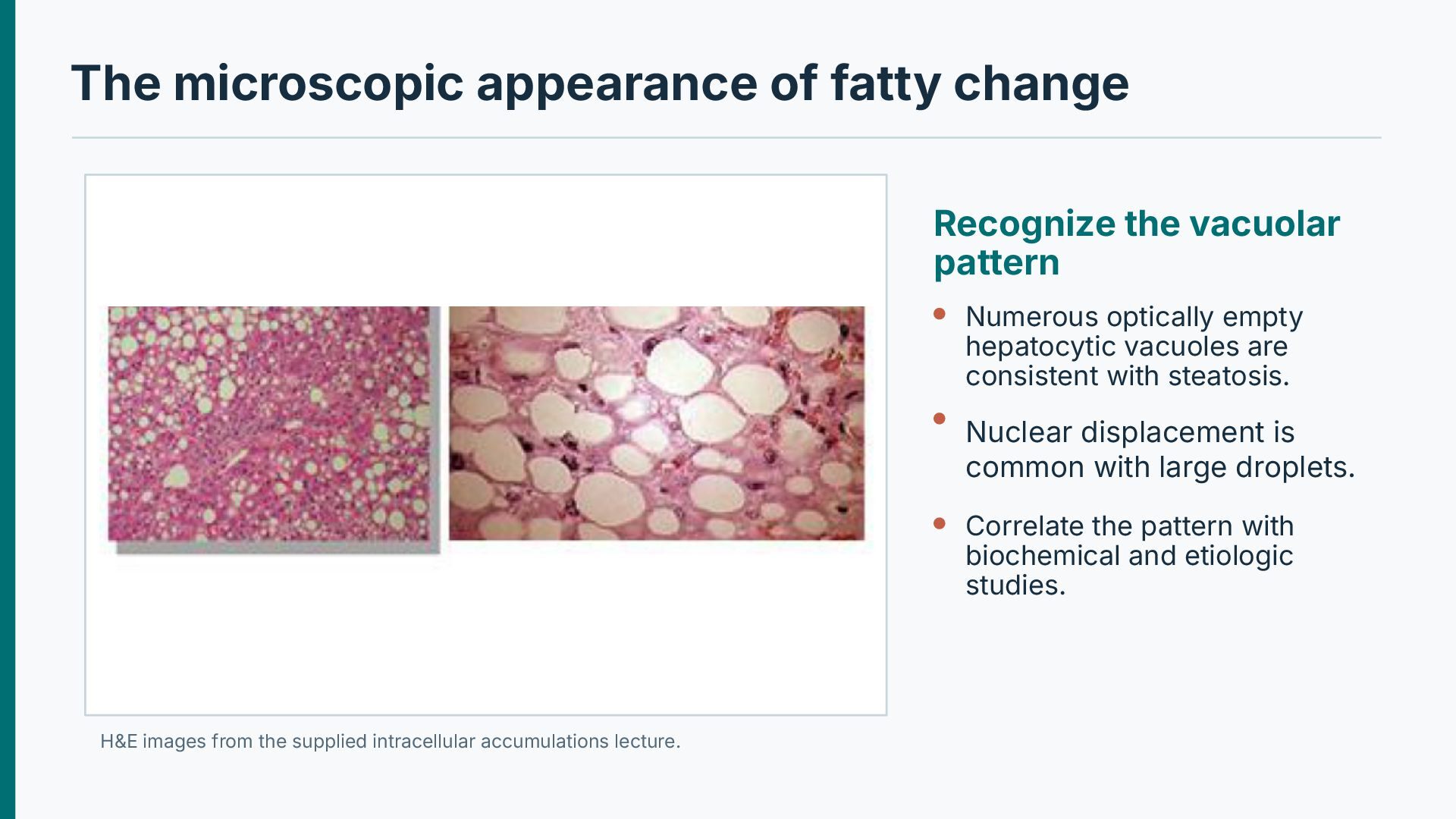
Numerous optically empty hepatocytic vacuoles are consistent with steatosis. Nuclear displacement is common with large droplets. Correlate the pattern with biochemical and etiologic studies. H&E images from the supplied intracellular accumulations lecture.
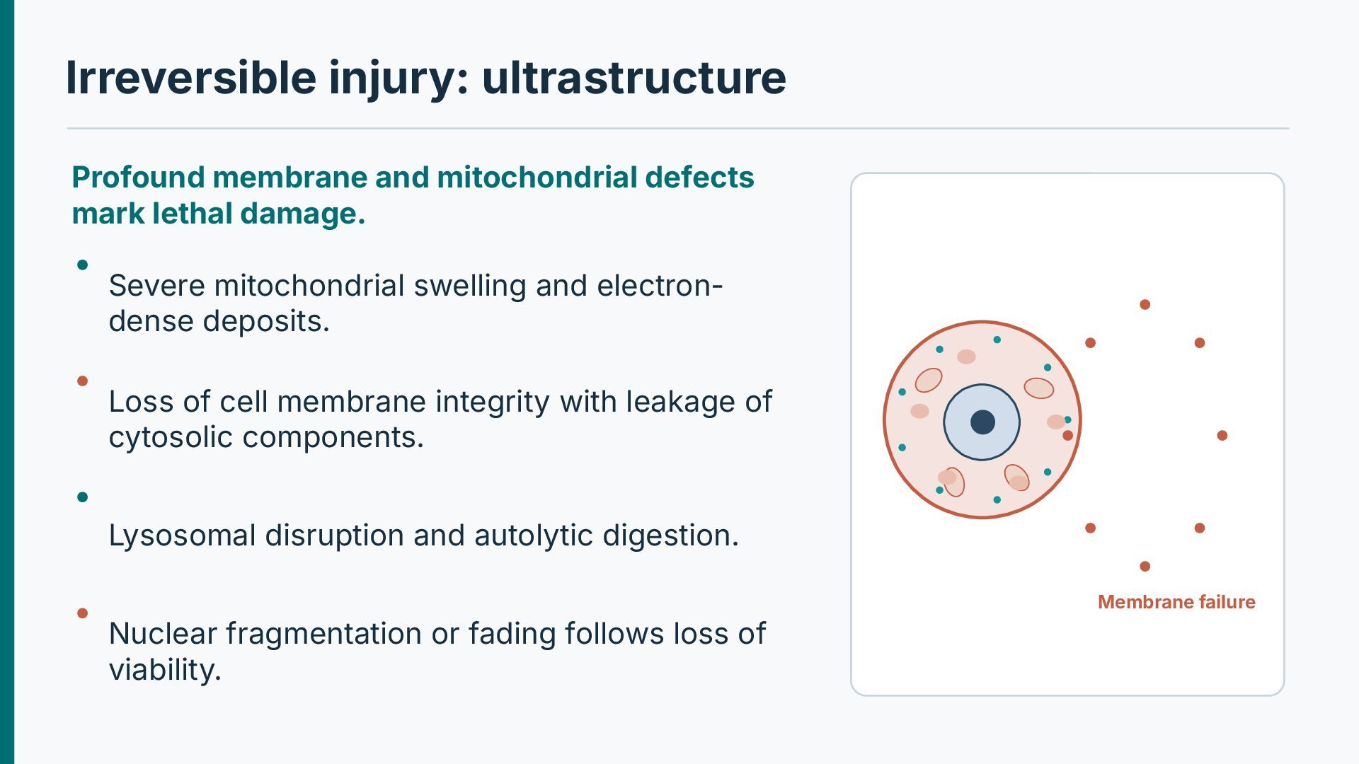
damage. Severe mitochondrial swelling and electrondense deposits. Loss of cell membrane integrity with leakage of cytosolic components. Lysosomal disruption and autolytic digestion. Membrane failure Nuclear fragmentation or fading follows loss of viability.
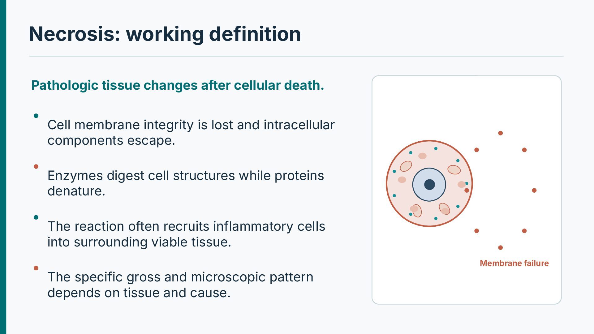
membrane integrity is lost and intracellular components escape. Enzymes digest cell structures while proteins denature. The reaction often recruits inflammatory cells into surrounding viable tissue. Membrane failure The specific gross and microscopic pattern depends on tissue and cause.
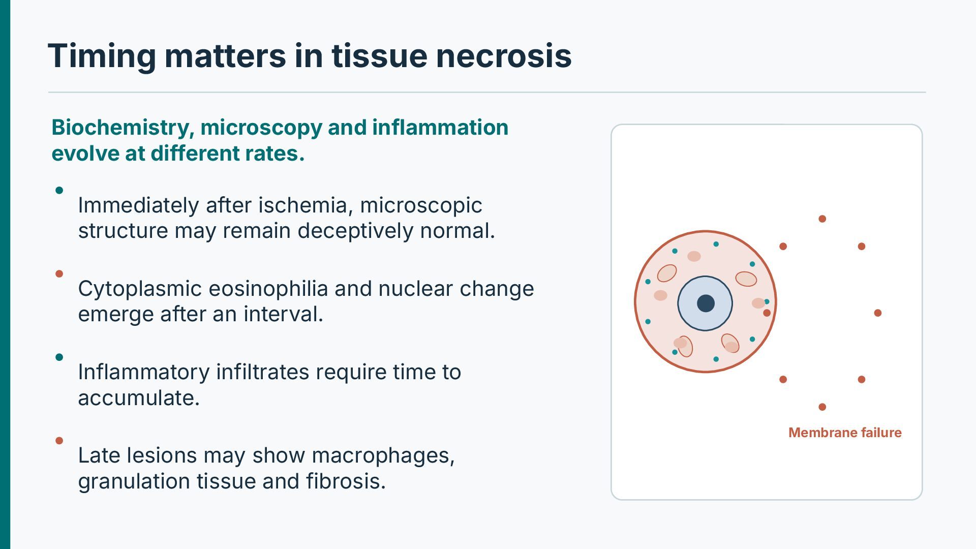
at different rates. Immediately after ischemia, microscopic structure may remain deceptively normal. Cytoplasmic eosinophilia and nuclear change emerge after an interval. Inflammatory infiltrates require time to accumulate. Membrane failure Late lesions may show macrophages, granulation tissue and fibrosis.
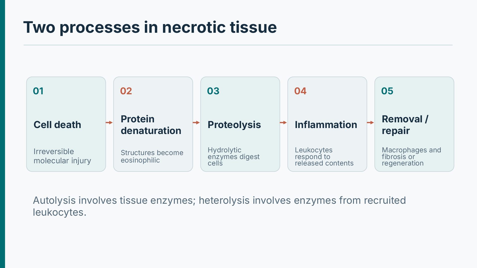
Cell death Protein denaturation Proteolysis Inflammation Removal / repair Irreversible molecular injury Structures become eosinophilic Hydrolytic enzymes digest cells Leukocytes respond to released contents Macrophages and fibrosis or regeneration Autolysis involves tissue enzymes; heterolysis involves enzymes from recruited leukocytes.
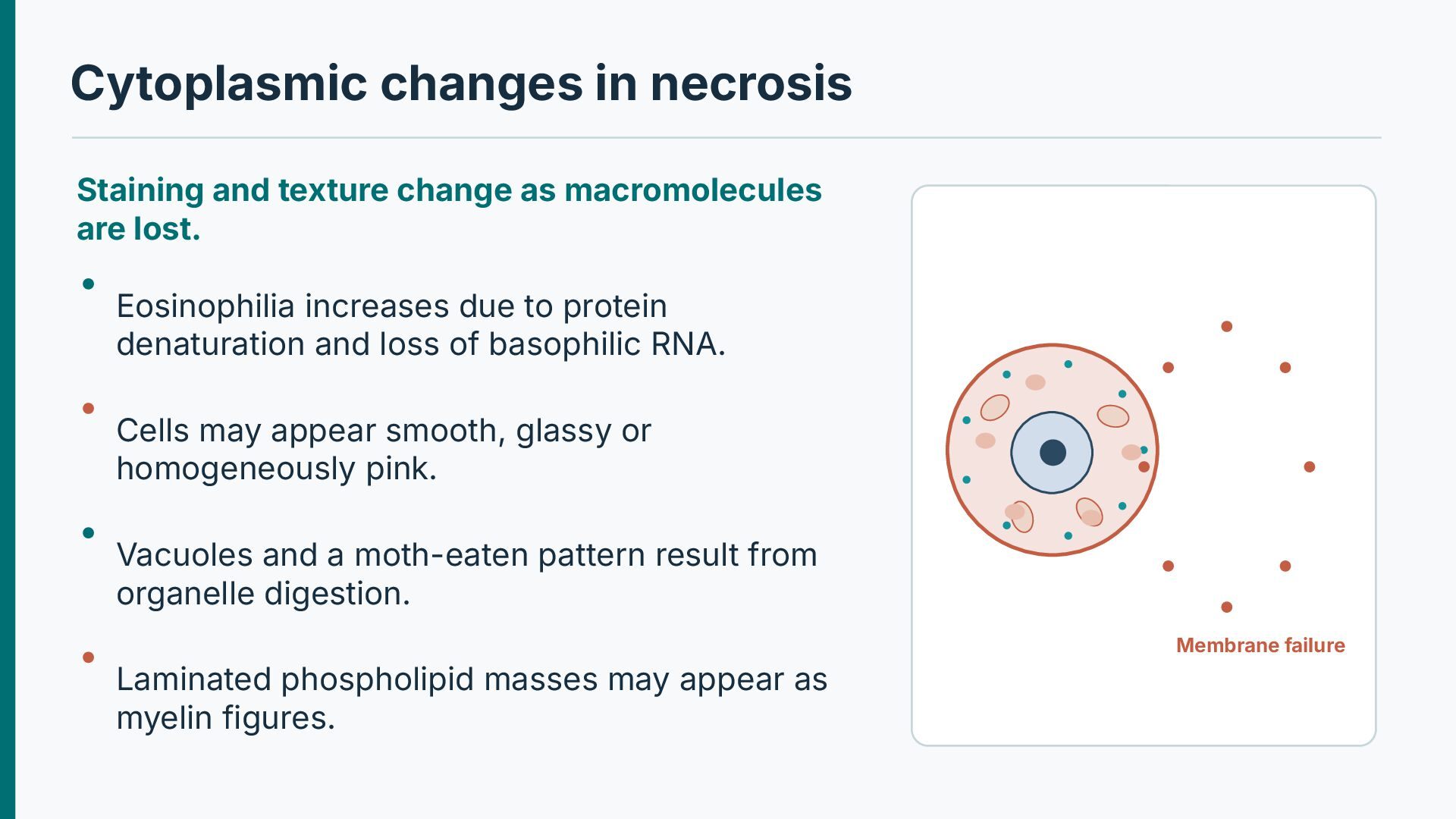
are lost. Eosinophilia increases due to protein denaturation and loss of basophilic RNA. Cells may appear smooth, glassy or homogeneously pink. Vacuoles and a moth-eaten pattern result from organelle digestion. Membrane failure Laminated phospholipid masses may appear as myelin figures.
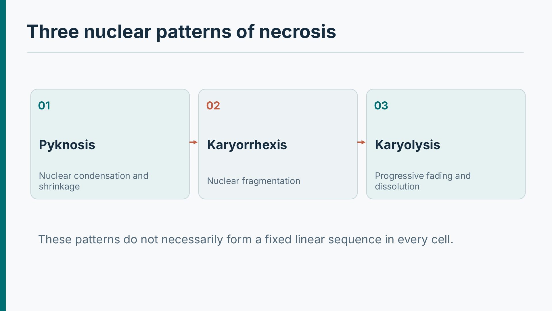
Karyolysis Nuclear condensation and shrinkage Nuclear fragmentation Progressive fading and dissolution These patterns do not necessarily form a fixed linear sequence in every cell.
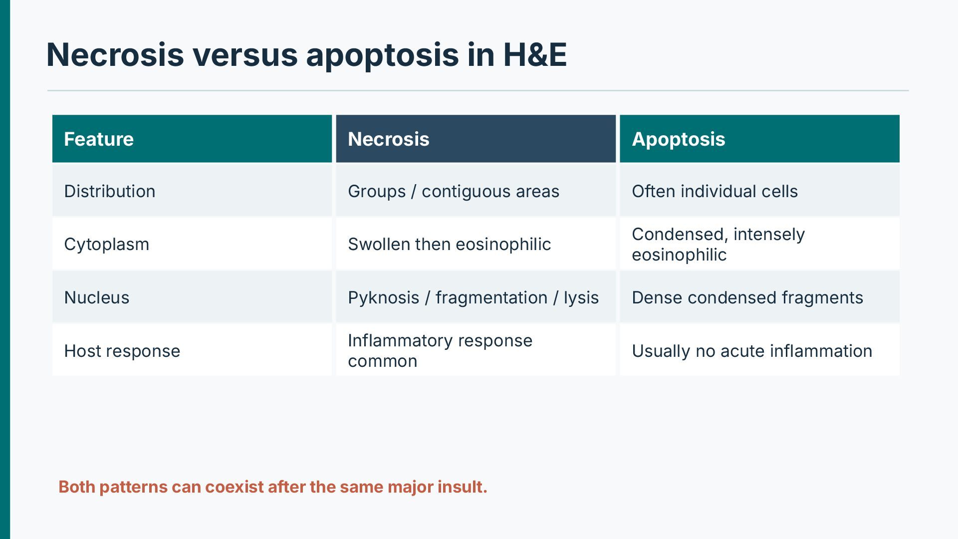
/ contiguous areas Often individual cells Cytoplasm Swollen then eosinophilic Condensed, intensely eosinophilic Nucleus Pyknosis / fragmentation / lysis Dense condensed fragments Host response Inflammatory response common Usually no acute inflammation Both patterns can coexist after the same major insult.
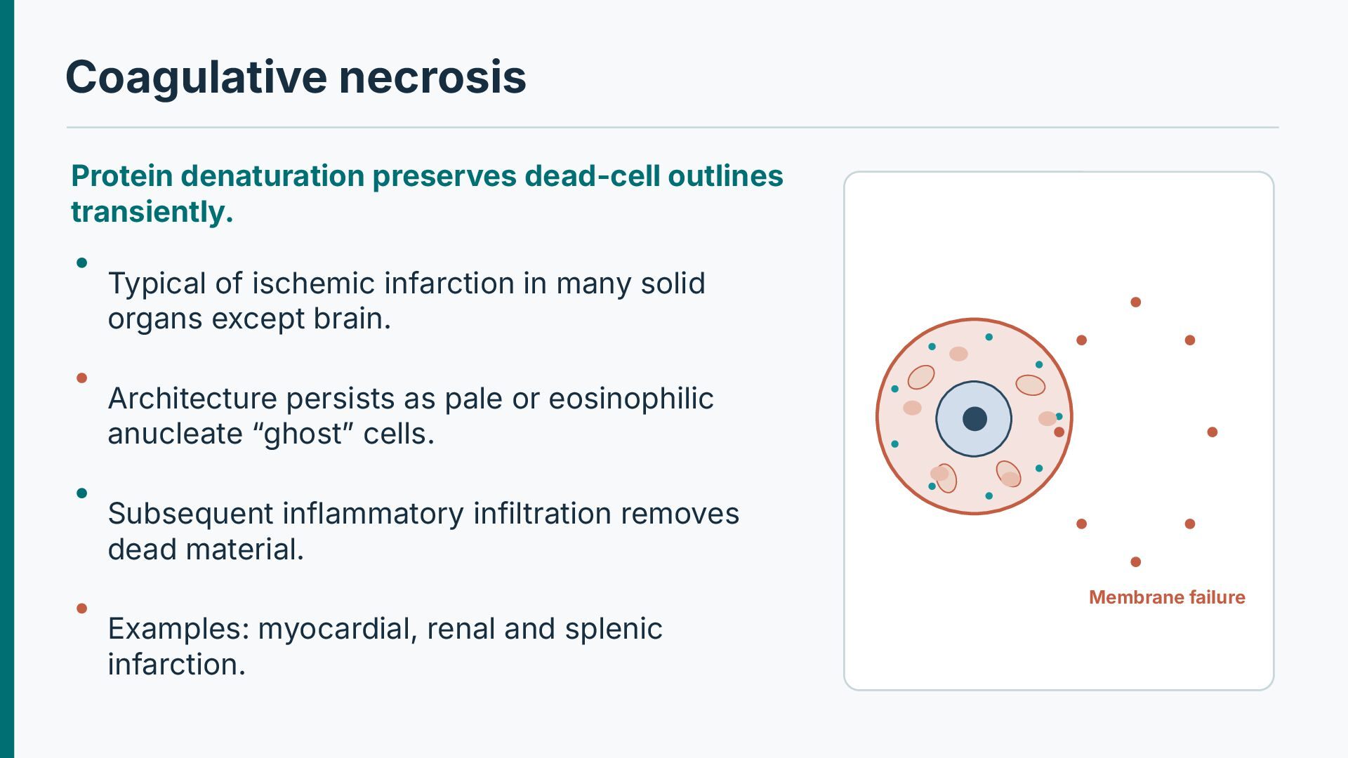
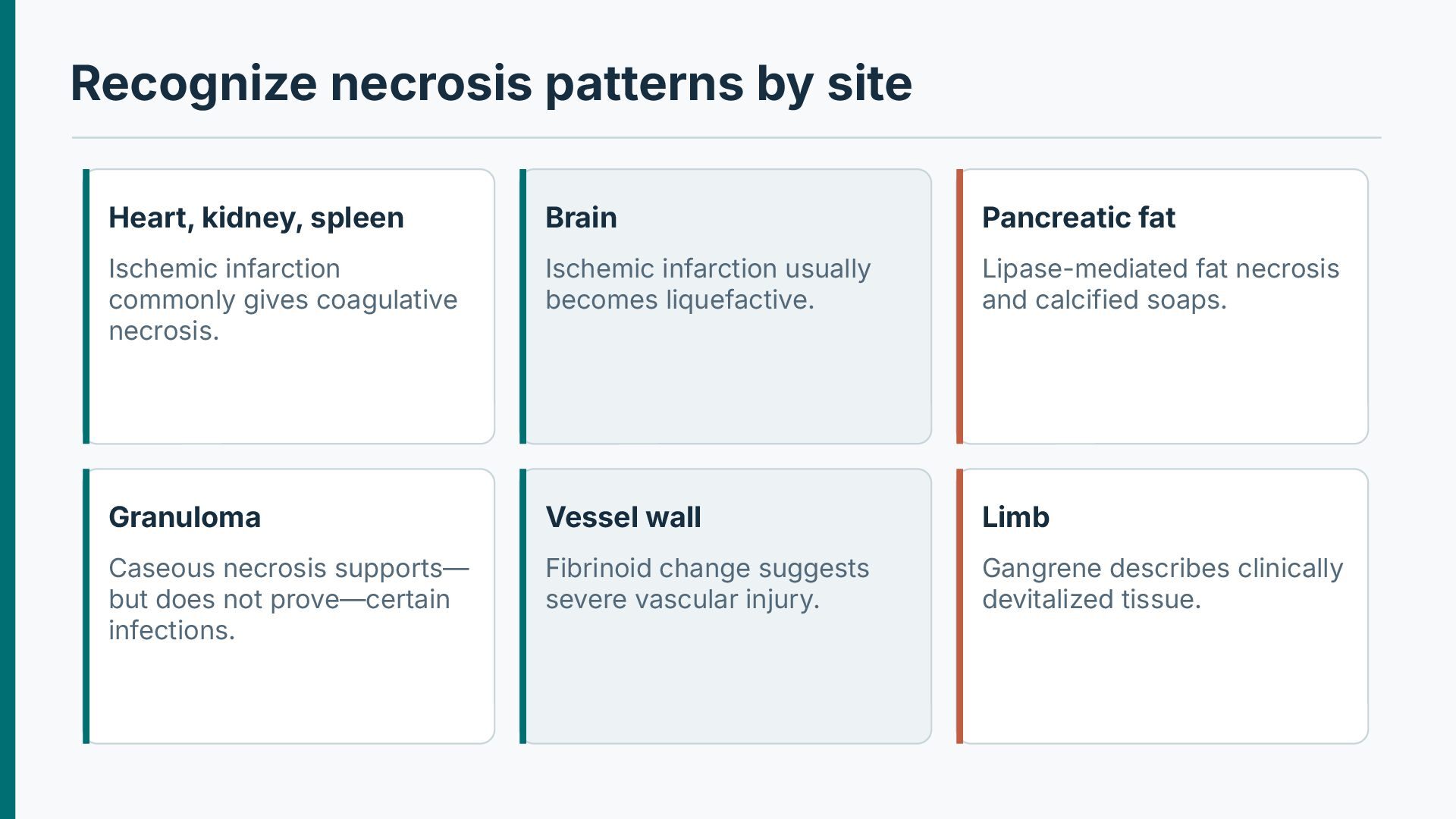
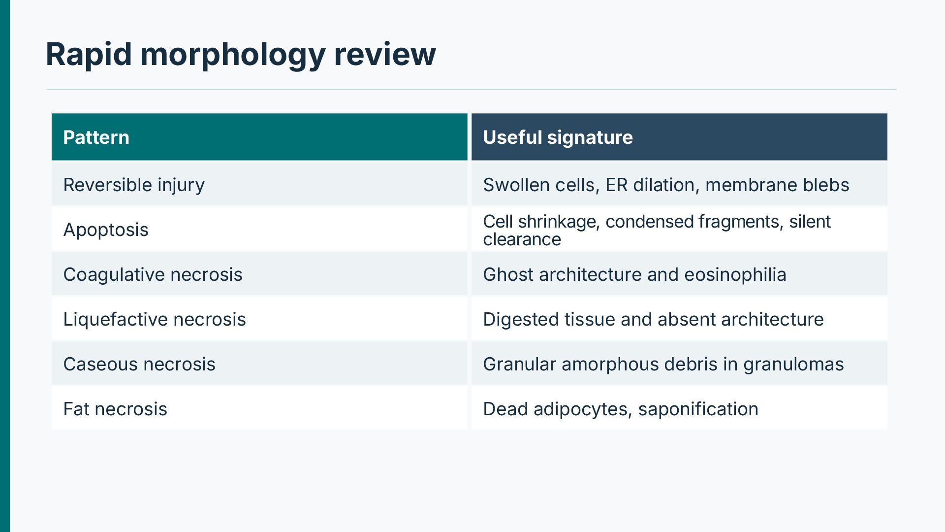
ischemic infarction in many solid organs except brain. Architecture persists as pale or eosinophilic anucleate “ghost” cells. Subsequent inflammatory infiltration removes dead material. Membrane failure Examples: myocardial, renal and splenic infarction.
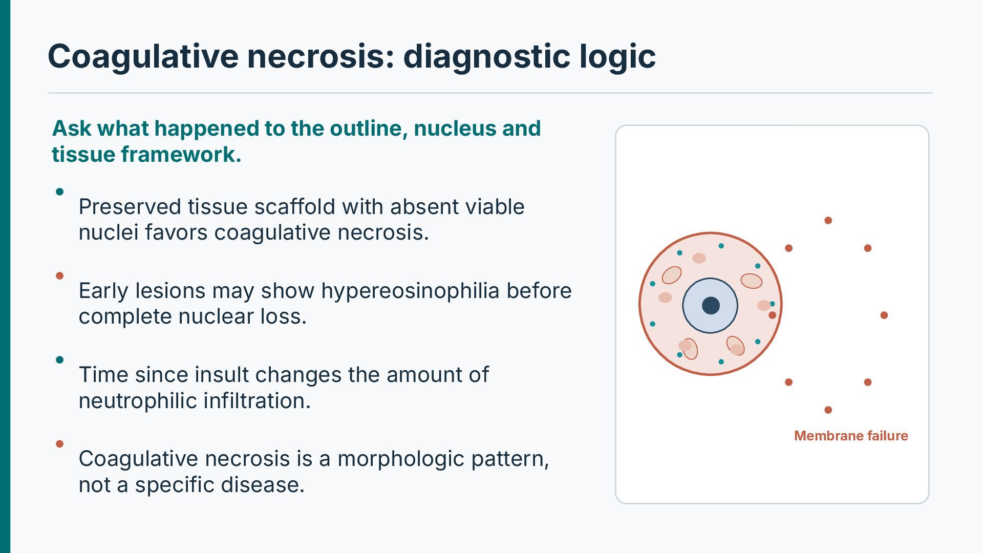
nucleus and tissue framework. Preserved tissue scaffold with absent viable nuclei favors coagulative necrosis. Early lesions may show hypereosinophilia before complete nuclear loss. Time since insult changes the amount of neutrophilic infiltration. Membrane failure Coagulative necrosis is a morphologic pattern, not a specific disease.
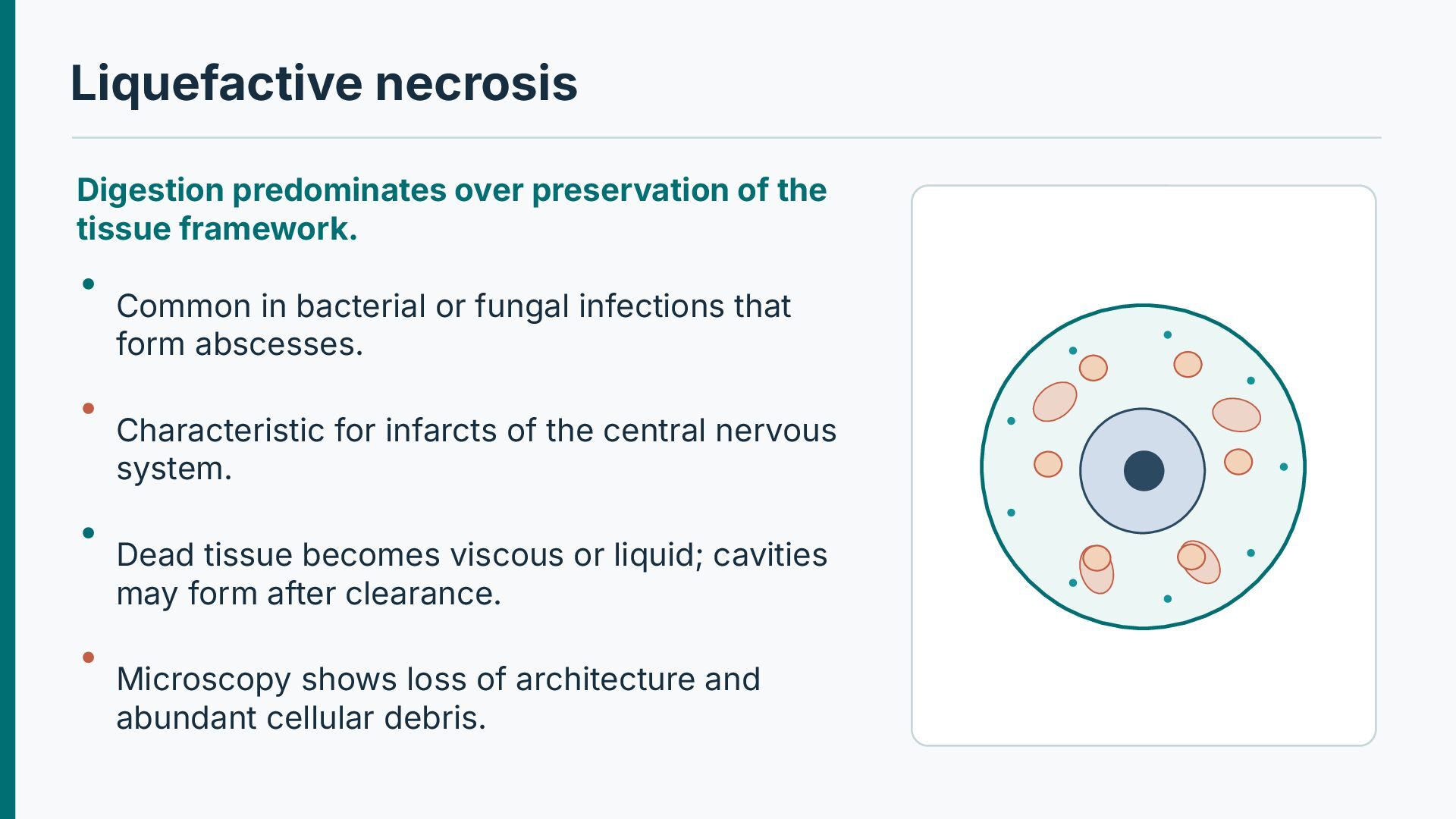
Common in bacterial or fungal infections that form abscesses. Characteristic for infarcts of the central nervous system. Dead tissue becomes viscous or liquid; cavities may form after clearance. Microscopy shows loss of architecture and abundant cellular debris.
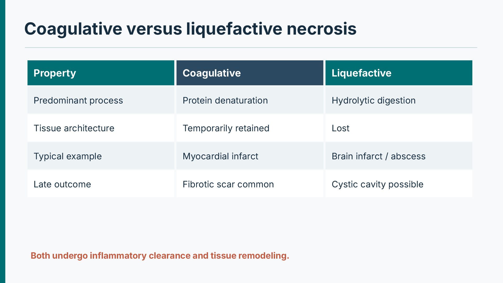
denaturation Hydrolytic digestion Tissue architecture Temporarily retained Lost Typical example Myocardial infarct Brain infarct / abscess Late outcome Fibrotic scar common Cystic cavity possible Both undergo inflammatory clearance and tissue remodeling.
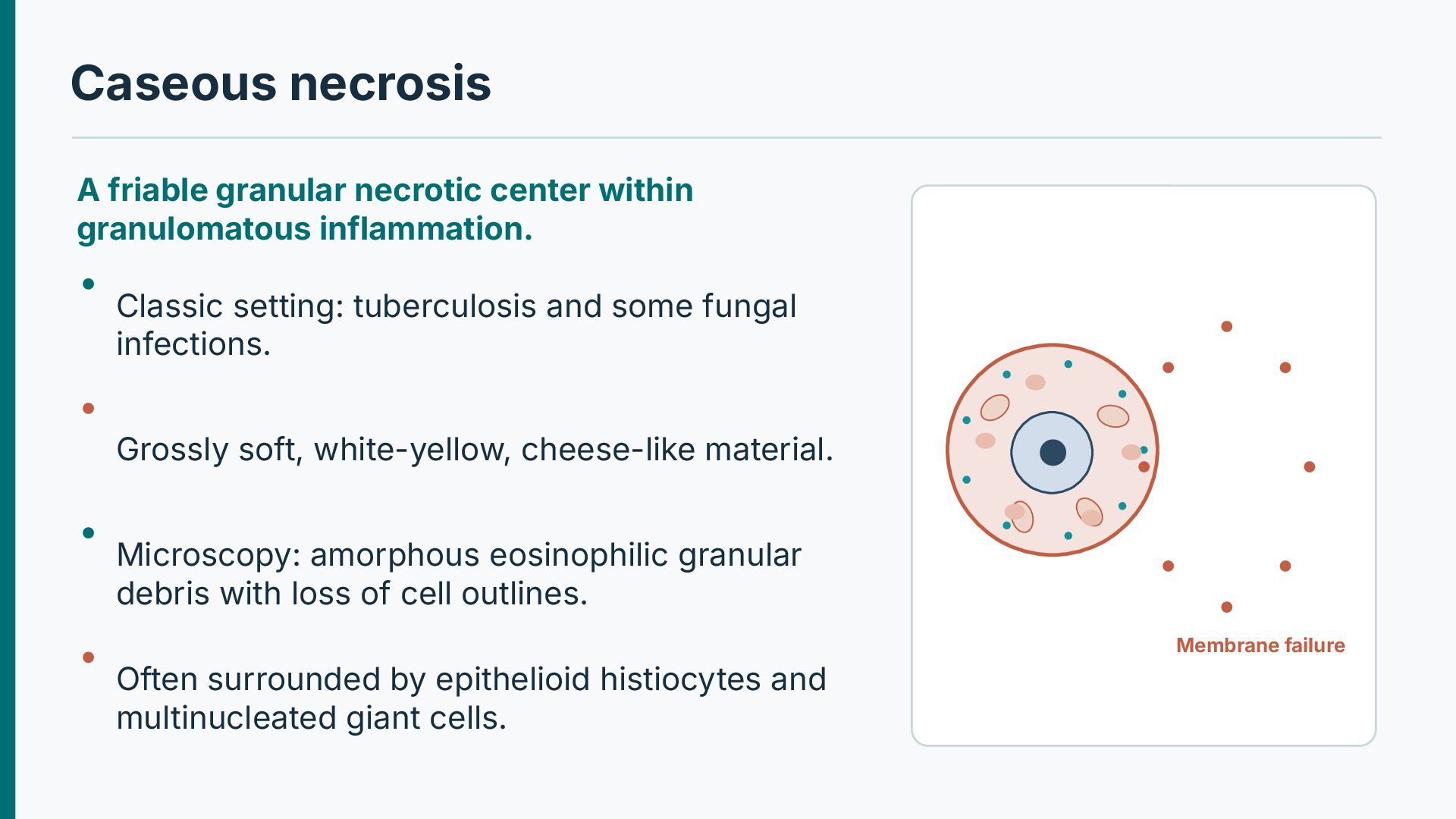
Classic setting: tuberculosis and some fungal infections. Grossly soft, white-yellow, cheese-like material. Microscopy: amorphous eosinophilic granular debris with loss of cell outlines. Membrane failure Often surrounded by epithelioid histiocytes and multinucleated giant cells.
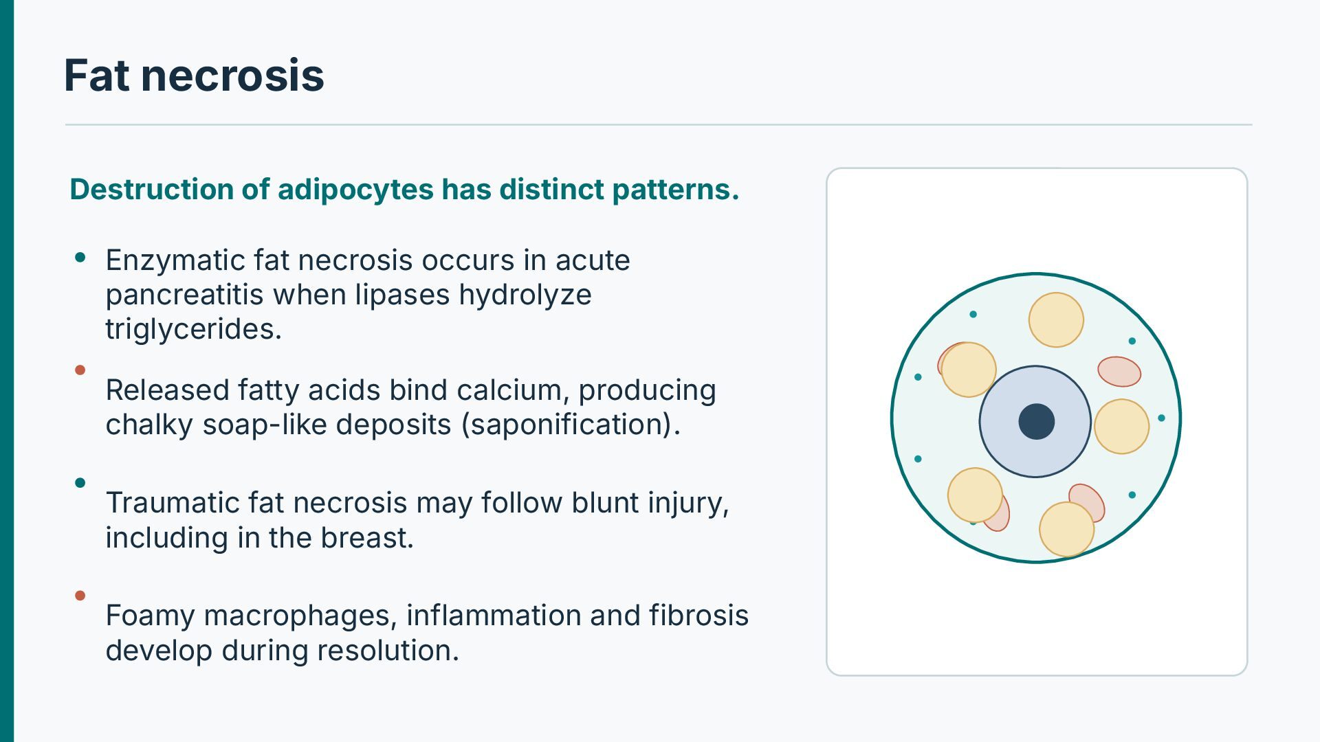
necrosis occurs in acute pancreatitis when lipases hydrolyze triglycerides. Released fatty acids bind calcium, producing chalky soap-like deposits (saponification). Traumatic fat necrosis may follow blunt injury, including in the breast. Foamy macrophages, inflammation and fibrosis develop during resolution.
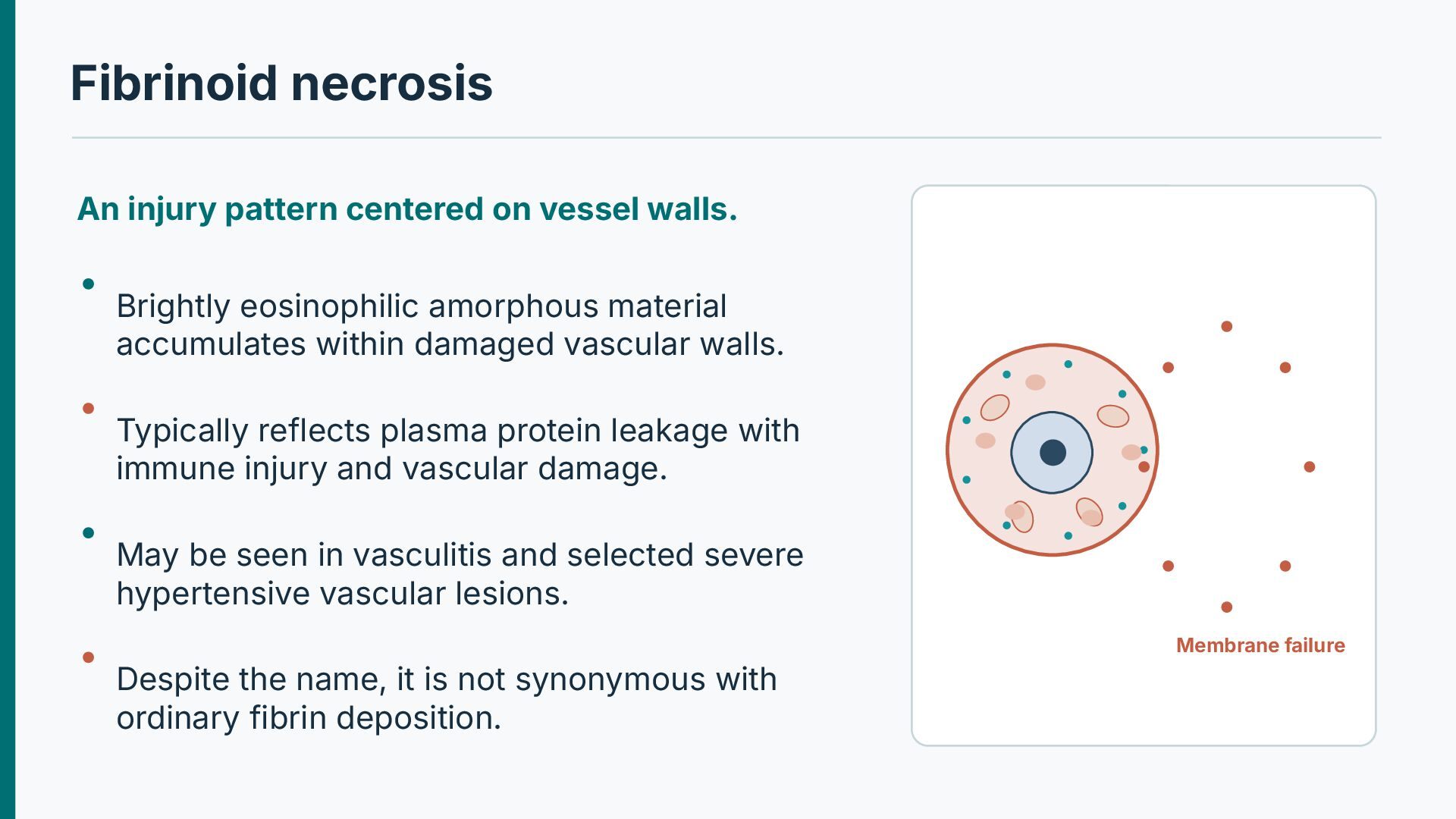
eosinophilic amorphous material accumulates within damaged vascular walls. Typically reflects plasma protein leakage with immune injury and vascular damage. May be seen in vasculitis and selected severe hypertensive vascular lesions. Membrane failure Despite the name, it is not synonymous with ordinary fibrin deposition.
more informative than the stain color. Evaluate whether eosinophilic material lies within a damaged arterial wall. Seek associated inflammation, vascular narrowing or thrombosis. Vessel-wall damage in immune-mediated disease can allow plasma proteins to enter the wall. Membrane failure Anatomic pattern and clinical tests are needed for etiologic classification.
unique microscopic necrosis mechanism. Dry gangrene is usually ischemic with predominant coagulative necrosis. Wet gangrene involves infection and prominent tissue liquefaction. Gas gangrene involves infection by gas-forming organisms, often clostridia. Membrane failure Extent and clinical features determine urgent management.
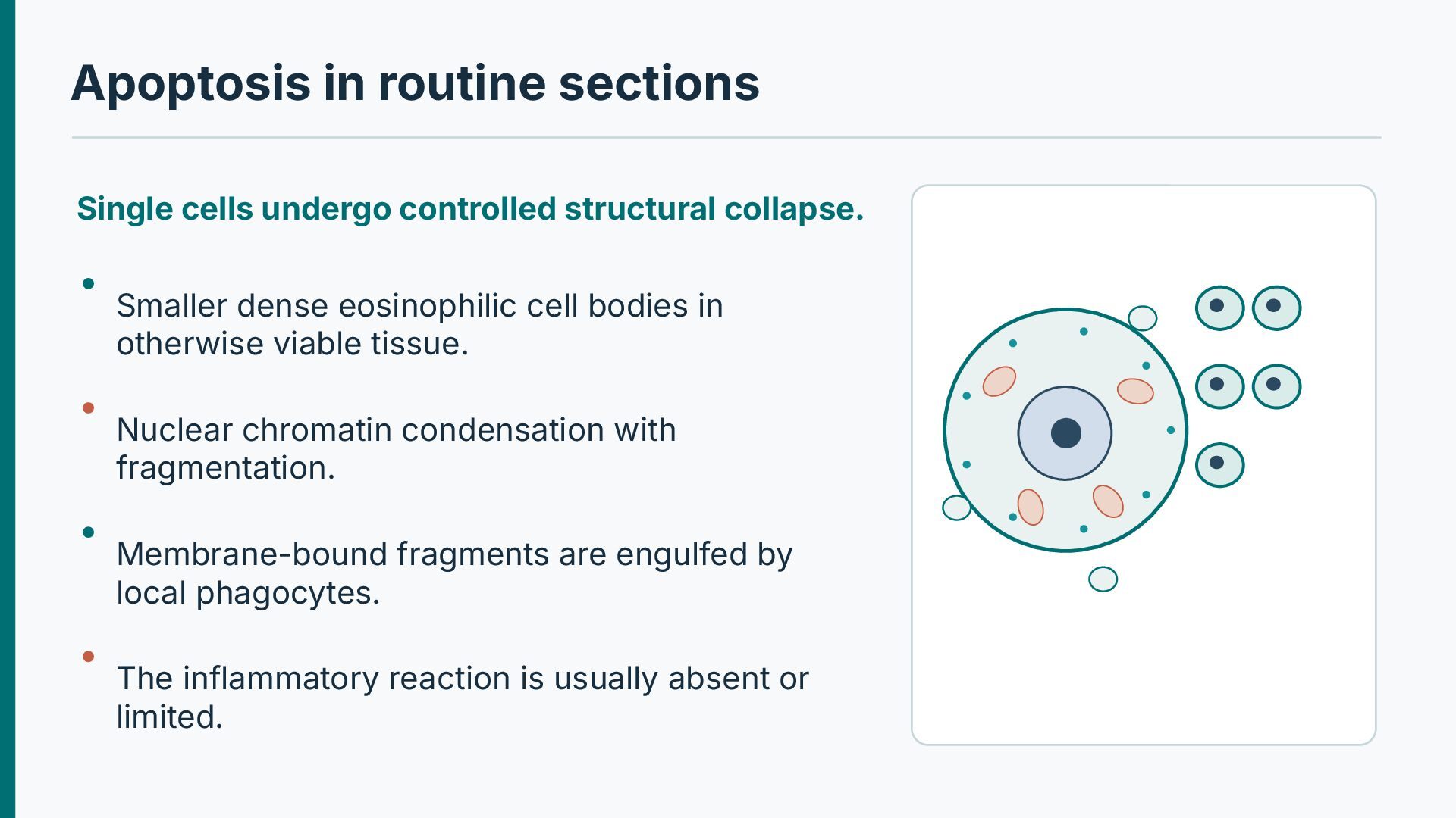
Smaller dense eosinophilic cell bodies in otherwise viable tissue. Nuclear chromatin condensation with fragmentation. Membrane-bound fragments are engulfed by local phagocytes. The inflammatory reaction is usually absent or limited.
alone. Autolysis after tissue removal can mimic or obscure cell injury. Nuclear pyknosis occurs in several contexts and is not by itself diagnostic of apoptosis. Dense inflammatory infiltrates may obscure the original necrosis pattern. Serial sections, organ context and suitable ancillary tests improve confidence.
brain lesion has lost recognizable parenchymal architecture and contains macrophages and fluid-filled spaces. Which morphologic process explains the cavitation?
· Chapter: Cell Injury, Cell Death, and Adaptations For further study: correlate the molecular mechanisms with gross and microscopic pathology. Illustrative pathways are original schematics; selected histology images are retained from the supplied teaching material.


 abusuraihsakhri
 syuchimu
 trycycle
 ykiyota
 salt2
 martinlippert
 ianozsvald
 konakalab
 hayataka88
 takusamar
 lara
 lfama
 aleyda
 thoeni
 codingconduct
 inesmontani
 thekraken
 marketingsoph
 reverentgeek
 michaelherold
 maggiecrowley
 keathley