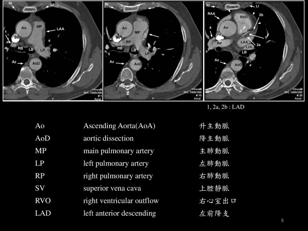
aortic dissection 降主動脈 MP main pulmonary artery 主肺動脈 LP left pulmonary artery 左肺動脈 RP right pulmonary artery 右肺動脈 SV superior vena cava 上腔靜脈 RVO right ventricular outflow 右心室出口 LAD left anterior descending 左前降支 8
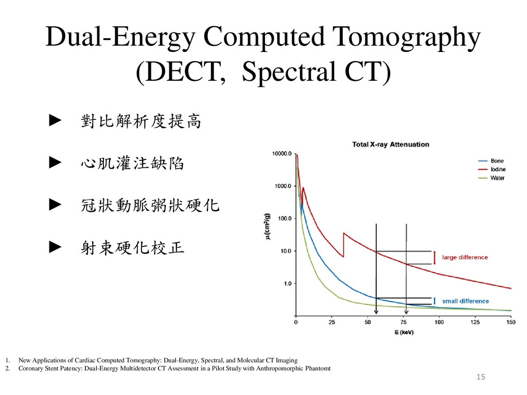
► 冠狀動脈粥狀硬化 ► 射束硬化校正 1. New Applications of Cardiac Computed Tomography: Dual-Energy, Spectral, and Molecular CT Imaging 2. Coronary Stent Patency: Dual-Energy Multidetector CT Assessment in a Pilot Study with Anthropomorphic Phantomt 15
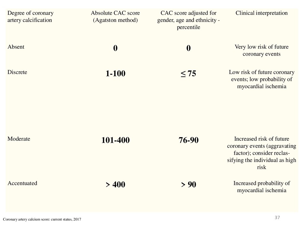
CAC score adjusted for gender, age and ethnicity - percentile Clinical interpretation Absent 0 0 Very low risk of future coronary events Discrete 1-100 ≤ 75 Low risk of future coronary events; low probability of myocardial ischemia Moderate 101-400 76-90 Increased risk of future coronary events (aggravating factor); consider reclas- sifying the individual as high risk Accentuated > 400 > 90 Increased probability of myocardial ischemia Coronary artery calcium score: current status, 2017 37
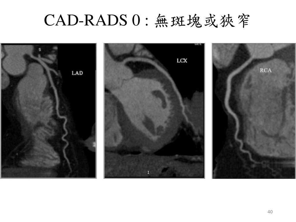
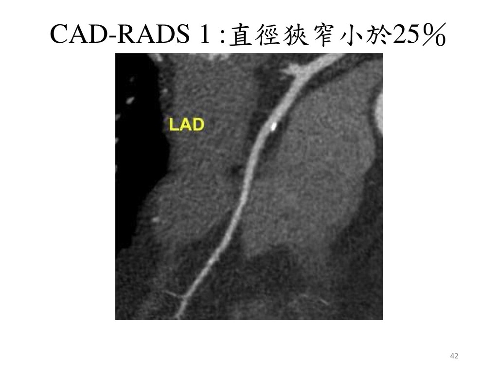
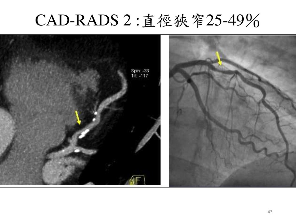
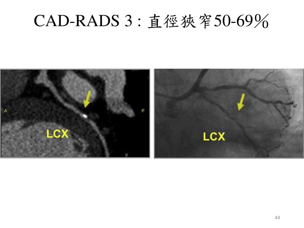
Cury, al. CAD-RADSTM Coronary Artery Disease – Reporting and Data System. An expert consensus document of the Society of Cardiovascular Computed Tomography (SCCT), the American College of Radiology (ACR) and the North American Society for Cardiovascular Imaging (NASCI). Endorsed by the American College of Cardiology, July, 2016. 38
Anatomy of the Heart at Multidetector CT: What the Radiologist Needs to Know, 2007. ► Lee W. Goldman. Principles of CT: Multislice CT, JNMT, 2008. ► J. of Cardiovasc, Trans. Res, Journal of CardiovascularTranslational Research, 2013. ► James K. Min, MD, al. Noninvasive Fractional Flow Reserve Derived From Coronary CT Angiography, 2015. ► Zahi A.FayadPhD, New Applications of Cardiac Computed Tomography: Dual-Energy, Spectral, and Molecular CT Imaging, 2015. ► Go Shirota, al. Pediatric 320-row cardiac computed tomography using electrocardiogram- gated model-based full iterative reconstruction, 2017. ► Matthias Renker, al. Evaluation of Heavily Calcified Vessels with Coronary CT Angiography: Comparison of Iterative and Filtered Back Projection Image Reconstruction, 2011. ► Ricardo C. Cury, al. CAD-RADSTM Coronary Artery Disease – Reporting and Data System. An expert consensus document of the Society of Cardiovascular Computed Tomography (SCCT), the American College of Radiology (ACR) and the North American Society for Cardiovascular Imaging (NASCI). Endorsed by the American College of Cardiology, July, 2016. 49