Upgrade to Pro — share decks privately, control downloads, hide ads and more …

The role of K+ channels in human articular chondrocyte electrophysiology

The role of K+ channels in human articular chondrocyte electrophysiology

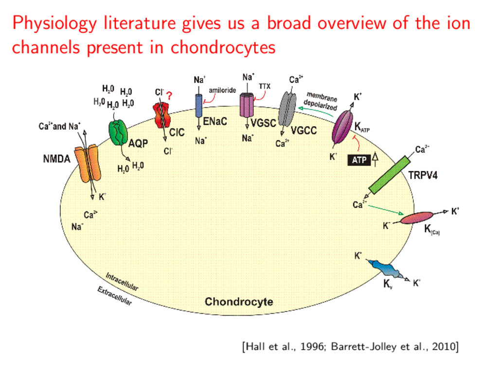
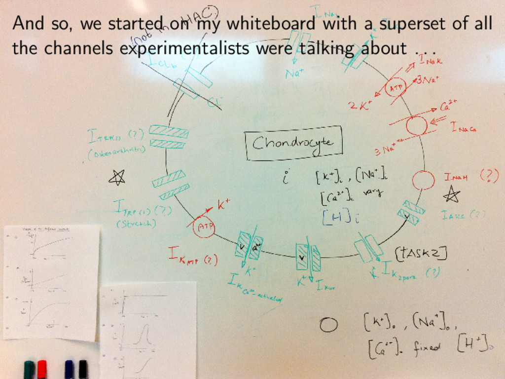
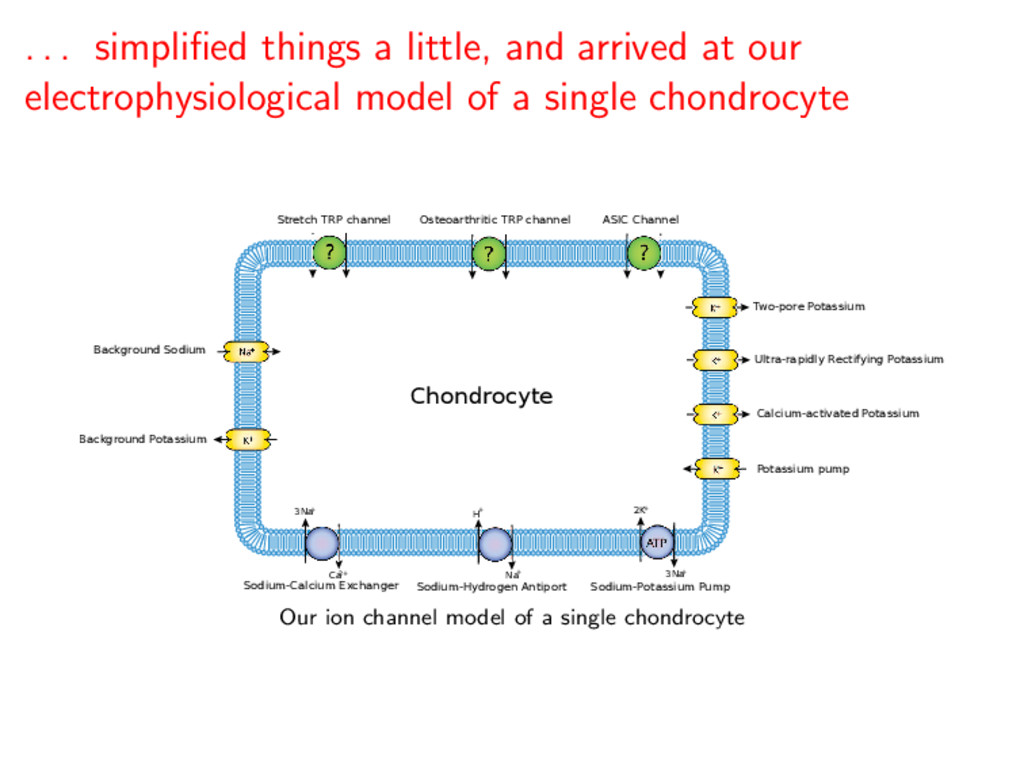
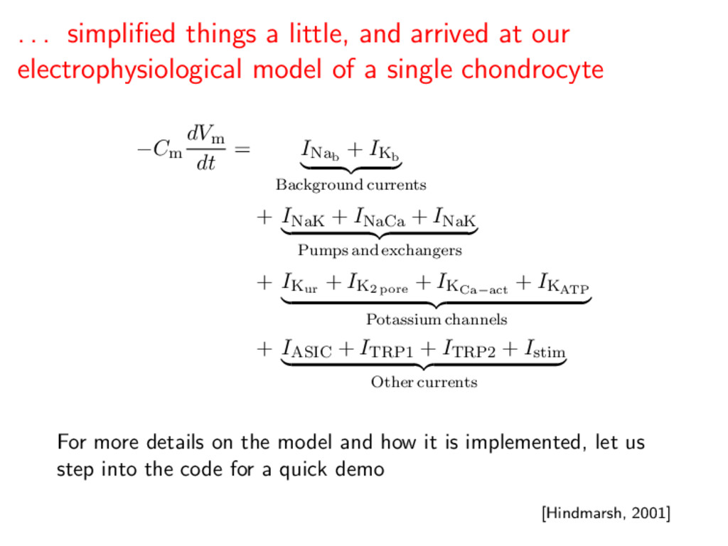
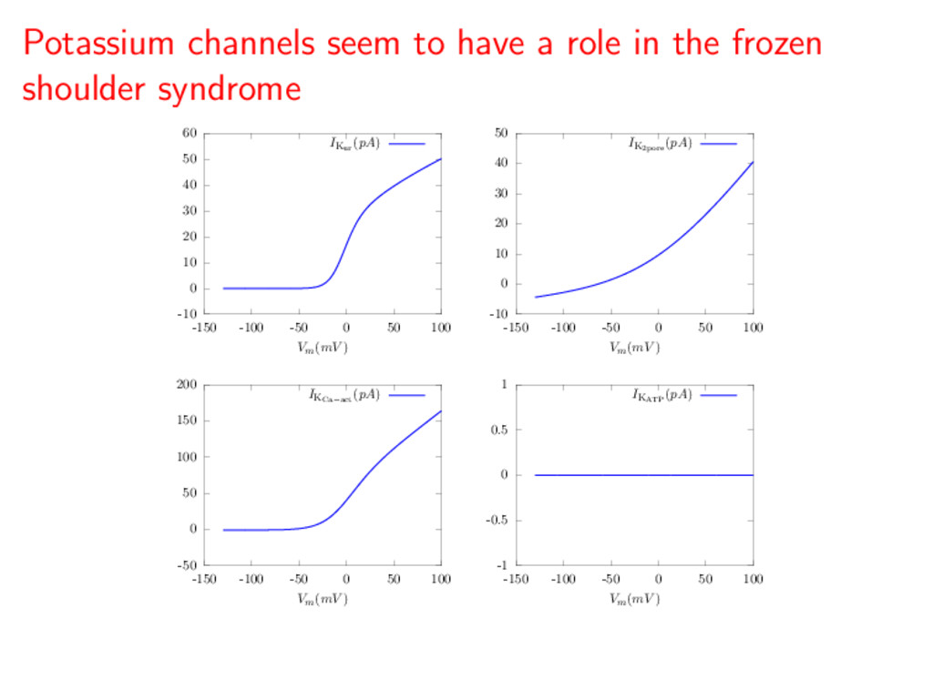
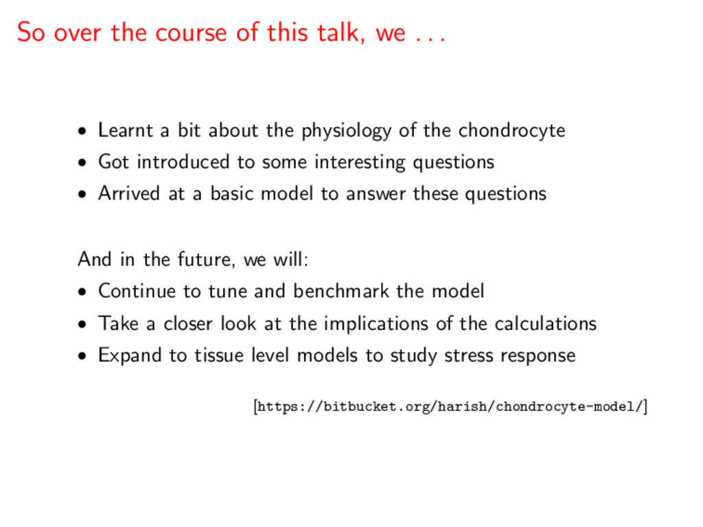
This talk presents the first computational model of the main electrophysiological characteristics of the human chondrocyte. This model is illustrated by an in silico study of the TRPV4 current and is based mainly on an initial experimental data set which identified the main K+ currents expressed in single chondrocytes isolated from knee joints of healthy adult human donors. This model is validated by illustrating the role of a novel 2-pore K+ current in regulating the chondrocyte resting potential, and presents the possibility for integrating available data from electrophysiological, PCR and gene array experiments. It is also an important tool for rationalization of working hypotheses, design of new experiments, and understanding the principles and limitations of patch clamp methods as applied to the isolated human chondrocyte.

Harish Narayanan

July 04, 2011
Tweet

More Decks by Harish Narayanan

Other Decks in Research

Transcript

  1. This talk will describe the status of our electrophysiological model

    of chondrocytes in human articular cartilage What is the chondrocyte, and why is it interesting? What are the specific questions we aim to answer? -5 0 5 10 15 20 25 30 -150 -100 -50 0 50 100 Vm(mV ) Ii/Cm(pA/pF) How does our cellular model help us answer these questions?
  2. Articular cartilage is the connective tissue that separates bones at

    joints, allowing them to slide past each other Articular cartilage as part of a synovial joint • Cartilage is composed of chondrocytes, an ECM (collagen and elastin) and proteoglycans • The tissue is aneural, avascular and alymphatic and is thus slow to grow and heal [Poole, 1997]
  3. Articular cartilage is the connective tissue that separates bones at

    joints, allowing them to slide past each other Articular cartilage as part of a synovial joint • Cartilage is composed of chondrocytes, an ECM (collagen and elastin) and proteoglycans • The tissue is aneural, avascular and alymphatic and is thus slow to grow and heal [Poole, 1997]
  4. Chondrocytes are the resident cells of articular cartilage and are

    responsible for maintaining the extracellular matrix Electron micrograph of typical chondrocyte • Chondrocytes are isolated within a voluminous ECM • Relies on diffusion from the articular surface for nutrient and metabolite exchange • Sensitive to extra-cellular pH, ionic environment, stress state • Arthritis is the associated degenerative pathology [Archer & Francis-West 2003]
  5. Chondrocytes are the resident cells of articular cartilage and are

    responsible for maintaining the extracellular matrix Electron micrograph of typical chondrocyte • Chondrocytes are isolated within a voluminous ECM • Relies on diffusion from the articular surface for nutrient and metabolite exchange • Sensitive to extra-cellular pH, ionic environment, stress state • Arthritis is the associated degenerative pathology [Archer & Francis-West 2003]
  6. We narrow our focus to a specific question to motivate

    our initial modelling effort What causes the “frozen shoulder syndrome” when tissue is exposed to the local anæsthetic bupivacaine? • How does the cell survive in an acidic environment and still continue to synthesise and maintain viable cartilage? • How does the chondrocyte “sense” and respond to loading? [Webb, 2009; Hall et al., 1996]
  7. We narrow our focus to a specific question to motivate

    our initial modelling effort What causes the “frozen shoulder syndrome” when tissue is exposed to the local anæsthetic bupivacaine? • How does the cell survive in an acidic environment and still continue to synthesise and maintain viable cartilage? • How does the chondrocyte “sense” and respond to loading? [Webb, 2009; Hall et al., 1996]
  8. In order to answer such questions, we need a model

    that helps us understand some fundamental things • The important important ion channels of the cell • The basis of the resting membrane potential • The role of cellular volume regulation [Lewis et al., 2011]
  9. Physiology literature gives us a broad overview of the ion

    channels present in chondrocytes [Hall et al., 1996; Barrett-Jolley et al., 2010]
  10. And so, we started on my whiteboard with a superset

    of all the channels experimentalists were talking about . . .
  11. . . . simplified things a little, and arrived at

    our electrophysiological model of a single chondrocyte Chondrocyte Ca 2+ 3Na + Sodium-Calcium Exchanger Background Sodium 2K + 3Na + Sodium-Potassium Pump H+ Na + Sodium-Hydrogen Antiport Ultra-rapidly Rectifying Potassium Two-pore Potassium Background Potassium Calcium-activated Potassium Osteoarthritic TRP channel Potassium pump Stretch TRP channel ASIC Channel Our ion channel model of a single chondrocyte
  12. . . . simplified things a little, and arrived at

    our electrophysiological model of a single chondrocyte −Cm dVm dt = INab + IKb Background currents + INaK + INaCa + INaK Pumps and exchangers + IKur + IK2 pore + IKCa−act + IKATP Potassium channels + IASIC + ITRP1 + ITRP2 + Istim Other currents For more details on the model and how it is implemented, let us step into the code for a quick demo [Hindmarsh, 2001]
  13. . . . simplified things a little, and arrived at

    our electrophysiological model of a single chondrocyte −Cm dVm dt = INab + IKb Background currents + INaK + INaCa + INaK Pumps and exchangers + IKur + IK2 pore + IKCa−act + IKATP Potassium channels + IASIC + ITRP1 + ITRP2 + Istim Other currents For more details on the model and how it is implemented, let us step into the code for a quick demo [Hindmarsh, 2001]
  14. At the moment, I am completing the implementation and starting

    to tune parameters -5 0 5 10 15 20 25 30 -150 -100 -50 0 50 100 Vm(mV ) Ii/Cm(pA/pF) [Clark et. al., 2011; Millward-Sadler et. al., 2000]
  15. Potassium channels seem to have a role in the frozen

    shoulder syndrome -1 -0.5 0 0.5 1 -150 -100 -50 0 50 100 Vm(mV ) -50 0 50 100 150 200 -150 -100 -50 0 50 100 Vm(mV ) -10 0 10 20 30 40 50 -150 -100 -50 0 50 100 Vm(mV ) -10 0 10 20 30 40 50 60 -150 -100 -50 0 50 100 Vm(mV ) IKATP (pA) IKCa−act (pA) IK2pore (pA) IKur (pA)
  16. So over the course of this talk, we . .

    . • Learnt a bit about the physiology of the chondrocyte • Got introduced to some interesting questions • Arrived at a basic model to answer these questions And in the future, we will: • Continue to tune and benchmark the model • Take a closer look at the implications of the calculations • Expand to tissue level models to study stress response [https://bitbucket.org/harish/chondrocyte-model/]
  17. So over the course of this talk, we . .

    . • Learnt a bit about the physiology of the chondrocyte • Got introduced to some interesting questions • Arrived at a basic model to answer these questions And in the future, we will: • Continue to tune and benchmark the model • Take a closer look at the implications of the calculations • Expand to tissue level models to study stress response [https://bitbucket.org/harish/chondrocyte-model/]