Upgrade to Pro — share decks privately, control downloads, hide ads and more …

General Embryology 2

General Embryology 2

This presentation includes the events occurring during third & fourth week of pregnancy and human development (formation of chorionic villi , trilaminar embryonic disc , notochord , and organogenesis

Dr.Sherif Fahmy

July 28, 2019
Tweet

More Decks by Dr.Sherif Fahmy

Other Decks in Science

Transcript

  1. 3rd Week of Pregnancy 1- Changes in the chorion: Formation

    of chorionic villi. 2- Changes in the embryonic disc: Formation of trilaminar embryonic disc (Gastrulation).
  2. Chorionic Villi They are projections from the chorion. Chorion: Wall

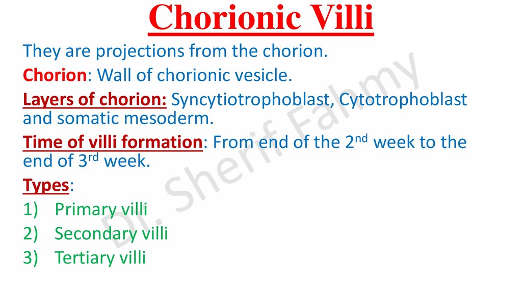
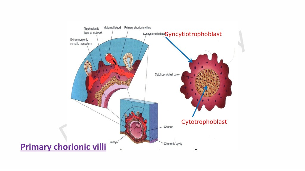
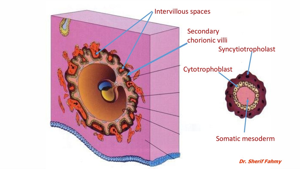
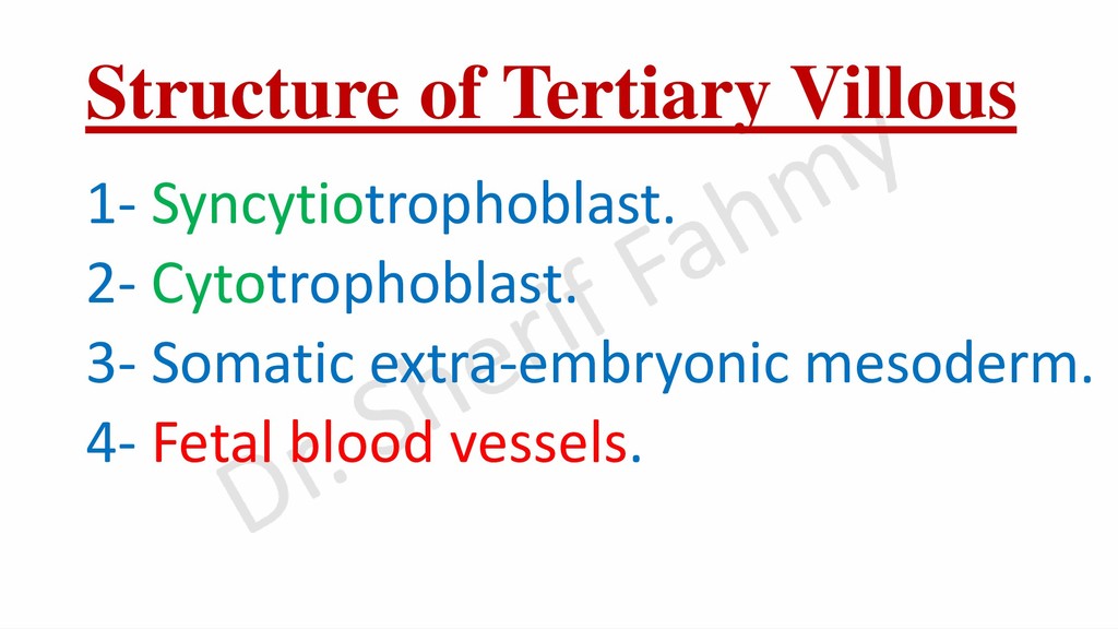
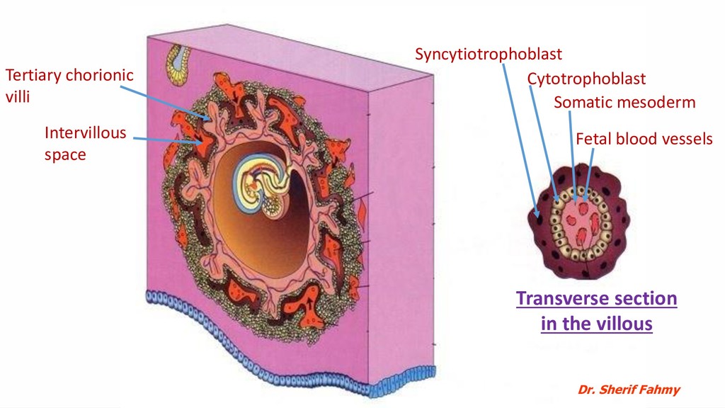
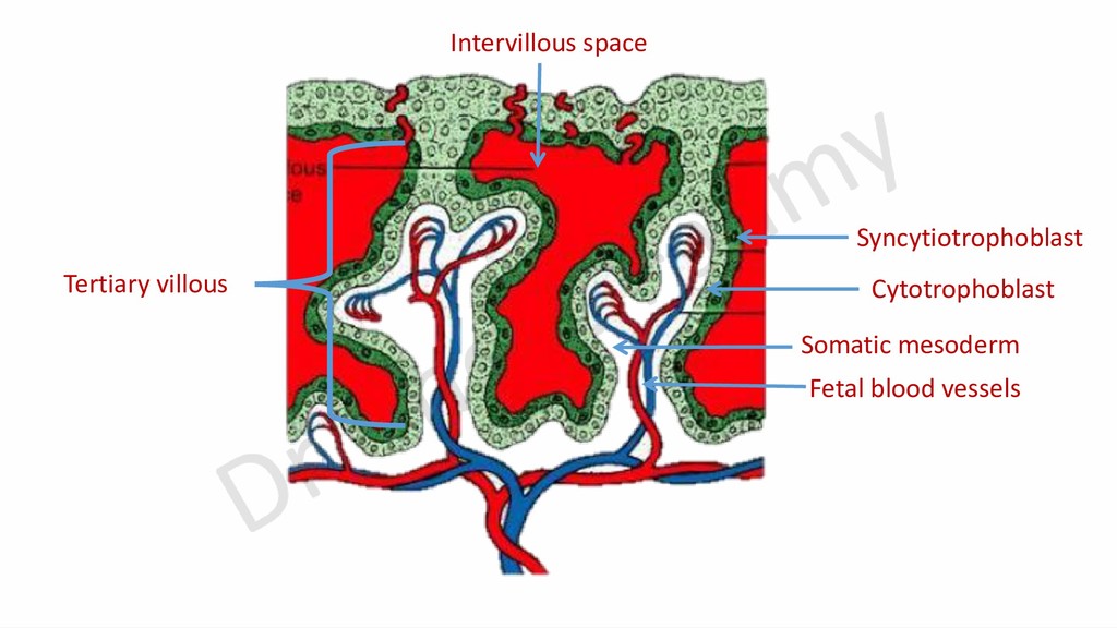
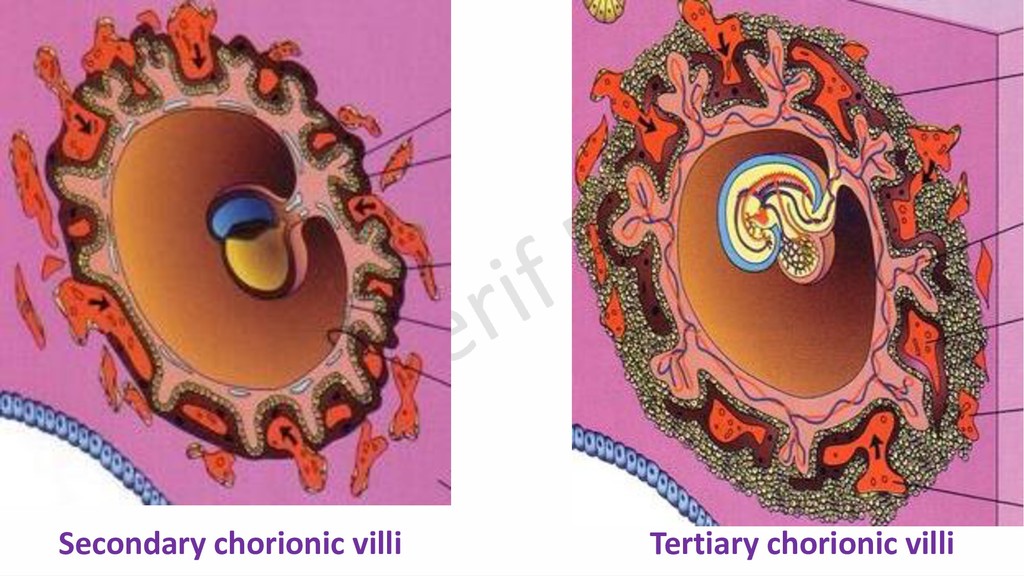
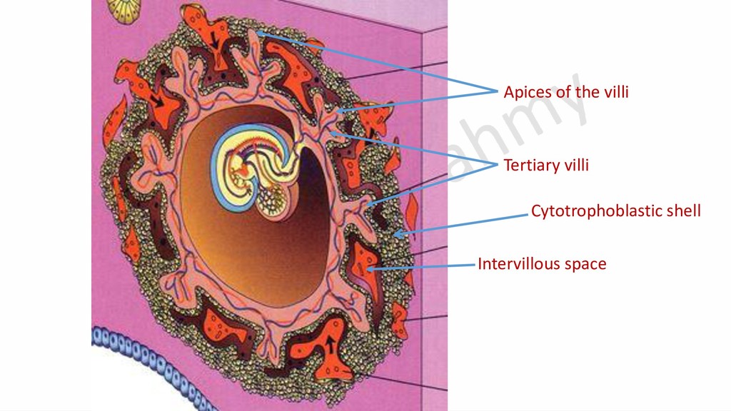
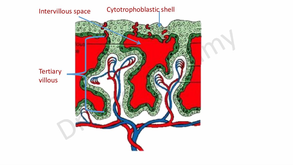
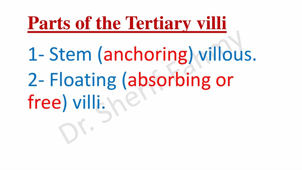
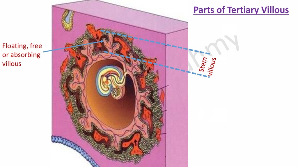
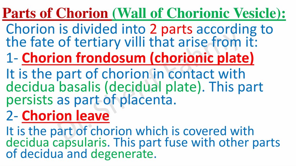
    of chorionic vesicle. Layers of chorion: Syncytiotrophoblast, Cytotrophoblast and somatic mesoderm. Time of villi formation: From end of the 2nd week to the end of 3rd week. Types: 1) Primary villi 2) Secondary villi 3) Tertiary villi
  3. Parts of Chorion (Wall of Chorionic Vesicle): Chorion is divided

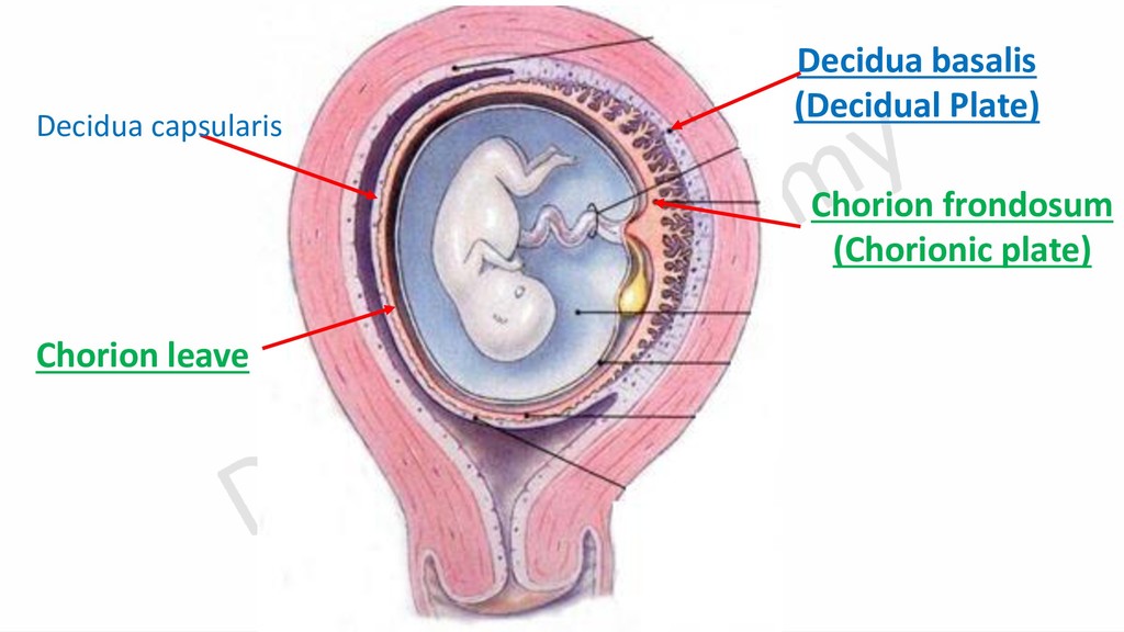
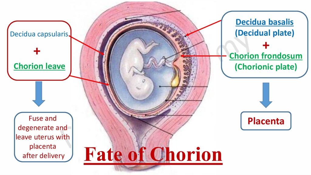
    into 2 parts according to the fate of tertiary villi that arise from it: 1- Chorion frondosum (chorionic plate) It is the part of chorion in contact with decidua basalis (decidual plate). This part persists as part of placenta. 2- Chorion leave It is the part of chorion which is covered with decidua capsularis. This part fuse with other parts of decidua and degenerate.
  4. Chorion frondosum (Chorionic plate) Chorion leave Decidua basalis (Decidual plate)

    Decidua capsularis Placenta Fuse and degenerate and leave uterus with placenta after delivery + +
  5. Gastrulation It is the transformation of bilaminar disc into trilaminar

    disc. It begins by formation of: 1- Primitive streak. 2- Primitive node. 3- Invagination. 4- Bucco-pharyngeal membrane. 5- Cloacal membrane.
  6. Amniotic cavity Yolk sac Bilaminar embryonic disc Primitive streak &

    groove Primitive node & pit Cloacal membrane Bucco-pharyngeal membrane Epiblast Hypoblast Amniotic cavity Cranial end Caudal end
  7. Primitive groove Epiblast Hypoblast Flask cells Endodermal cells Invagination Intra-embryonic

    mesoderm Primitive pit Transverse section of embryonic disc during invagination
  8. Bucco- pharyngeal membrane Cloacal membrane Primitive streak Primitive node Epiblast

    cells that invaginate through primitive groove Notochord Intra-embryonic mesoderm Epiblast cells
  9. Amniotic cavity Embryonic disc Secondary yolk sac Amniotic cavity Buccopharyngeal

    membrane Primitive pit Cloacal membrane Connecting stalk Connecting stalk Notochord under ectoderm Primitive streak LS
  10. Amniotic cavity Ectoderm 1-Pre-notochordal process Primitive pit Primitive streak Connecting

    stalk Secondary yolk sac Endoderm Bucco-pharyngeal membrane LS Cloacal membrane Dr. Sherif Fahmy
  11. Amniotic cavity Yolk sac Degeneration of the fused median region

    of endoderm and floor of notochordal canal Roof of notochordal canal LS Dr. Sherif Fahmy
  12. TS

  13. Importance of Notochord 1-It is a temporary axial skeleton of

    the embryonic disc. 2-During folding, its firmness limits head fold. 3-Vertebral column is formed around it.
  14. Fate of Notochord •It is the primitive axial skeleton around

    which the vertebral column is formed. •It remains in intervertebral disc as nucleus pulposus.
  15. Organogenesis (4th to 8th Week) Fate of embryonic disc: 1-

    Fate of Ectoderm. 2- Fate of Endoderm. 3- Fate of Mesoderm.
  16. Transverse section in the region of neurulation Neural fold Neural

    crest Surface ectoderm Notochord Dr. Sherif Fahmy
  17. Fate of the neural tube •The part of the tube

    in the vertebral canal forms the spinal cord. •The part of the tube in the skull forms the brain.
  18. Fate of neural crest •Ganglia: Sensory, sympathetic and parasympathetic. •Cells:

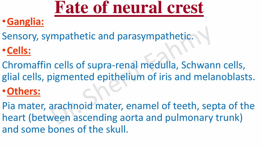
    Chromaffin cells of supra-renal medulla, Schwann cells, glial cells, pigmented epithelium of iris and melanoblasts. •Others: Pia mater, arachnoid mater, enamel of teeth, septa of the heart (between ascending aorta and pulmonary trunk) and some bones of the skull.
  19. Derivatives of surface ectoderm -Epidermis layer of skin. -Otic placodes

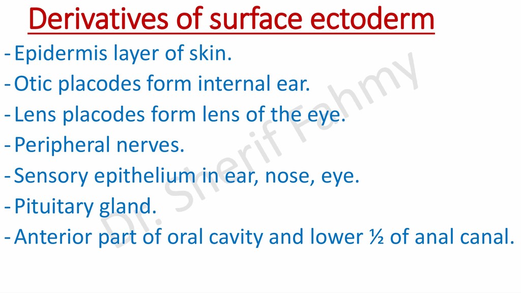
    form internal ear. -Lens placodes form lens of the eye. -Peripheral nerves. -Sensory epithelium in ear, nose, eye. -Pituitary gland. -Anterior part of oral cavity and lower ½ of anal canal.