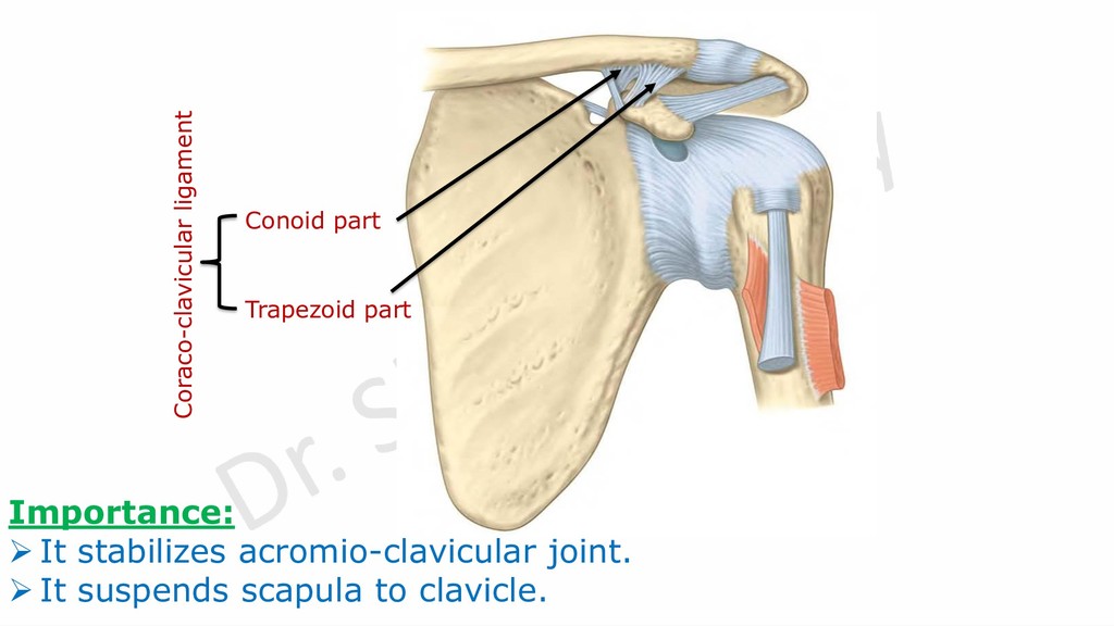
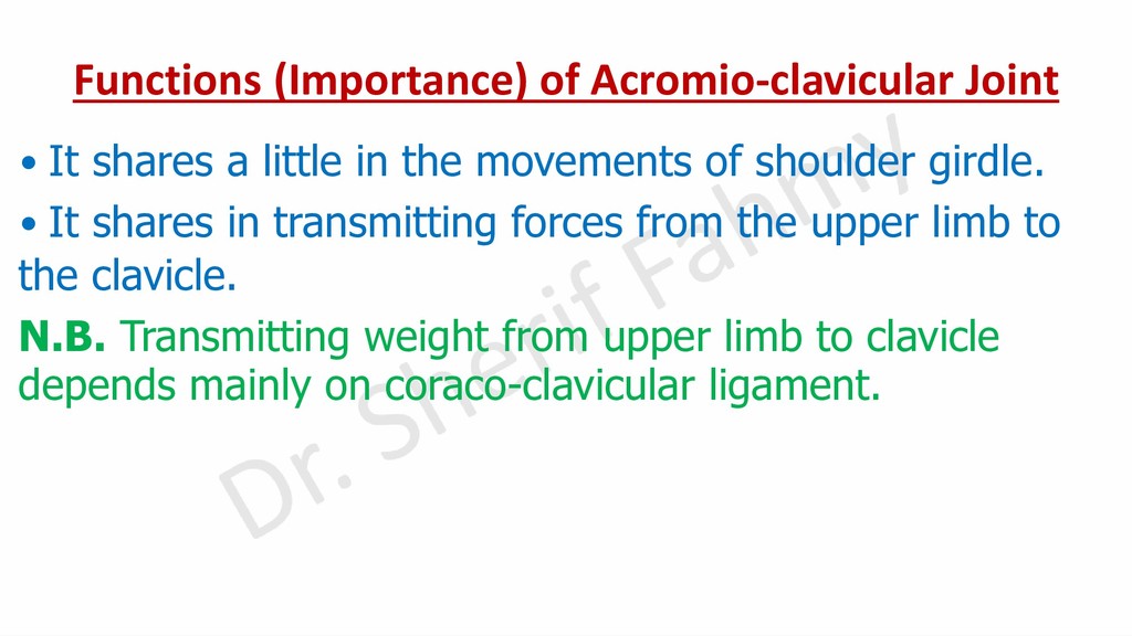
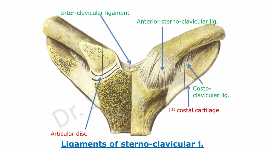
in the movements of shoulder girdle. • It shares in transmitting forces from the upper limb to the clavicle. N.B. Transmitting weight from upper limb to clavicle depends mainly on coraco-clavicular ligament.
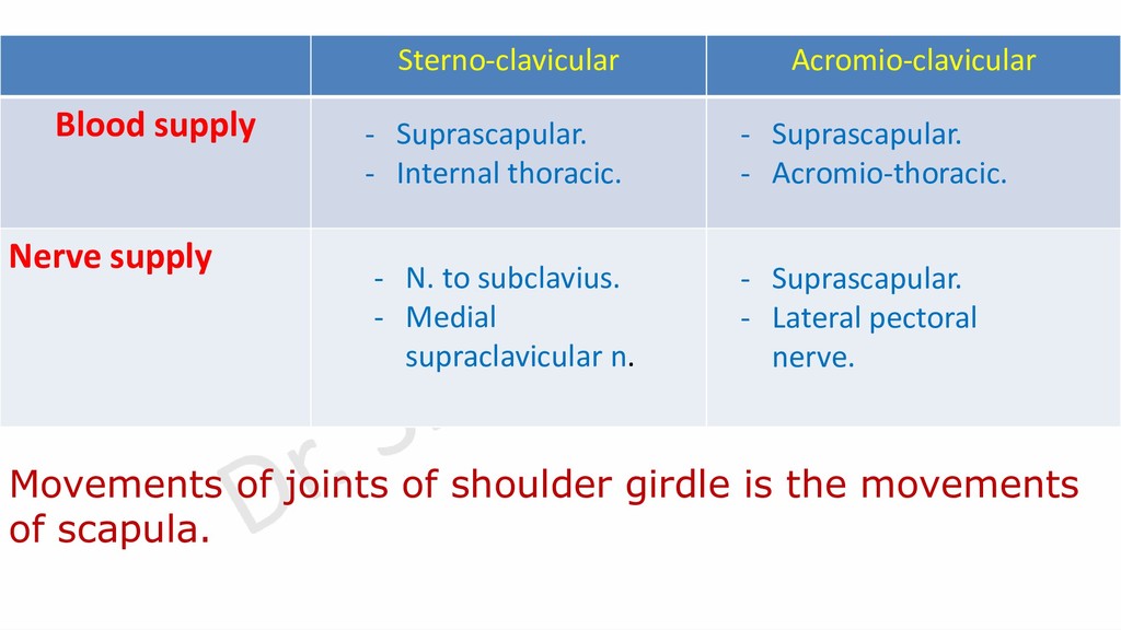
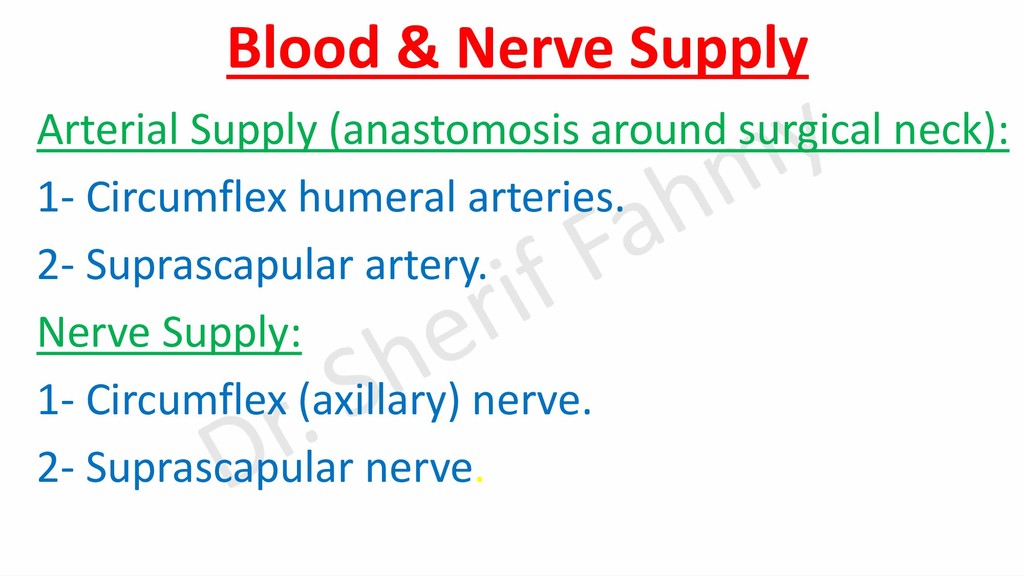
thoracic. - Suprascapular. - Acromio-thoracic. - N. to subclavius. - Medial supraclavicular n. - Suprascapular. - Lateral pectoral nerve. Movements of joints of shoulder girdle is the movements of scapula.
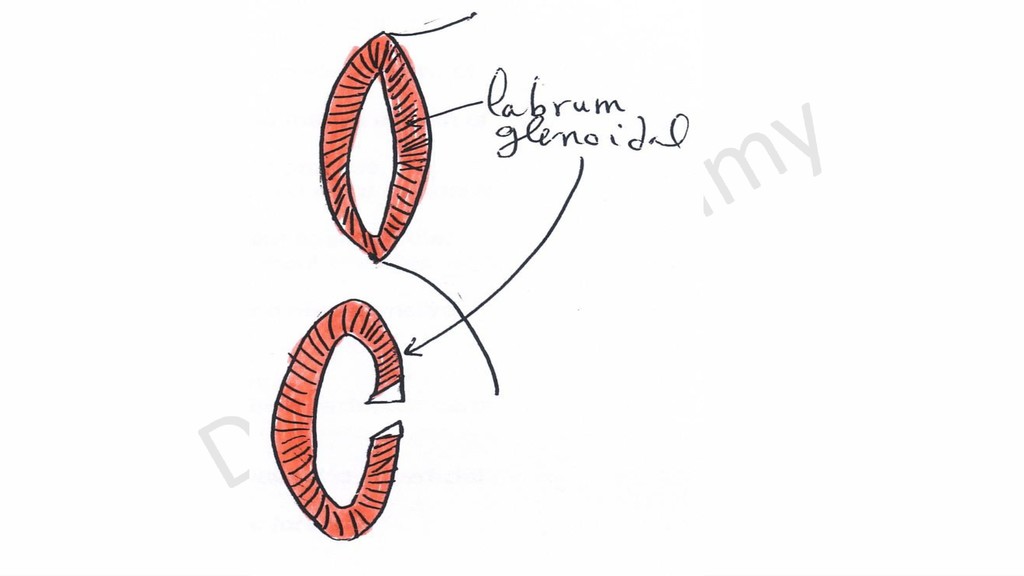
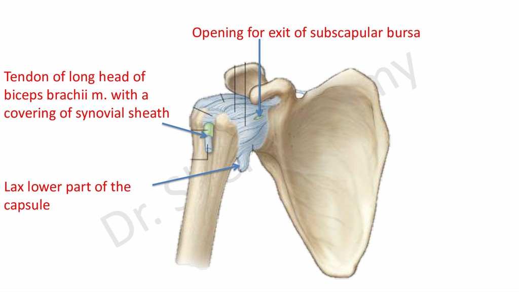
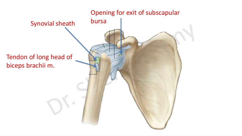
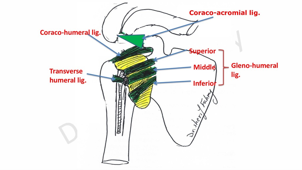
supra-glenoid tubercle. -Attached laterally to anatomical neck and medially it descends for 1 cm below anatomical neck till surgical neck. -It is weak because it has two anterior openings and its lower part is lax.
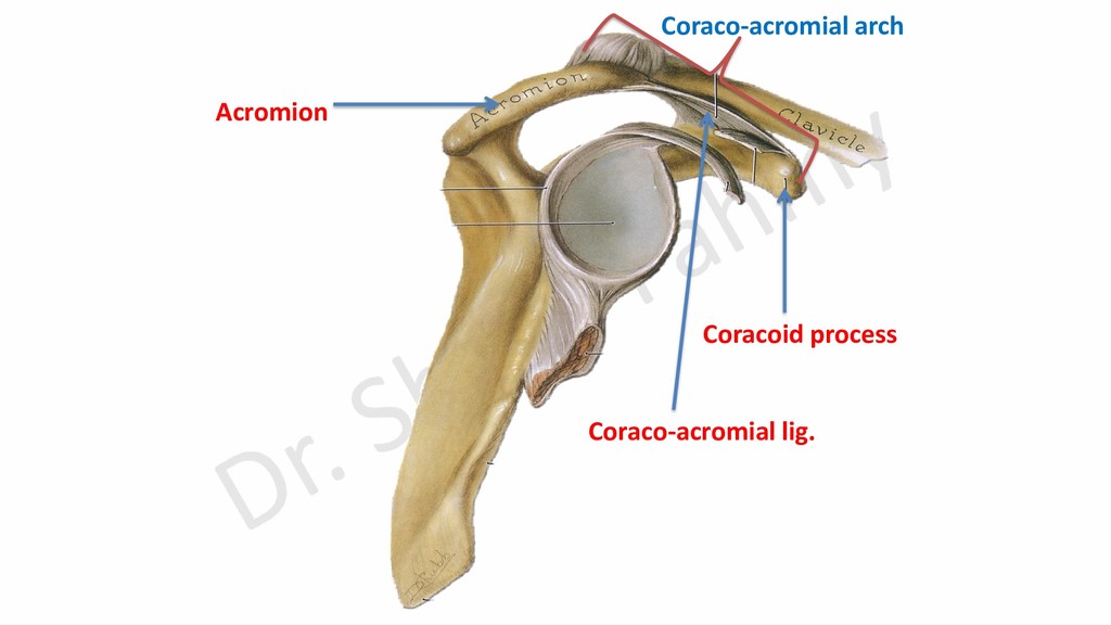
the joint. Above: Supraspinatus & tendon of long head of biceps. In front: Subscapularis m. Behind: Infraspinatus and teres minor ms. Below: Long head of triceps only during abduction. 2- Coraco-acromial arch.
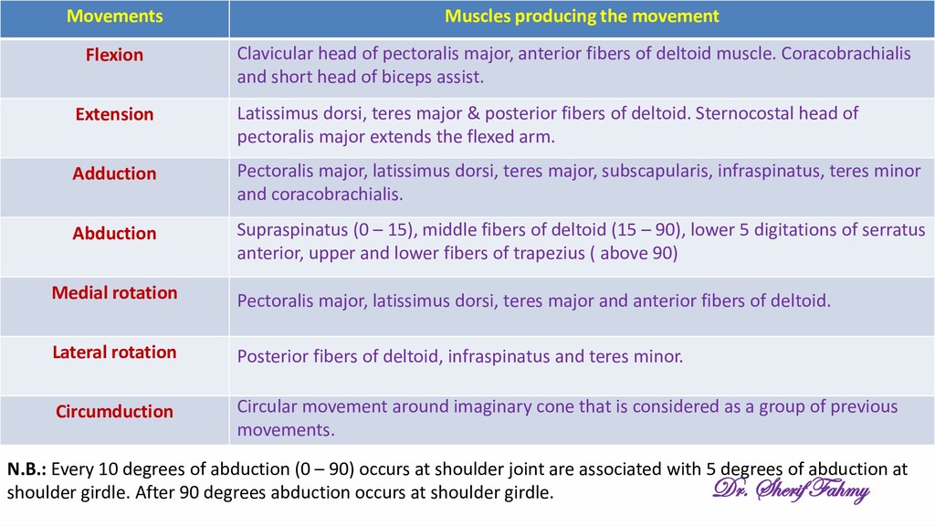
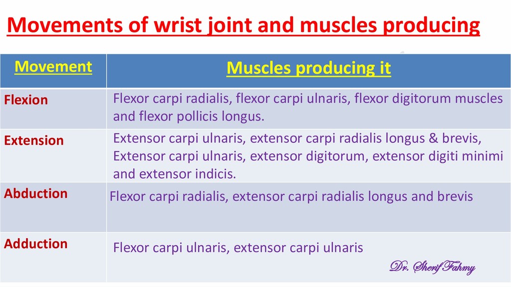
rotation Lateral rotation Circumduction N.B.: Every 10 degrees of abduction (0 – 90) occurs at shoulder joint are associated with 5 degrees of abduction at shoulder girdle. After 90 degrees abduction occurs at shoulder girdle. Clavicular head of pectoralis major, anterior fibers of deltoid muscle. Coracobrachialis and short head of biceps assist. Latissimus dorsi, teres major & posterior fibers of deltoid. Sternocostal head of pectoralis major extends the flexed arm. Pectoralis major, latissimus dorsi, teres major, subscapularis, infraspinatus, teres minor and coracobrachialis. Supraspinatus (0 – 15), middle fibers of deltoid (15 – 90), lower 5 digitations of serratus anterior, upper and lower fibers of trapezius ( above 90) Pectoralis major, latissimus dorsi, teres major and anterior fibers of deltoid. Posterior fibers of deltoid, infraspinatus and teres minor. Circular movement around imaginary cone that is considered as a group of previous movements. Dr. Sherif Fahmy
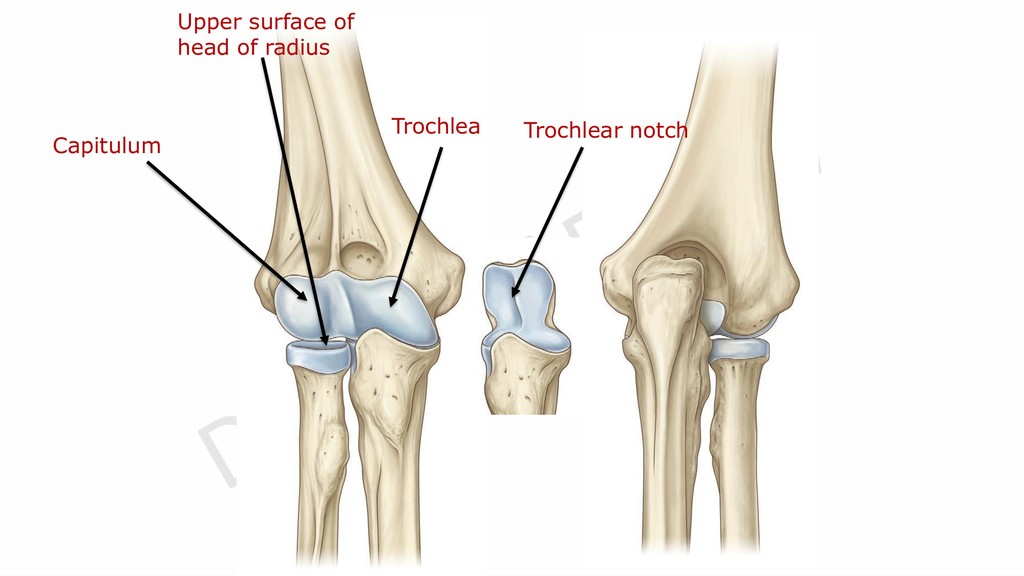
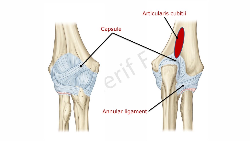
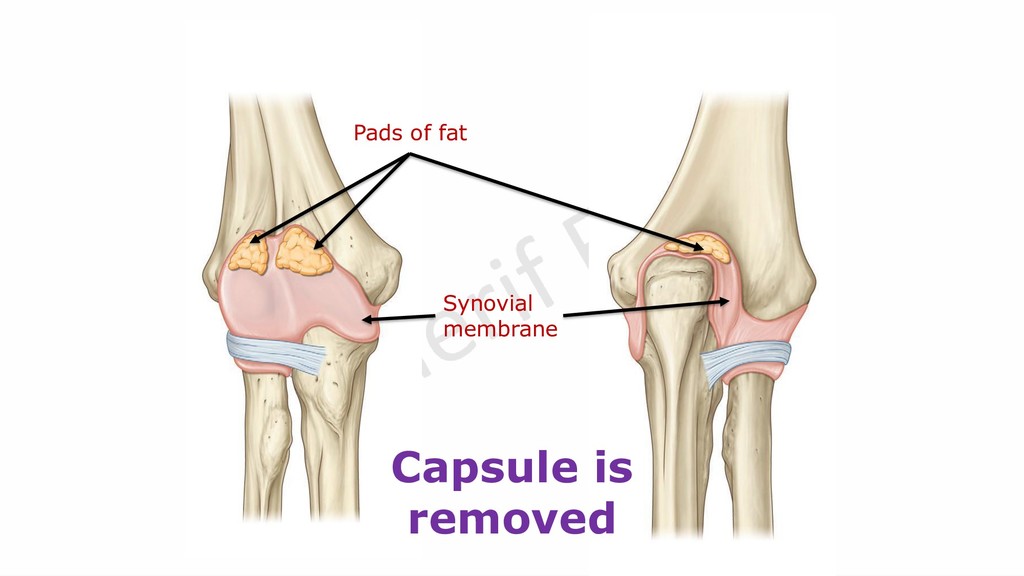
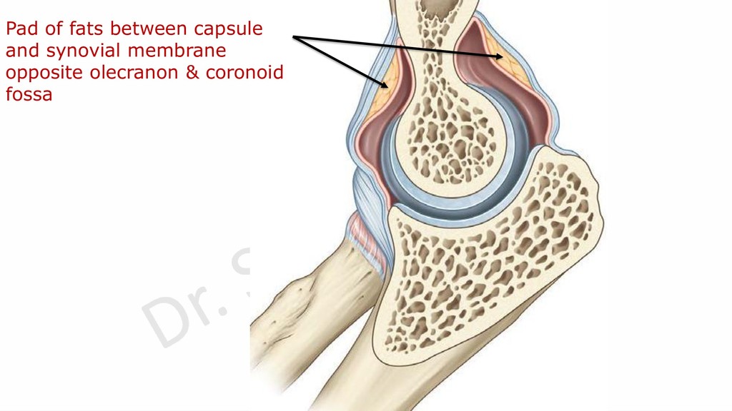
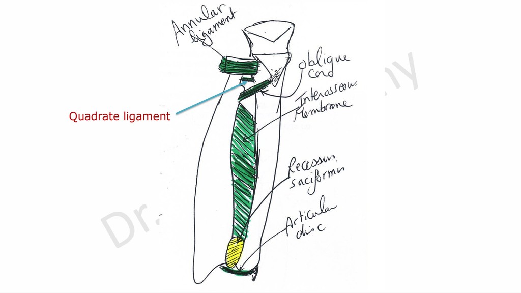
lower end of humerus. ➢3 pads of fat are present opposite each fossa between capsule and synovial membrane. ➢It is continuous below with synovial membrane of superior radio-ulnar joint.
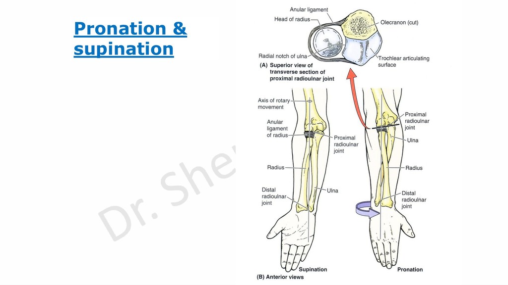
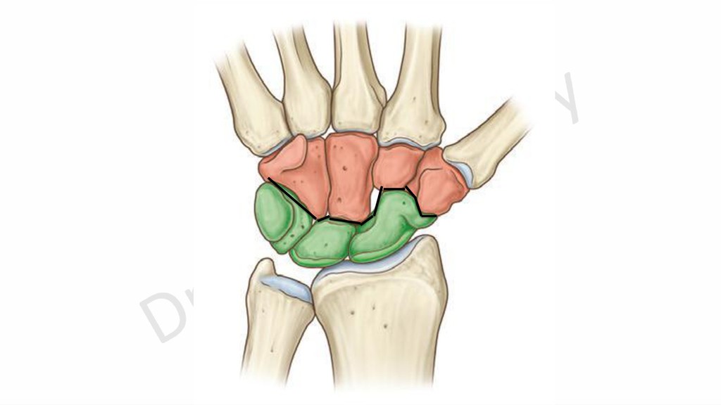
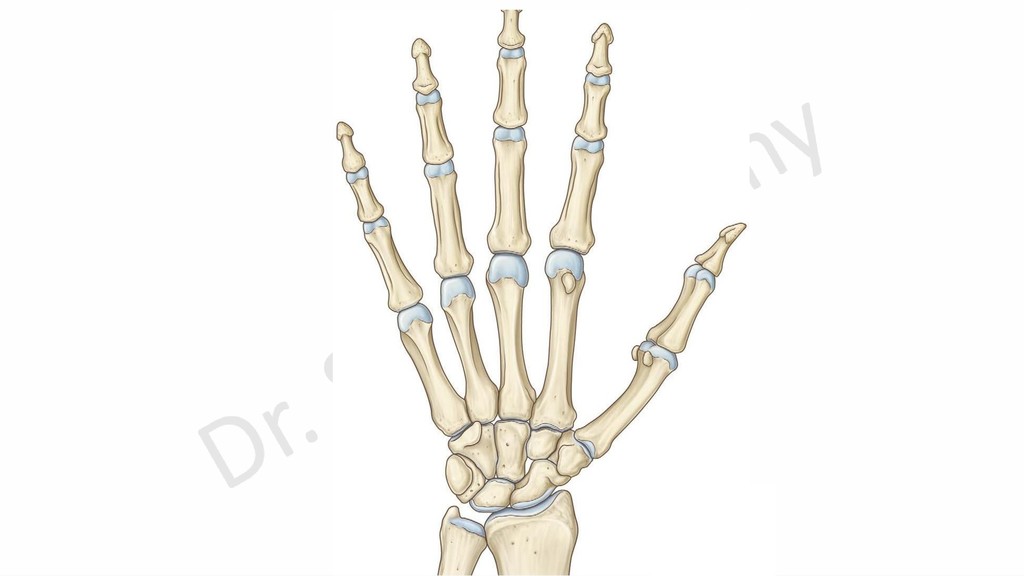
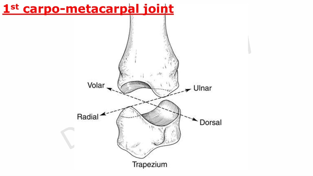
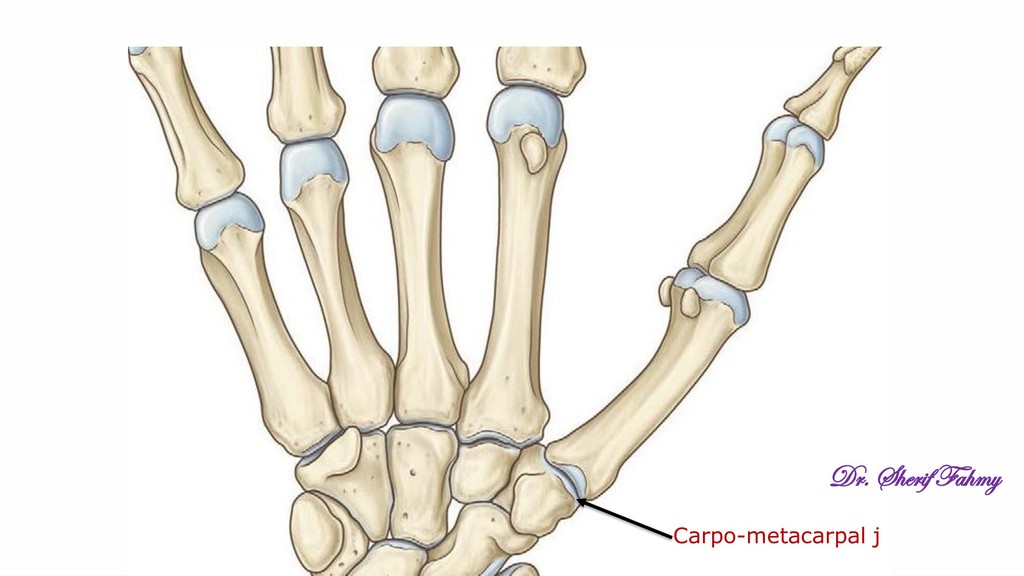
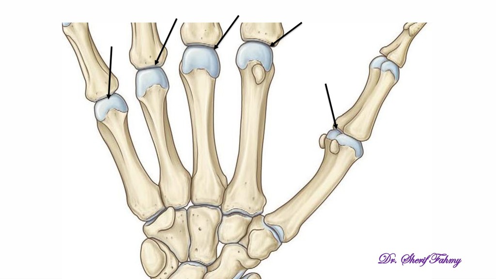
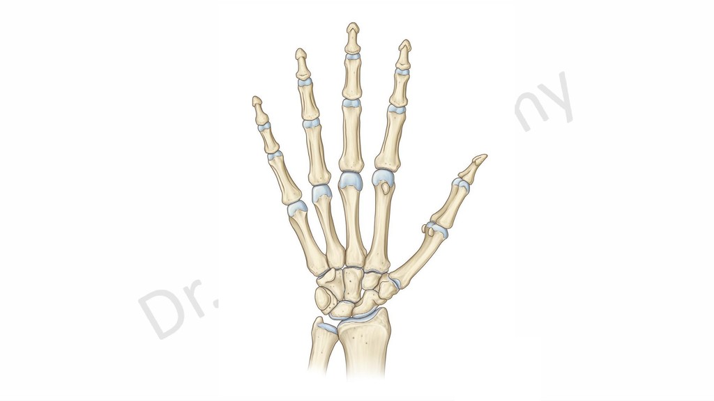
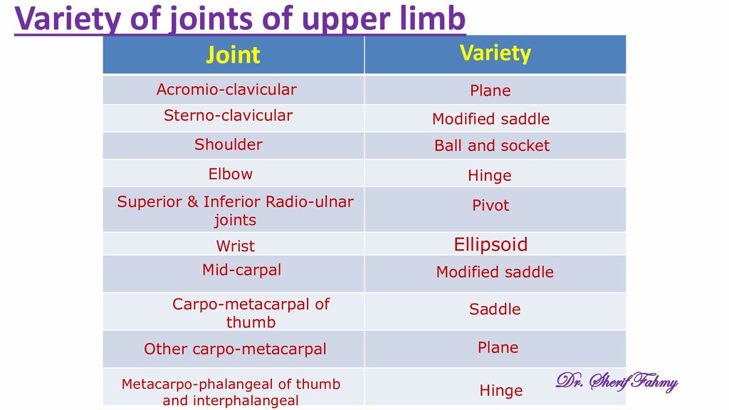
socket Elbow Hinge Superior & Inferior Radio-ulnar joints Pivot Wrist Ellipsoid Mid-carpal Modified saddle Carpo-metacarpal of thumb Saddle Other carpo-metacarpal Plane Metacarpo-phalangeal of thumb and interphalangeal Hinge Variety of joints of upper limb Dr. Sherif Fahmy