Frequency – numbers of cycles per second 1 Hertz = 1 cycle / second Velocity – wavelength x frequency Velocity of sound is dependent upon density and compressibility of the medium
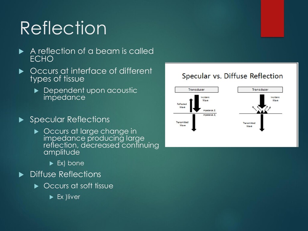
Occurs at interface of different types of tissue Dependent upon acoustic impedance Specular Reflections Occurs at large change in impedance producing large reflection, decreased continuing amplitude Ex) bone Diffuse Reflections Occurs at soft tissue Ex )liver
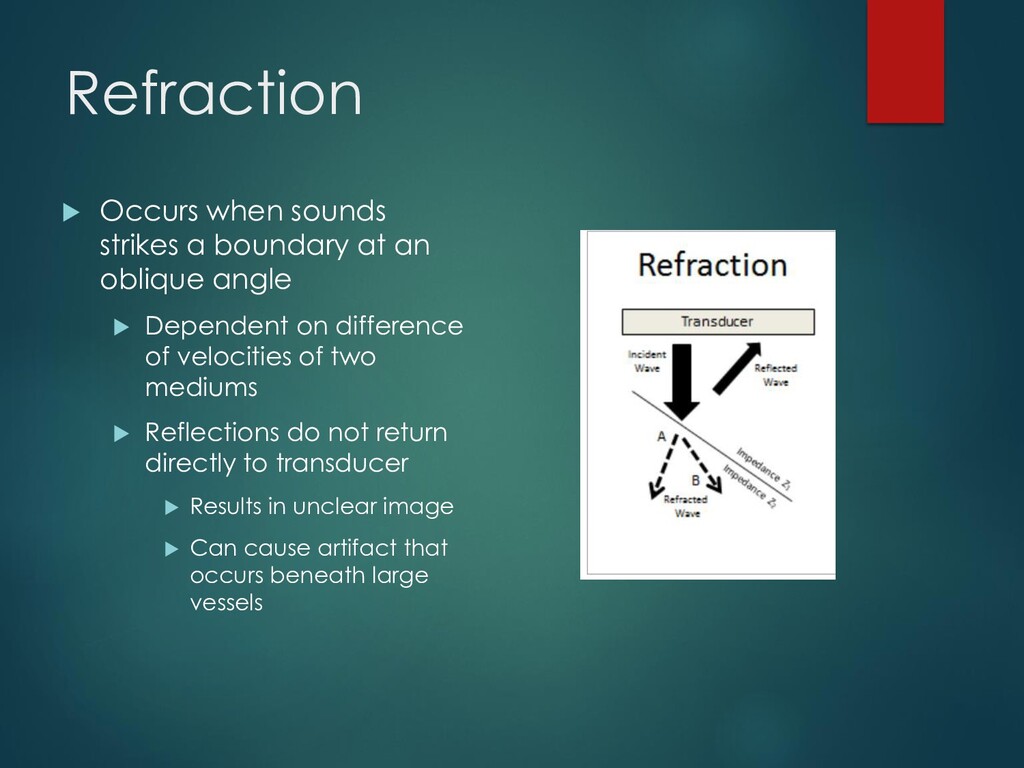
oblique angle Dependent on difference of velocities of two mediums Reflections do not return directly to transducer Results in unclear image Can cause artifact that occurs beneath large vessels
heat Higher frequencies absorbed faster Better axial resolution Lower frequency US may be needed to increase penetration for deeper structures Lower resoluton
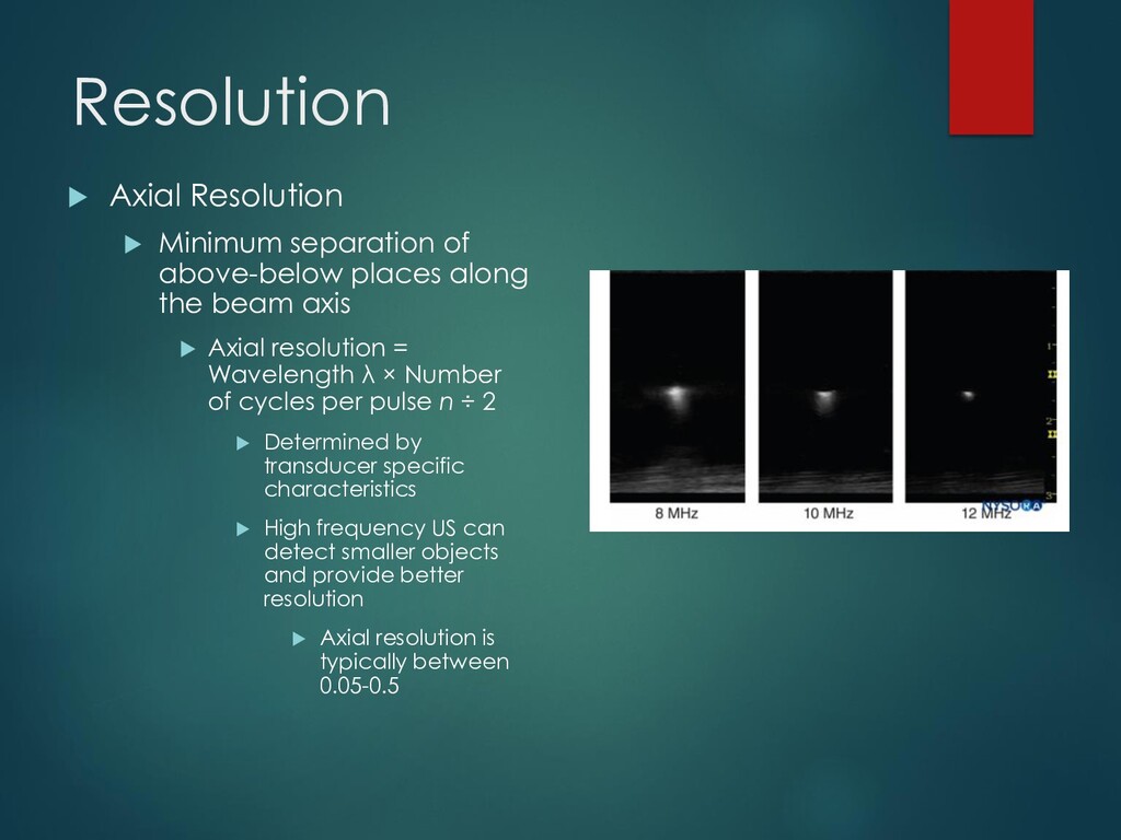
along the beam axis Axial resolution = Wavelength λ × Number of cycles per pulse n ÷ 2 Determined by transducer specific characteristics High frequency US can detect smaller objects and provide better resolution Axial resolution is typically between 0.05-0.5
minimum side by side distance between two objects Determined by frequency and beam width High frequency transducers = narrow focus Better lateral resolution
cross-sectional image through the area of interest Horizontal and vertical directions represent real distances Intensity of grey scale indicated echo strength
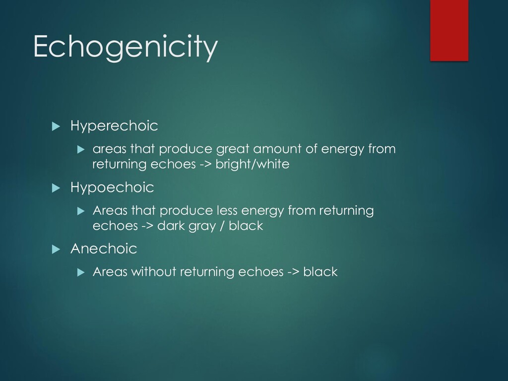
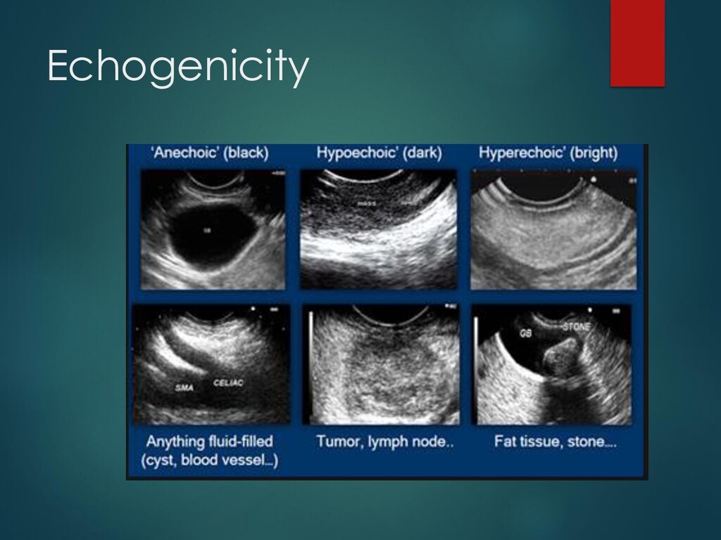
energy from returning echoes -> bright/white Hypoechoic Areas that produce less energy from returning echoes -> dark gray / black Anechoic Areas without returning echoes -> black
ratio of output to input electric power Amplifies returning signals as well as background noise Depth – start deep and decrease as needed Leave small area behind to observe useful artifacts Low frequency probes = increased depth w/ lower resolution High frequency probes = decreased depth w/ higher resolution