Upgrade to Pro — share decks privately, control downloads, hide ads and more …

Examination of Blood | Hematology Practical | D...

Sponsored · Your Podcast. Everywhere. Effortlessly. Share. Educate. Inspire. Entertain. You do you. We'll handle the rest. →

Examination of Blood | Hematology Practical | DMLT 1st Year

This presentation covers Examination of Blood for DMLT 1st Year Hematology.Topics Covered:Complete Blood Count (CBC)Hemoglobin Estimation - Sahli's MethodTotal RBC, WBC, Platelet CountDifferential Leukocyte Count (DLC)ESR, PCV, Blood Grouping & Rh TypingPeripheral Blood Smear PreparationIncludes sample collection, order of draw (EDTA tube - Lavender cap), normal values and clinical significance.Useful for DMLT, BMLT, CMS, DOTT practical exams and viva.Subject: Hematology & Introduction to MLT
Teacher: Annu Ma'am

Avatar for Er. Annu maurya

Er. Annu maurya

September 11, 2026

More Decks by Er. Annu maurya

Other Decks in Education

Transcript

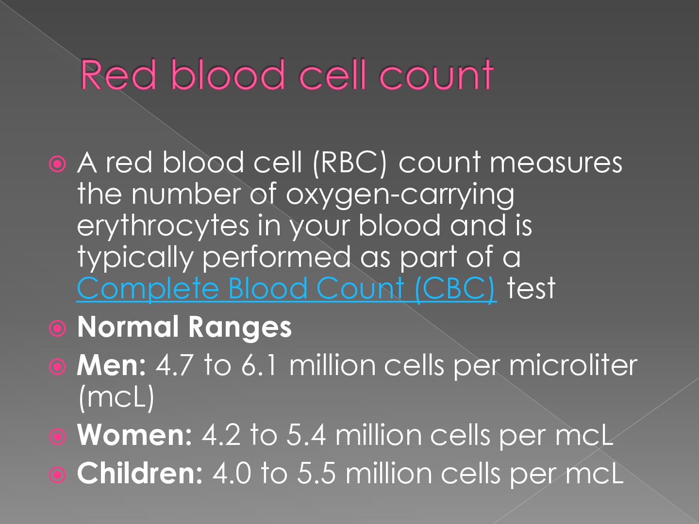
  1.  A red blood cell (RBC) count measures the number

    of oxygen-carrying erythrocytes in your blood and is typically performed as part of a Complete Blood Count (CBC) test  Normal Ranges  Men: 4.7 to 6.1 million cells per microliter (mcL)  Women: 4.2 to 5.4 million cells per mcL  Children: 4.0 to 5.5 million cells per mcL
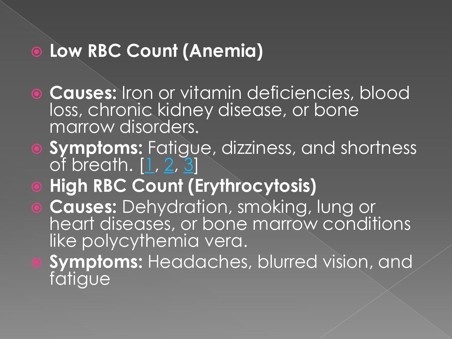
  2.  Low RBC Count (Anemia)  Causes: Iron or vitamin

    deficiencies, blood loss, chronic kidney disease, or bone marrow disorders.  Symptoms: Fatigue, dizziness, and shortness of breath. [1, 2, 3]  High RBC Count (Erythrocytosis)  Causes: Dehydration, smoking, lung or heart diseases, or bone marrow conditions like polycythemia vera.  Symptoms: Headaches, blurred vision, and fatigue
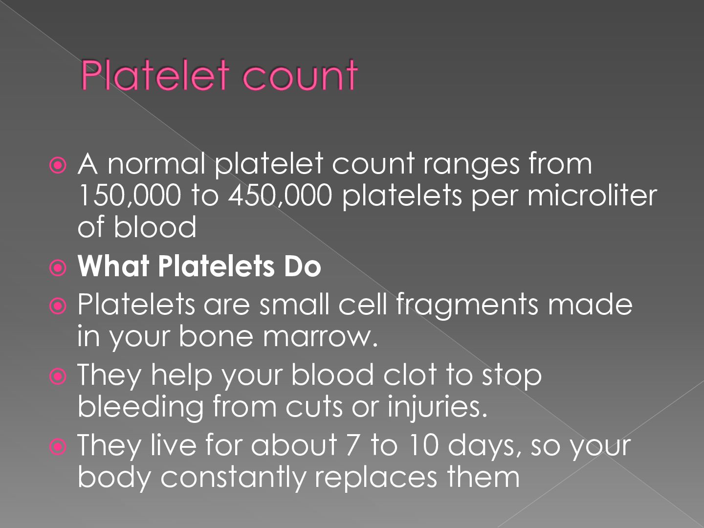
  3.  A normal platelet count ranges from 150,000 to 450,000

    platelets per microliter of blood  What Platelets Do  Platelets are small cell fragments made in your bone marrow.  They help your blood clot to stop bleeding from cuts or injuries.  They live for about 7 to 10 days, so your body constantly replaces them
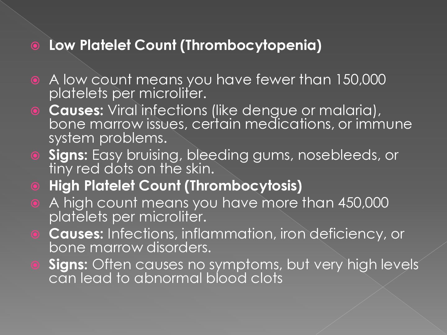
  4.  Low Platelet Count (Thrombocytopenia) A low count means you

    have fewer than 150,000 platelets per microliter.  Causes: Viral infections (like dengue or malaria), bone marrow issues, certain medications, or immune system problems.  Signs: Easy bruising, bleeding gums, nosebleeds, or tiny red dots on the skin.  High Platelet Count (Thrombocytosis)  A high count means you have more than 450,000 platelets per microliter.  Causes: Infections, inflammation, iron deficiency, or bone marrow disorders.  Signs: Often causes no symptoms, but very high levels can lead to abnormal blood clots 
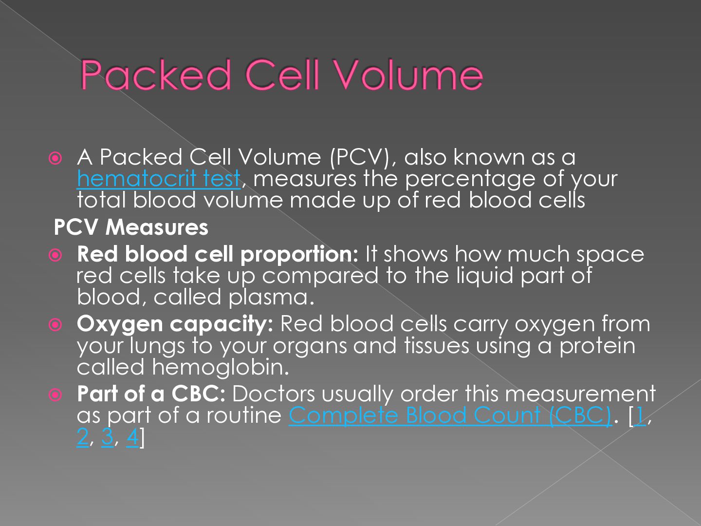
  5. A Packed Cell Volume (PCV), also known as a hematocrit

    test, measures the percentage of your total blood volume made up of red blood cells PCV Measures  Red blood cell proportion: It shows how much space red cells take up compared to the liquid part of blood, called plasma.  Oxygen capacity: Red blood cells carry oxygen from your lungs to your organs and tissues using a protein called hemoglobin.  Part of a CBC: Doctors usually order this measurement as part of a routine Complete Blood Count (CBC). [1, 2, 3, 4] 
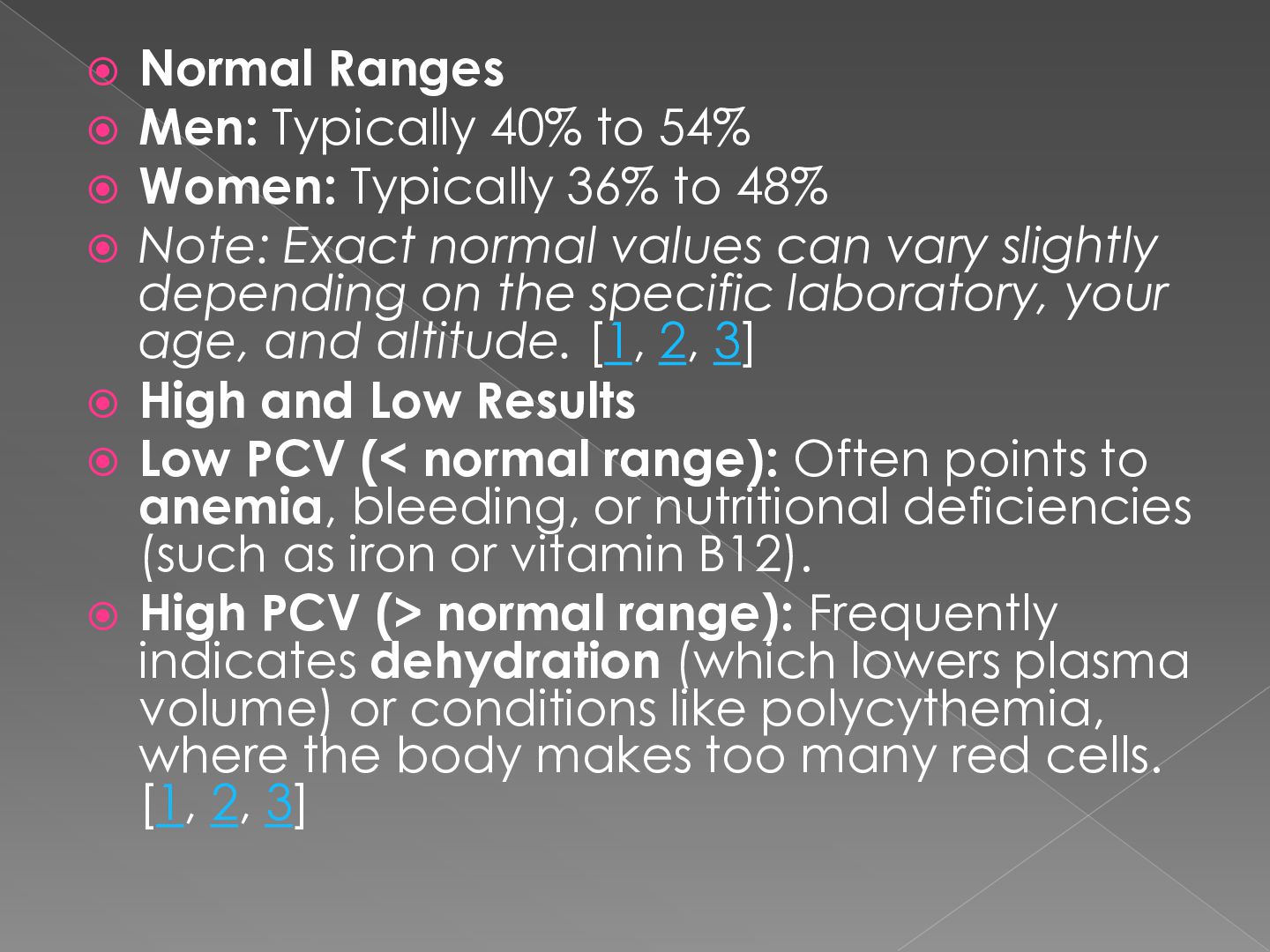
  6.  Normal Ranges  Men: Typically 40% to 54% 

    Women: Typically 36% to 48%  Note: Exact normal values can vary slightly depending on the specific laboratory, your age, and altitude. [1, 2, 3]  High and Low Results  Low PCV (< normal range): Often points to anemia, bleeding, or nutritional deficiencies (such as iron or vitamin B12).  High PCV (> normal range): Frequently indicates dehydration (which lowers plasma volume) or conditions like polycythemia, where the body makes too many red cells. [1, 2, 3]
  7.  ESR (erythrocyte sedimentation rate, or sed rate) blood test

    measures how quickly red blood cells settle to the bottom of a test tube in one hour to detect inflammation in the body.
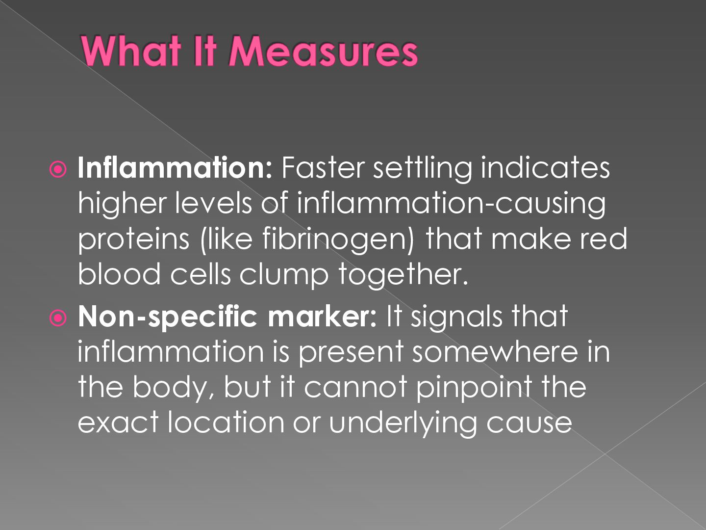
  8.  Inflammation: Faster settling indicates higher levels of inflammation-causing proteins

    (like fibrinogen) that make red blood cells clump together.  Non-specific marker: It signals that inflammation is present somewhere in the body, but it cannot pinpoint the exact location or underlying cause
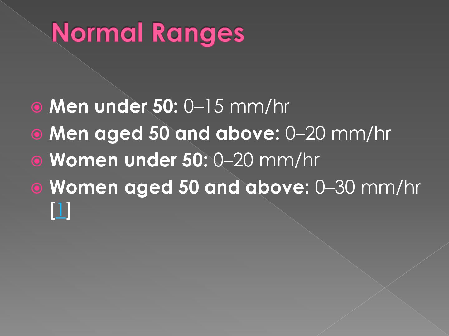
  9.  Men under 50: 0–15 mm/hr  Men aged 50

    and above: 0–20 mm/hr  Women under 50: 0–20 mm/hr  Women aged 50 and above: 0–30 mm/hr [1]
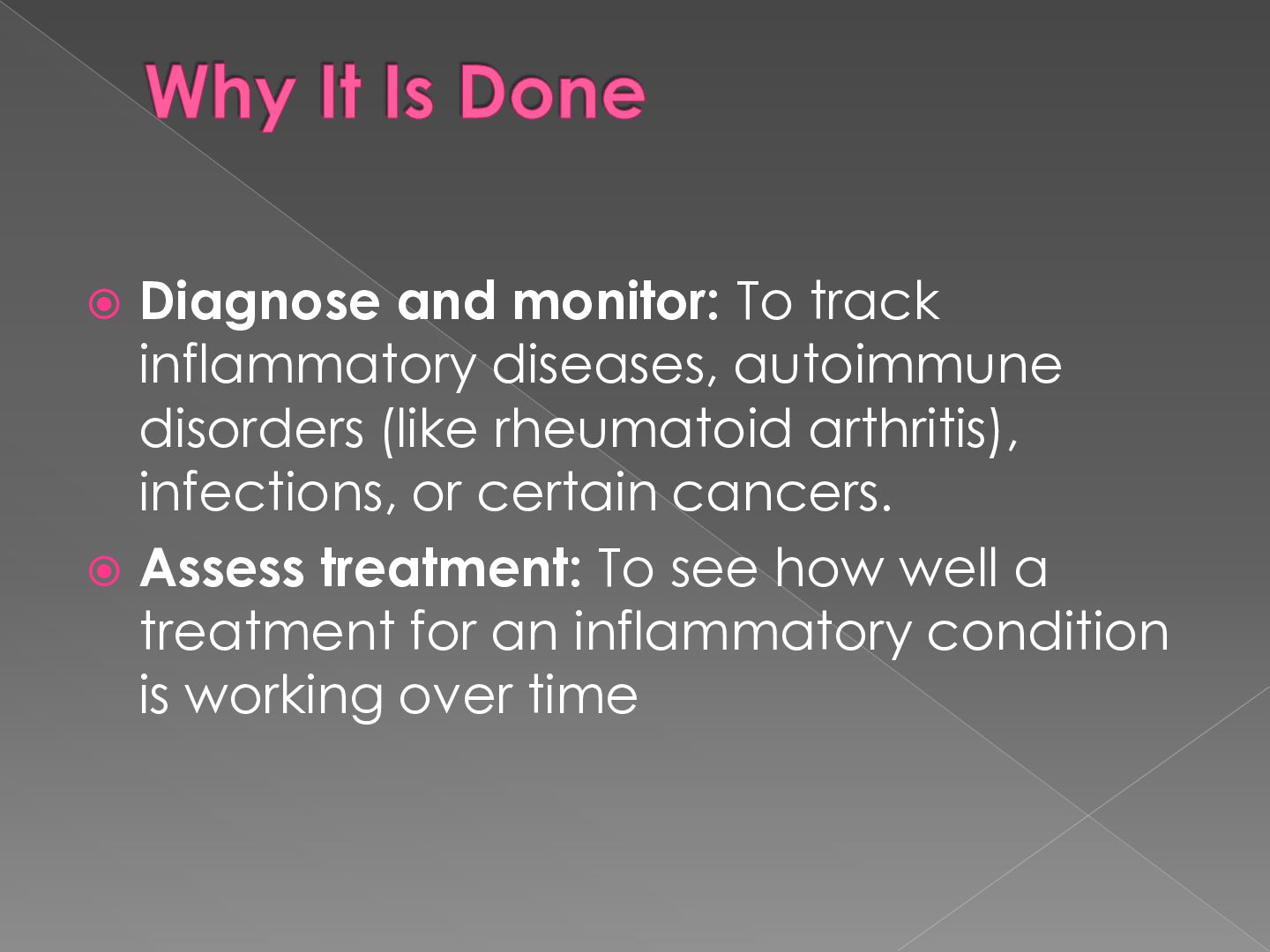
  10.  Diagnose and monitor: To track inflammatory diseases, autoimmune disorders

    (like rheumatoid arthritis), infections, or certain cancers.  Assess treatment: To see how well a treatment for an inflammatory condition is working over time
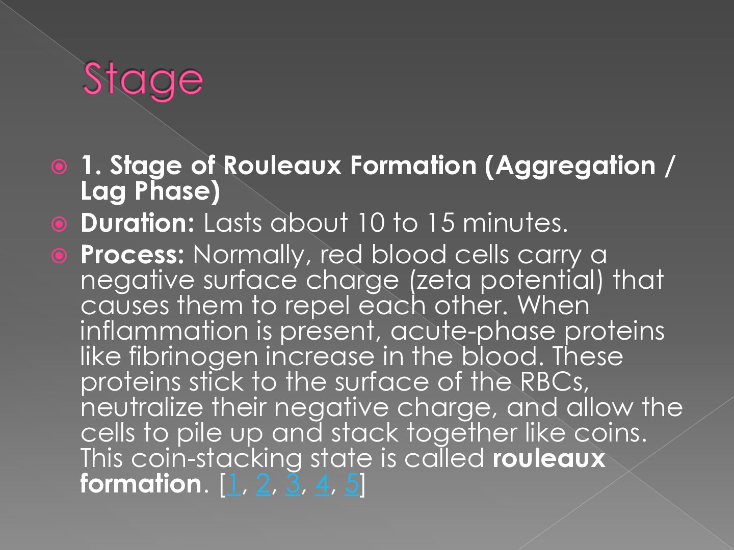
  11.  1. Stage of Rouleaux Formation (Aggregation / Lag Phase)

     Duration: Lasts about 10 to 15 minutes.  Process: Normally, red blood cells carry a negative surface charge (zeta potential) that causes them to repel each other. When inflammation is present, acute-phase proteins like fibrinogen increase in the blood. These proteins stick to the surface of the RBCs, neutralize their negative charge, and allow the cells to pile up and stack together like coins. This coin-stacking state is called rouleaux formation. [1, 2, 3, 4, 5]
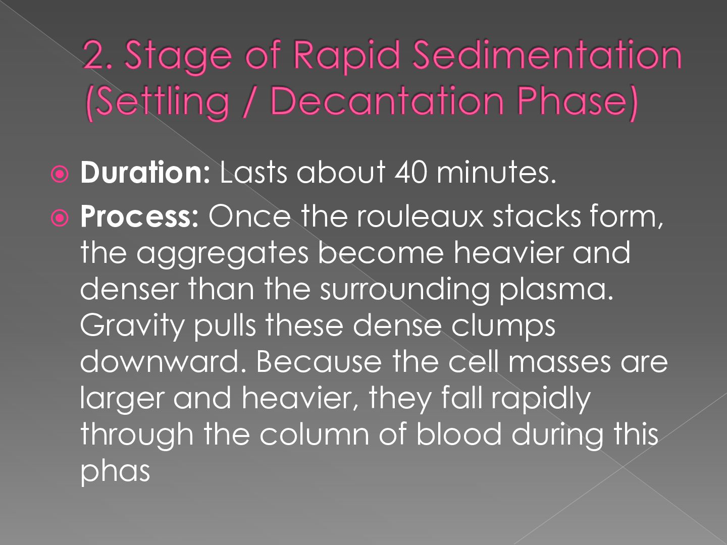
  12.  Duration: Lasts about 40 minutes.  Process: Once the

    rouleaux stacks form, the aggregates become heavier and denser than the surrounding plasma. Gravity pulls these dense clumps downward. Because the cell masses are larger and heavier, they fall rapidly through the column of blood during this phas
  13.  Duration: Lasts about 10 minutes.  Process: The falling

    red blood cells reach the bottom of the tube and begin to crowd and pack tightly together. The accumulation slows down and stops as the sedimented cells form a dense layer at the base, leaving clear, yellowish plasma at the top.
  14.  Placement: The anticoagulated blood is mixed and drawn into

    a tall, vertical Westergren tube up to the 200 mm mark.  Incubation: The tube is left completely undisturbed in a vertical position at room temperature for exactly one hour.  Measurement: After one hour, the distance from the top of the blood column to the top of the settled red blood cell layer is measured in millimeters (mm).  Reporting: The final result is recorded in millimeters per hour (mm/hr)
  15.  The Wintrobe method measures the erythrocyte sedimentation rate (ESR)

    by recording how far red blood cells sink in a 100 mm long, 2.5 mm wide glass tube in one hour. Normal values are 0–9 mm/hr for males and 0–20 mm/hr for females
  16. Equipment and Setup Wintrobe Tube: 11 cm long glass tube

    with a 2.5 mm internal bore marked from 0 to 100 mm in both descending and ascending order.  Anticoagulant: EDTA-treated venous blood is used directly without extra liquid dilution.  Test Steps  Fill Tube: Mix the blood well and draw it into a Pasteur or capillary pipette. Fill the Wintrobe tube precisely to the zero mark at the top, avoiding any air bubbles.  Position Stand: Place the tube upright in a vertical rack on a steady, vibration-free surface away from direct heat or sun.  Read Result: Leave the setup undisturbed for exactly one hour, then read the depth in millimeters of clear plasma at the top  
  17.  Blood indices are measurements from a Complete Blood Count

    (CBC) that describe the size, shape, and hemoglobin content of your red blood cells to help diagnose conditions like anemia
  18. 1. MCV (Mean Corpuscular Volume): Measures the average size of

    your red blood cells.Normal range: 80 to 100 femtoliters (fL).  High: Large cells (macrocytic anemia).  Low: Small cells (microcytic anemia).
  19.  Mean Corpuscular Hemoglobin (MCH): Measures the average weight or

    amount of hemoglobin inside a single red blood cell.Normal range: 27 to 31 picograms (pg) per cell.  Clinical use: Low MCH indicates less hemoglobin, often making cells look pale (hypochromic)
  20.  Mean Corpuscular Hemoglobin Concentration (MCHC): Measures the average concentration

    or percentage of hemoglobin relative to the cell's size.Normal range: 32 to 36 grams per deciliter (g/dL).  Clinical use: Low MCHC means hemoglobin is spread too thinly inside the cell.
  21.  Red Cell Distribution Width (RDW): Measures the variation in

    the size and volume of your red blood cells.Clinical use: A high RDW shows a wide mix of cell sizes (anisocytosis), which often points to early nutritional deficiencies
  22.  To prepare a standard thin peripheral blood smear, place

    a small drop of blood on one end of a clean glass slide. Touch a second spreader slide to the drop at a 35–45° angle, let the blood spread across the edge, and push the spreader swiftly forward to create a thin film with a feathered edge
  23.  Materials Needed  Clean glass microscope slides (free of
    dust and grease)  Fresh or anticoagulated (EDTA) blood sample  Spreader slide (a slide with a smooth, clean edge
  24.       Clean slides: Wipe glass

    slides clean using lens paper to remove dust or glass shards. Position the drop: Place a small, medium-sized drop of blood about 1 cm from one end of the primary slide. Set the angle: Place the edge of the spreader slide in front of the blood drop at a 35 to 45-degree angle against the main slide. Contact the blood: Pull the spreader slide backward gently until it touches the drop of blood, allowing the blood to spread out along the width of the edge. Push forward: Push the spreader slide forward in a rapid, smooth, and steady motion across the slide. Dry: Allow the blood film to air-dry completely at room temperature before any staining process
  25.  Reticuocyte (immature red blood cells) and malaria parasites (Plasmodium

    species) are both identified using blood smear microscopy stained with Romanowski stains like Giemsa.
  26.  Definition: Reticulocytes are young, newly released red blood cells

    containing residual ribonucleic acid (RNA) network remnants.  Staining: Seen using special supravital stains (like New Methylene Blue) which precipitate RNA into a distinctive dark blue reticular network or dots.
  27.  Giemsa Appearance: On standard Giemsa-stained thin films, they appear

    slightly larger than mature red blood cells (normocytes) and may display a faint bluish-gray tint (polychromasia).  Malaria Connection: Certain malaria species (Plasmodium vivax and Plasmodium ovale) show a strict biological preference or tropism for invading these immature reticulocytes rather than mature cells
  28.  Thick Smears: Prepared by lysing red blood cells; used

    as the high-sensitivity gold standard to rapidly detect the presence of parasites and quantify overall parasite burden relative to white blood cells.  Thin Smears: Prepared with a fixed single cell layer; used to preserve cell morphology for identifying specific Plasmodium species and calculating percent parasitemi
  29.  Species Morphology: 1. Plasmodium falciparum: Infects red blood cells

    of any maturity, showing delicate ring forms, multiple chromatin dots, and distinct bananashaped (crescentic) gametocytes. 2. Plasmodium vivax: Selectively targets reticulocytes, causing infected cells to enlarge significantly with amoeboid trophozoites and Schuffner's dots. 3. Plasmodium ovale: Infects reticulocytes, presenting oval-shaped host cells with fimbriated (spiky/feathery) edges and Schuffner's dots. 4. Plasmodium malariae: Infects mature cells with characteristic compact ring forms, heavypigmented band or basket-shaped trophozoites, and rosette schizonts.
  30.  It is a laboratory procedure used to measure the

    amount of hemoglobin— the iron-rich protein in red blood cells that carries oxygen—to diagnose conditions like anemia. [1, 2]
  31. Cyanmethemoglobin Method: The gold standard and most accurate colorimetric method,

    where blood is mixed with Drabkin's reagent to form a stable cyanmethemoglobin pigment measured via spectrophotometry at 540 nm.  Sahli's Acid Hematin Method: A manual, inexpensive technique where blood is mixed with hydrochloric acid (N/10 HCl) to form an acid hematin solution, which is then diluted with distilled water until its brown color matches a standard comparator.  Automated Analyzers: Modern electrical impedance or flow cytometry systems that measure hemoglobin alongside complete blood counts quickly and with high precision 
  32.  A Total Leukocyte Count (TLC) test measures the number

    of white blood cells in your blood to evaluate your immune system's health
  33.  What the Test Measures 1. White blood cells: Cells

    that defend your body against infections, viruses, and bacteria. 2. Part of CBC: It is usually included in a standard Complete Blood Count test. 3. Sample collection: A phlebotomist draws a small amount of blood from a vein in your arm
  34.  Normal Range  Standard adult range: 4,000 to 11,000

    cells per microliter (µL) of blood.  No fasting needed: You can take the test at any time without fasting
  35.  Interpreting Results  High count (Leukocytosis): A count above

    11,000/µL usually signals an infection, inflammation, stress, or an immune reaction.  Low count (Leukopenia): A count below 4,000/µL can mean a weakened immune system, viral infection, or bone marrow issue
  36.  A Differential Leukocyte Count (DLC) test measures the percentage

    of each of the five main types of white blood cells in your blood to help doctors evaluate your immune health and detect infections or inflammatio
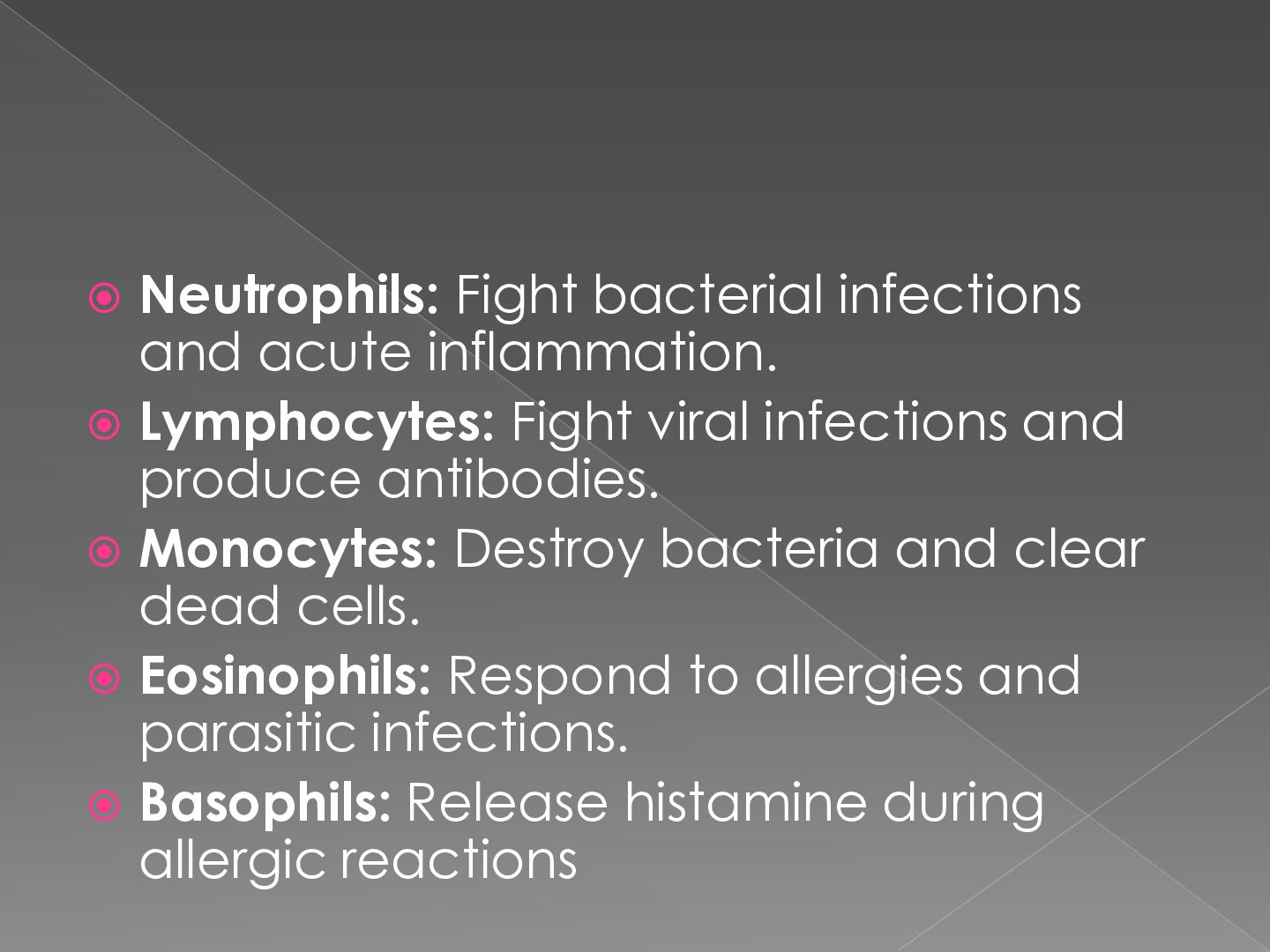
  37.  Neutrophils: Fight bacterial infections and acute inflammation.  Lymphocytes:

    Fight viral infections and produce antibodies.  Monocytes: Destroy bacteria and clear dead cells.  Eosinophils: Respond to allergies and parasitic infections.  Basophils: Release histamine during allergic reactions
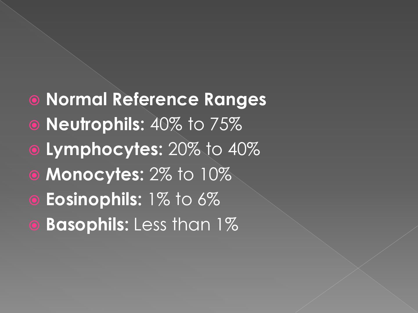
  38.  Normal Reference Ranges  Neutrophils: 40% to 75% 

    Lymphocytes: 20% to 40%  Monocytes: 2% to 10%  Eosinophils: 1% to 6%  Basophils: Less than 1%