Upgrade to Pro — share decks privately, control downloads, hide ads and more …

Components of Radiographic Equipment - X-ray Tu...

Sponsored · Your Podcast. Everywhere. Effortlessly. Share. Educate. Inspire. Entertain. You do you. We'll handle the rest. →

Components of Radiographic Equipment - X-ray Tube, Generator, Collimator, Grid, CR, DR | DMLT 1st Year Notes

Complete and easy notes on Components of Radiographic Equipment for DMLT 1st Year and BSc Radiology students.

Topics Covered:
1. X-Ray Tube - Cathode, Anode, Glass Envelope
2. Operating Console - kVp, mA, Timer, AEC
3. High Tension Generator - Types and Function
4. Collimator - Light Beam Diaphragm and Uses
5. Filters and Grids - Types of Grids
6. Potter-Bucky Diaphragm
7. Image Receptor - Film, Cassette, CR Imaging Plate, DR Flat Panel Detector
8. Portable and Mobile X-ray Machine

Prepared by Annu Maurya for DMLT students. Useful for exam preparation, viva and practical.

Keywords: radiographic equipment notes, x-ray tube diagram, DMLT 1st year notes, radiology notes pdf, CR DR system, collimator, bucky grid

Er. Annu maurya

September 10, 2026

Transcript

23 slides
  1. X- ray machine parts  An X-ray machine consists of

    three main hardware sections: the operating console, the highfrequency generator, and the X-ray tube head
  2. System Components Operating Console: The control panel where the operator

    sets exposure factors like voltage (kVp), current (mA), and time.  High-Frequency Generator: Steps up regular voltage into the high voltage required to power and accelerate electrons across the system.  X-ray Tube Head: The protective housing holding the internal vacuum tube that generates the radiation beam. 
  3. Collimator: A lead shutter device attached below the tube window

    to restrict and shape the X-ray beam, reducing unnecessary patient exposure.  Bucky and Grid: A moving or stationary grid placed near the patient to absorb scatter radiation, paired with a Bucky mechanism holding the image receptor.  Image Receptor / Detector: The digital flat panel detector or film cassette that captures the remaining X-ray pattern to form the image 
  4. Inside the X-Ray Tube Cathode: The negative electrode containing a

    heated tungsten filament that emits electrons through thermionic emission, plus a focusing cup to direct the beam.  Anode: The positive target electrode (often rotating) made of tungsten that absorbs high-speed electrons and converts their energy into X-ray photons.  Glass or Metal Envelope: A vacuum-sealed housing enclosing the cathode and anode so electrons can travel without air resistance.  Protective Housing & Oil: Lead-lined outer casing filled with cooling oil to absorb stray radiation and dissipate heat 
  5. Image receptors  An image receptor is a device that

    captures X-ray or radiation beams passing through a patient's body to create a visual picture of internal body parts.
  6. Types of Image Receptors Film-Screen Systems (Analog): Uses photographic film

    and special luminescent screens. The screen glows when hit by X-rays, which helps expose the film. It needs chemical processing to show the final picture.  Computed Radiography (CR): Uses a photostimulable phosphor plate inside a plastic cassette. A laser scanner reads the stored energy from the plate to make a digital image  Digital Radiography (DR): Uses flat-panel detectors or electronic sensors like CCD (charge-coupled device) and CMOS (complementary metal-oxide-semiconductor). It turns X-rays directly or indirectly into digital data for instant viewing on a computer 
  7. How They Work Radiation Exposure: Invisible X-ray beams pass through

    body tissues. Dense tissues like bone absorb more rays, while soft tissues absorb less.  Latent Image Creation: The pattern of remaining radiation hits the receptor. This forms a hidden, temporary image called a latent image.  Processing: The latent image turns into a visible picture. Analog film needs chemical baths, while digital systems use computers to process and display the file immediately 
  8. Grids  A radiographic grid is a device placed between

    the patient and the image receptor to absorb scatter radiation and improve image contrast
  9. Purpose and Function Reduces Scatter: Absorbs off-direction X-ray photons caused

    by Compton scattering inside the patient.  Improves Contrast: Prevents foggy, low-contrast images and sharpens anatomical visibility.  Increases Dose: Requires higher exposure settings (mAs) because it blocks some of the primary beam, increasing patient radiation dose.  Anatomy Size: Used generally when body part thickness is greater than 10 cm (such as the abdomen, skull, or spine) 
  10. Design Terms Grid Ratio: The height of the lead strips

    divided by the width of the radiolucent space between them (common ratios range from 5:1 to 16:1). Higher ratios clean up more scatter but require higher radiation output.  Grid Frequency: The number of lead lines per inch or centimeter 
  11. Common Types of Grids Linear / Parallel: Lead strips run

    parallel to each other; best for specific focal ranges.  Focused: Lead strips angle outward to match the divergent X-ray beam.  Crossed: Two linear grids stacked on top of each other for maximum scatter cleanup.  Moving (Bucky): Reciprocates or moves during exposure to blur out grid lines on the final image 
  12. Bucky  In radiology, a Bucky (named after inventor Gustav

    Bucky) is a device that holds the X-ray film cassette or digital detector and houses an antiscatter grid. It controls scattered radiation to make X-ray images sharper and clearer
  13. What a Bucky Does Holds the receptor: It keeps the

    X-ray plate or digital detector steady.  Blocks scatter: It uses a grid of thin lead strips to block scatter radiation from blurring the image.  Moves during exposure: Many modern Buckys move (reciprocate) slightly while the X-ray is taken so that grid lines do not show up on the picture 
  14. Where It Is Found Table Bucky: Built into the bottom

    of an X-ray examination table for patients lying down.  Wall Bucky (Upright Stand): Mounted on a wall for standing or sitting chest and spine X-rays 
  15. Portable x ray machine A portable X-ray machine is a

    compact, lightweight medical imaging device that brings diagnostic radiography directly to the patient's bedside, in emergency rooms, or during remote field visits  A portable X-ray machine is a compact, lightweight, or wheeled medical device designed to take X-ray images at a patient's bedside rather than transporting them to a radiology department 
  16. Main Components X-Ray Tube & Collimator: Generates and focuses the

    Xray beam toward the target area  High-Frequency Generator: Supplies and controls the electrical energy needed, adjusting parameters like kVp (kilovoltage peak) and mAs (milliampere-seconds).  Control Panel: Digital interface used by the radiographer to set exposure levels and select patient anatomy profiles  Power Source: Operates using rechargeable battery packs, capacitor discharge systems, or standard wall outlets.  Image Receptor / Detector: Uses digital radiography (DR) flat-panel detectors or computed radiography (CR) cassettes to capture and instantly transmit images to the hospital network. 
  17. Common Uses Intensive care units (ICUs) and emergency rooms (ERs)

    for critical patients.  Bedside chest X-rays for immobile or severely ill patients.  Nursing homes, field hospitals, and remote or mobile health camps 
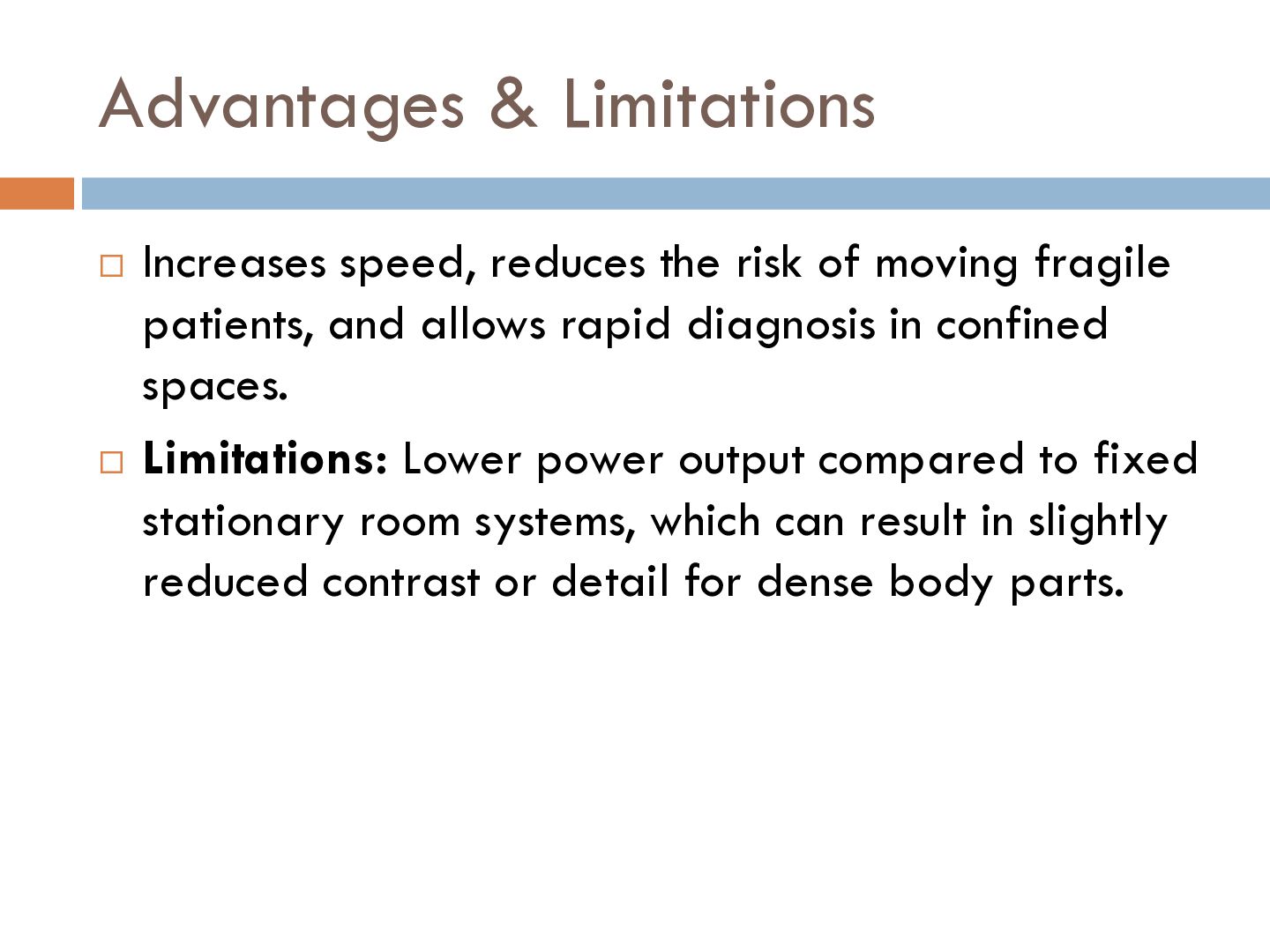
  18. Advantages & Limitations Increases speed, reduces the risk of moving

    fragile patients, and allows rapid diagnosis in confined spaces.  Limitations: Lower power output compared to fixed stationary room systems, which can result in slightly reduced contrast or detail for dense body parts. 
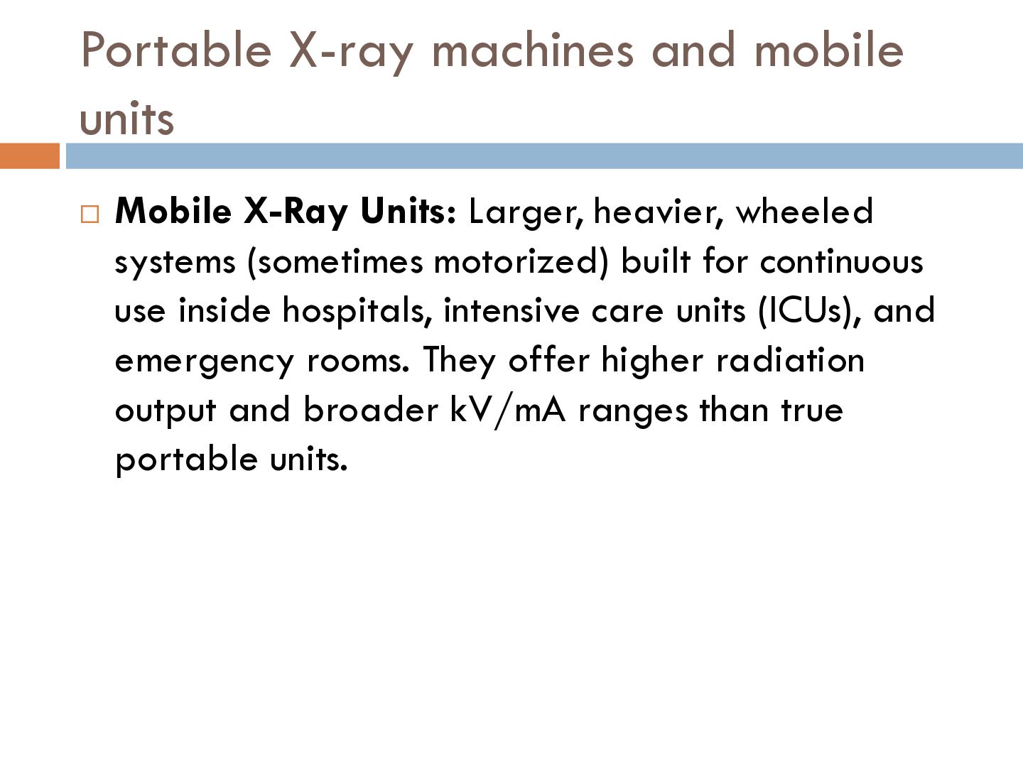
  19. Portable X-ray machines and mobile units  Mobile X-Ray Units:

    Larger, heavier, wheeled systems (sometimes motorized) built for continuous use inside hospitals, intensive care units (ICUs), and emergency rooms. They offer higher radiation output and broader kV/mA ranges than true portable units.
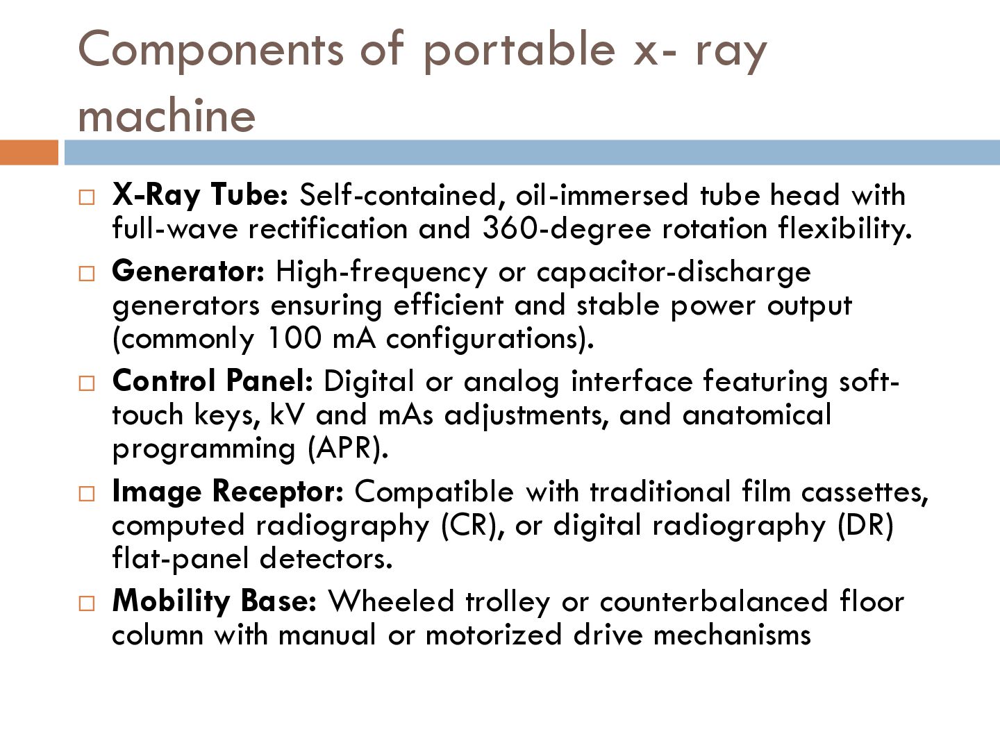
  20. Components of portable x- ray machine X-Ray Tube: Self-contained, oil-immersed

    tube head with full-wave rectification and 360-degree rotation flexibility.  Generator: High-frequency or capacitor-discharge generators ensuring efficient and stable power output (commonly 100 mA configurations).  Control Panel: Digital or analog interface featuring softtouch keys, kV and mAs adjustments, and anatomical programming (APR).  Image Receptor: Compatible with traditional film cassettes, computed radiography (CR), or digital radiography (DR) flat-panel detectors.  Mobility Base: Wheeled trolley or counterbalanced floor column with manual or motorized drive mechanisms 
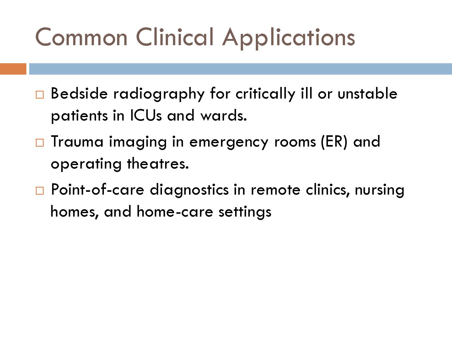
  21. Common Clinical Applications Bedside radiography for critically ill or unstable

    patients in ICUs and wards.  Trauma imaging in emergency rooms (ER) and operating theatres.  Point-of-care diagnostics in remote clinics, nursing homes, and home-care settings 

More Decks by Er. Annu maurya

Other Decks in Education