Upgrade to PRO for Only $50/Year—Limited-Time Offer! 🔥

FY 2024: MDC 8 - Musculosystem

Avatar for e4health e4health PRO
April 03, 2024
480

FY 2024: MDC 8 - Musculosystem

Avatar for e4health

e4health PRO

April 03, 2024
Tweet

Transcript

  1. H I M | C O D I N G

    & C D I | H E A LT H I T | R E V C Y C L E Empowering Better Health e4health tackles healthcare’s data, quality and revenue challenges empowering your providers to focus on better care.
  2. Objectives • Review MDC 8- Diseases and disorders of the

    musculoskeletal system and connective tissue with a focus on selected diagnoses and procedures • Learner will acquire a basic understanding of the diagnoses and procedures included in MDC-8 • Discuss Query opportunities in MDC-8 • Review coding clinics relevant to the chosen topics in each DRG
  3. MDC 8-MS- DRGs (Medical) • 533-534 FRACTURES OF FEMUR WITH/WITHOUT

    MCC • 535-536 FRACTURES OF HIP AND PELVIS WITH/WITHOUT MCC • 537 SPRAINS, STRAINS, AND DISLOCATIONS OF HIP, PELVIS AND THIGH WITH CC/MCC • 538 SPRAINS, STRAINS, AND DISLOCATIONS OF HIP, PELVIS AND THIGH WITHOUT CC/MCC • 539-541 OSTEOMYELITIS WITH/WITHOUT CC/MCC • 542-544 PATHOLOGICAL FRACTURES AND MUSCULOSKELETAL AND CONNECTIVE TISSUE MALIGNANCY WITH/WITHOUT CC/MCC • 545-547 CONNECTIVE TISSUE DISORDERS WITH/WITHOUT CC/MCC • 548-550 SEPTIC ARTHRITIS WITH/WITHOUT CC/MCC • 551-552 MEDICAL BACK PROBLEMS WITH/WITHOUT MCC • 553-554 BONE DISEASES AND ARTHROPATHIES WITH/WITHOUT MCC • 555-556 SIGNS AND SYMPTOMS OF MUSCULOSKELETAL SYSTEM AND CONNECTIVE TISSUE WITH/WITHOUT MCC • 557-558 TENDONITIS, MYOSITIS AND BURSITIS WITH/WITHOUT MCC • 559-561 AFTERCARE, MUSCULOSKELETAL SYSTEM AND CONNECTIVE TISSUE WITH/WITHOUT CC/MCC • 562-563 FRACTURE, SPRAIN, STRAIN AND DISLOCATION EXCEPT FEMUR, HIP, PELVIS AND THIGH WITH/WITHOUT MCC • 564-566 OTHER MUSCULOSKELETAL SYSTEM AND CONNECTIVE TISSUE DIAGNOSES WITH/WITHOUT CC/MCC
  4. MDC 8-MS- DRGs (Surgical) • 453-455 COMBINED ANTERIOR AND POSTERIOR

    SPINAL FUSION WITH/WITHOUT CC/MCC • 456-458 SPINAL FUSION EXCEPT CERVICAL WITH SPINAL CURVATURE, MALIGNANCY, INFECTION OR EXTENSIVE FUSIONS WITH/WITHOUT CC/MCC • 459-460 SPINAL FUSION EXCEPT CERVICAL WITH/WITHOUT MCC • 461-462 BILATERAL OR MULTIPLE MAJOR JOINT PROCEDURES OF LOWER EXTREMITY WITH/WITHOUT MCC • 463-465 WOUND DEBRIDEMENT AND SKIN GRAFT EXCEPT HAND FOR MUSCULOSKELETAL AND CONNECTIVE TISSUE DISORDERS WITH/WITHOUT CC/MCC • 466-468 REVISION OF HIP OR KNEE REPLACEMENT WITH/WITHOUT CC/MCC • 521-522 HIP REPLACEMENT WITH PRINCIPAL DIAGNOSIS OF HIP FRACTURE WITH/WITHOUT MCC • 469 MAJOR HIP AND KNEE JOINT REPLACEMENT OR REATTACHMENT OF LOWER EXTREMITY WITH MCC OR TOTAL ANKLE REPLACEMENT • 470 MAJOR HIP AND KNEE JOINT REPLACEMENT OR REATTACHMENT OF LOWER EXTREMITY WITHOUT MCC • 471-473 CERVICAL SPINAL FUSION WITH/WITHOUT CC/MCC
  5. MDC 8-MS- DRGs (Surgical) • 474-476 AMPUTATION FOR MUSCULOSKELETAL SYSTEM

    AND CONNECTIVE TISSUE DISORDERS WITH/WITHOUT CC/MCC • 477-479 BIOPSIES OF MUSCULOSKELETAL SYSTEM AND CONNECTIVE TISSUE WITH/WITHOUT CC/MCC • 480-482 HIP AND FEMUR PROCEDURES EXCEPT MAJOR JOINT WITH/WITHOUT CC/MCC • 483 MAJOR JOINT OR LIMB REATTACHMENT PROCEDURES OF UPPER EXTREMITIES • 485-487 KNEE PROCEDURES WITH PRINCIPAL DIAGNOSIS OF INFECTION WITH/WITHOUT CC/MCC • 488 KNEE PROCEDURES WITHOUT PRINCIPAL DIAGNOSIS OF INFECTION WITH CC/MCC • 489 KNEE PROCEDURES WITHOUT PRINCIPAL DIAGNOSIS OF INFECTION WITHOUT CC/MCC • 518 BACK AND NECK PROCEDURES EXCEPT SPINAL FUSION WITH MCC OR DISC DEVICE OR NEUROSTIMULATOR • 519 BACK AND NECK PROCEDURES EXCEPT SPINAL FUSION WITH CC • 520 BACK AND NECK PROCEDURES EXCEPT SPINAL FUSION WITHOUT CC/MCC • 492-494 LOWER EXTREMITY AND HUMERUS PROCEDURES EXCEPT HIP, FOOT AND FEMUR WITH/WITHOUT CC/MCC • 495-497 LOCAL EXCISION AND REMOVAL OF INTERNAL FIXATION DEVICES EXCEPT HIP AND FEMUR WITH/WITHOUT CC/MCC
  6. MDC 8-MS- DRGs (Surgical) • 498 LOCAL EXCISION AND REMOVAL

    OF INTERNAL FIXATION DEVICES OF HIP AND FEMUR WITH CC/MCC • 499 LOCAL EXCISION AND REMOVAL OF INTERNAL FIXATION DEVICES OF HIP AND FEMUR WITH/WITHOUT CC/MCC • 500-502 SOFT TISSUE PROCEDURES WITH/WITHOUT CC/MCC • 503-505 FOOT PROCEDURES WITH/WITHOUT CC/MCC • 506 MAJOR THUMB OR JOINT PROCEDURES • 507 MAJOR SHOULDER OR ELBOW JOINT PROCEDURES WITH CC/MCC • 508 MAJOR SHOULDER OR ELBOW JOINT PROCEDURES WITHOUT CC/MCC • 509 ARTHROSCOPY • 510-512 SHOULDER, ELBOW OR FOREARM PROCEDURES, EXCEPT MAJOR JOINT PROCEDURES WITH/WITHOUT CC/MCC • 513 HAND OR WRIST PROCEDURES, EXCEPT MAJOR THUMB OR JOINT PROCEDURES WITH CC/MCC • 514 HAND OR WRIST PROCEDURES, EXCEPT MAJOR THUMB OR JOINT PROCEDURES WITHOUT CC/MCC • 515-517 OTHER MUSCULOSKELETAL SYSTEM AND CONNECTIVE TISSUE O.R. PROCEDURES WITH/WITHOUT CC/MCC
  7. Site and laterality • Most of the codes within Chapter

    13 have site and laterality designations • The site represents the bone, joint or the muscle involved • For some conditions where more than one bone, joint or muscle is usually involved, such as osteoarthritis, there is a “multiple sites” code available • For categories where no multiple site code is provided and more than one bone, joint or muscle is involved, multiple codes should be used to indicate the different sites involved
  8. Bone vs. joint For certain conditions, the bone may be

    affected at the upper or lower end, (e.g., avascular necrosis of bone, M87, Osteoporosis, M80, M81) Though the portion of the bone affected may be at the joint, the site designation will be the bone, not the joint
  9. Acute traumatic vs. chronic or recurrent conditions • Many musculoskeletal

    conditions are a result of previous injury or trauma to a site, or are recurrent conditions • Bone, joint or muscle conditions that are the result of a healed injury are usually found in chapter 13 • Recurrent bone, joint or muscle conditions are also usually found in chapter 13 • Any current, acute injury should be coded to the appropriate injury code from chapter 19 • Chronic or recurrent conditions should generally be coded with a code from chapter 13 • If it is difficult to determine from the documentation in the record which code is best to describe a condition, query the provider
  10. Femur fractures • Closely review the imaging for the location

    of the fracture on the bone • Closely review for documentation of the type of fracture: • Transverse • Oblique • Spiral • Torus • Segmental • Comminuted https://www.orthopedic-institute.org/wp-content/uploads/2015/03/fractures.jpg
  11. Femoral Head/Neck Fractures • Femur head and neck fractures •

    Review radiology results for location of the fracture • Review documentation for bone and laterality • Hip not otherwise specified • Intracapsular • Midcervical • Pertrochanteric • Physeal of upper end of femur • Subcapital • Subtrochanteric • Transcervical • Trochanter Hip Fractures - OrthoInfo - AAOS
  12. Pelvic fractures • Includes: • Open and Closed Fractures of

    the acetabulum, ilium, ischium and Pubis • Review for location of an acetabular fracture (can be taken from radiology reports) • Anterior and posterior wall • Anterior and posterior column • Transverse • Transverse posterior • Medial wall • Dome
  13. Femur, Hip and Pelvic fractures • Query opportunities • Type

    of fracture • Clarification of periprosthetic fracture • Acute blood loss anemia • Fat embolism • Rhabdomyolysis • Additional pelvic fractures in imaging that could be missed • Retroperitoneal hemorrhage in the presence of a pelvic fracture • Query if only noted in imaging and not documented by the physician • Occurs frequently in severe pelvic fractures
  14. Sprains, strains and dislocations of hip, pelvis and thigh Coding

    guidelines, clinical criteria and query opportunities
  15. Definitions • Sprain – a stretch or tear in a

    ligament • Strain – a stretch or tear of a muscle or tendon • Sprains and strains of the hip and thigh • Iliofemoral or ischiocapsular ligament sprain, sprain of hip, strain of quadriceps, adductor, posterior muscle group of hip and thigh • Unlikely to require admission as a principal diagnosis, but may be present along with other injuries that require hospitalization and intervention
  16. Dislocation of hip • Dislocation of the hip • Subluxation

    or dislocation of the anterior, central obturator, posterior or unspecified hip join • Review for an additional fracture – impacts coding – careful review of imaging reports • If a prosthetic Joint is present, it is coded as a complication of the prosthesis even if caused by trauma Anterior Hip Dislocation (Case courtesy of Dr Sajoscha Sorrentino, Radiopaedia.org. From the case rID: 14836)
  17. Sprains, strains and dislocations of hip, pelvis and thigh •

    Query opportunities • Laterality • Complication of prosthesis • Presence of arthritis • Presence of stress fracture
  18. Osteomyelitis • Osteomyelitis is reported by site • Is usually

    the result of an infection elsewhere that spreads through the blood and infects weakened or diseased bone • Can be acute or chronic. Chronic is rare • Osteomyelitis is automatically associated with diabetes, and the principal diagnosis will be the diabetes code
  19. Osteomyelitis • Query opportunities • Sepsis/Bacteremia • Other abscesses near

    the area of the osteomyelitis • Clarification of the underlying cause if unclear • Clarification of location, stage and POA status of pressure ulcers • Clarification of type of ulcer if unclear • Underlying bacteria if known and not documented as the underlying cause • Other bone disease, metastatic sites making the patient susceptible • Any associated implants (fusion hardware, internal fixation devices, joint prosthesis) • If related, coded as a complication of an implant
  20. Guidelines • 7th character A is for use as long

    as the patient is receiving active treatment for the fracture • While the patient may be seen by a new or different provider over the course of treatment for a pathological fracture, assignment of the 7th character is based on whether the patient is undergoing active treatment and not whether the provider is seeing the patient for the first time • 7th character D is to be used for encounters after the patient has completed active treatment for the fracture and is receiving routine care for the fracture during the healing or recovery phase • The other 7th characters, listed under each subcategory in the Tabular List, are to be used for subsequent encounters for treatment of problems associated with the healing, such as malunions, nonunions, and sequelae • Care for complications of surgical treatment for fracture repairs during the healing or recovery phase should be coded with the appropriate complication codes
  21. Pathological fractures • Pathological fractures • Physician must document a

    relationship between any condition except osteoporosis and the pathological fracture • Use appropriate documentation to link the conditions such as due to, caused by, related to • Review for evidence of bone disease such as • Fosamax, Aredia, Didronel indicating osteoporosis, Paget’s disease • Renal disease with osteodystrophy • History of cancer with bone metastasis • Dietary conditions such as malnutrition • Requires the same reporting as traumatic fractures related to location, location on the bone, laterality and encounter type
  22. Pathological fractures Etiologies: • Review that all sites are documented

    as primary or metastatic Malignant neoplasm of the bone • Includes gastrointestinal stromal tumor, stromal tumors sarcoma • Includes connective and soft tissue such as blood vessels, lymphatic vessels, peripheral nerves • Query the specific location of GIST if not documented Malignant neoplasm of connective and other soft tissue
  23. Pathological fractures • Etiologies: • Gastrointestinal Stromal Tumor (GIST) •

    Arise from interstitial cells of Cajal or the precursors to these cells • Sporadic GIST tumor • Only one tumor • Found mostly in patients without a family history • Familial GIST • Frequently multiple tumors • Overgrowth of other cells in the GI tract • Patches of dark skin on other areas • Urticaria Pigmentosa – raised patches of brown skin that stings or itches when touched • SDH deficient GIST • Occurs in children and young adults, more female than male • High risk of developing other types of noncancerous tumors of the nervous system
  24. Pathological fractures • Query opportunities • Review imaging and PMH

    for evidence of undocumented cancer sites such as the lung, breast or prostate • Evidence of malnutrition and severity • Diagnosis associated with medications being administered • Chemotherapy associated pancytopenia • Anemia due to malignancy • Documentation of all metastatic sites • Cancer related pain
  25. Stress Fracture Different from pathological fractures and can be known

    as “fatigue fracture,” “March fracture,” or “stress reaction fracture” Due to repetitive force applied before the bone and its supporting tissues have had enough time to absorb such force that results in damage to the bone Initially test negative in an X-ray display, and days or weeks may pass before the fracture line is visible on an X-ray Stress fractures are classified to subcategory M84.3 • Additional External cause of morbidity codes are used to identify the cause of the stress fracture (for example, code Y93.01, Activity, walking, marching and hiking)
  26. Atypical Femoral Fracture • A form of stress fracture that

    is associated with osteoporosis and usually occur in the subtrochanteric region of hip or femoral shaft • Occur in patients receiving long-term bisphosphonate therapy or other medications such as glucocorticoids • These fractures can: • Have an appearance of transverse or short oblique orientation • Have minimal or no trauma associated with these fractures • Have a lack of comminution • Have a cortical thickening that is either generalized or localized at the lateral cortex of the fracture site • Have a periosteal reaction of the lateral cortex • Have a medial spike when the fracture is complete • Classified to subcategory M84.75, Atypical femoral fracture and the entire term “atypical femoral fracture” must be documented to assign a code from subcategory M84.75 • Seventh characters are required for atypical femoral fractures to indicate initial encounter, subsequent encounter, or sequela • Followed by the code for long-term use of bisphosphonates, Z79.83, Long term (current) use of bisphosphonates
  27. Stress fractures • Query opportunities • Laterality • Presence of

    osteoporosis • Non-compliance to medical advice
  28. Osteoporosis Osteoporosis is a systemic condition, meaning that all bones

    of the musculoskeletal system are affected. Therefore, site is not a component of the codes under category M81, Osteoporosis without current pathological fracture. The site codes under category M80, Osteoporosis with current pathological fracture, identify the site of the fracture, not the osteoporosis • Category M81, Osteoporosis without current pathological fracture, is for use for patients with osteoporosis who do not currently have a pathologic fracture due to the osteoporosis, even if they have had a fracture in the past • For patients with a history of osteoporosis fractures, status code Z87.310, Personal history of (healed) osteoporosis fracture, should follow the code from M81 • Category M80, Osteoporosis with current pathological fracture, is for patients who have a current pathologic fracture at the time of an encounter. The codes under M80 identify the site of the fracture • A code from category M80, not a traumatic fracture code, should be used for any patient with known osteoporosis who suffers a fracture, even if the patient had a minor fall or trauma, if that fall or trauma would not usually break a normal, healthy bone
  29. Osteoporosis • Query opportunities • Laterality • If trauma, clarify

    if due to osteoporosis (non-traumatic) vs. a traumatic event • Presence of stress fracture • Review radiology for presence of osteopenia vs. osteoporosis vs. osteomyelitis • Associated conditions, such as: • Electrolyte imbalance, such as hypocalcemia or hypomagnesemia • Thyroid conditions
  30. Arthritis • Variety of conditions that affects joints, muscles, and

    connective tissue • Symptoms include inflammation, swelling, pain, stiffness, and mobility • Can occur independently or due to another condition • If applicable, combination codes should be assigned when available
  31. Rheumatoid arthritis • Rheumatoid arthritis • Autoimmune disease • Systemic

    disease that can have manifestations in other organs • Code a combination code that links the site of arthritis with laterality and other manifestations such as organ involvement • Coded as with or without Rheumatoid factor • Can be responsible for bone weakness, and inflammatory changes that cause deformities in the hands and feet
  32. Rheumatoid arthritis • Rheumatoid arthritis – Extraarticular Manifestations • Skin

    • Nodules – 50% of people with RA. Lumps that form over the skin, over bony areas. Treated with disease modifying antirheumatic drugs. • Rashes caused by vasculitis. Small red dots that can result in skin ulcers. • Drug effects from corticosteroids NSAIDS • Bones • Generalized loss of bone density from chronic inflammation • Consider clarification if there is a fracture in a patient with RA for pathological fracture
  33. • Rheumatoid arthritis – Extraarticular Manifestations • Eyes • Scleritis

    that can lead to scarring and may cause permanent damage • Dryness • Glaucoma, cataracts and pigment changes with vision loss related to medication. • Mouth • Dryness • Oral sores and ulcers from methotrexate Rheumatoid arthritis
  34. • Rheumatoid arthritis – Extraarticular Manifestations • Heart disease –

    Primary cause of morbidity and mortality in patients with RA • Premature atherosclerosis with a higher plaque burden • Myocarditis, dilated cardiomyopathy, rheumatoid nodules and rarely amyloidosis • Pericarditis • Valvular disease • Pulmonary hypertension • Aortic Aneurysm • Liver and kidneys • Impacted by medications taken for RA Rheumatoid arthritis
  35. SLE- Systemic Lupus Erythematosus • Systemic lupus erythematosus (SLE) •

    Autoimmune condition with similar manifestation to RA • Can have flare ups • Treatment toward inflammation and to decrease chances of organ damage This Photo by Unknown author is licensed under CC BY.
  36. • Query Opportunities • Diagnosis relationship with systemic disease such

    as RA and Lupus • Cardiomyopathy • Encephalitis • Encephalopathy • Pleural effusion • Renal failure specificity (glomerulonephritis) in patients with RA and Lupus • Adverse effects of medications, such as dehydration or oral thrush Connective Tissue Disorders
  37. Septic arthritis • Can lead to sepsis if left untreated

    An infection in the joint – not truly sepsis • If present and there is no documented association query the provider Review for the presence of a joint prosthesis Presentation may vary depending on the cause and underlying conditions Query for a recent history of joint injection, or drainage of fluids
  38. • Query Opportunities • Complication of joint injection or drainage

    of fluid • Causative organism and nature of mechanism • Laterality • Non-compliance • Adverse effects of medications, such as dehydration, acute kidney injury Septic Arthritis
  39. Vertebral fractures Fracture of vertebral column without mention of spinal

    cord injury These are traumatic fractures of all levels of the vertebra •Stable and unstable burst fracture •Traumatic compression fracture of spine •Wedge compression fracture Review record for evidence of pathological fracture Evidence of spinal cord injury changes the code and DRG Review for specific involved specificity to include if the fracture was displaced or nondisplaced Query the physician/provider whenever these specifics are lacking in the medical record
  40. Intravertebral disc disorders • Intravertebral disc disorders • Includes disc

    displacement, herniated nucleus pulposus or post-laminectomy syndrome • Are also reported with myelopathy or radiculopathy • Use the code for the most superior level when there are overlapping sites
  41. Intravertebral disc disorders •Degenerative spine conditions •Disc herniation •RA •Tumors,

    hematomas Myelopathy- A compression injury to the spinal cord caused by: (not all inclusive) •Difficulty with fine motor skills •Changes in reflexes •Pain, numbness, tingling and weakness in extremities •Lower back pain •Incontinence of bowel or bladder Symptoms include:
  42. Intravertebral disc disorders • Terminology of disc degeneration is not

    the same as disc displacement (herniation) • Presence or absence of myelopathy is important to determine the assignment of codes for certain back order • For example: Myelopathy = functional disorder and/or pathologic change in the spinal cord due to compression • Spondylosis and herniation of intervertebral disc needs to be differentiated between with or without myelopathy • Paresthesia (“pins and needless”) is not the same as paralysis as well when it comes to herniation of disc
  43. Intravertebral disc disorders • Radiculopathy – compression and inflammation of

    the nerve root • Lumbar – (sciatica) • Cervical • Thoracic
  44. Back disorders • Query Opportunities • Review carefully for evidence

    of spinal cord injury. If not documented, query the provider • Specificity of displaced or non-displaced fracture exact vertebra that is fractured • Specificity of traumatic vs. pathological fracture in a patient with bone disease and a compression type fracture, or minimal trauma • If signs and symptoms indicate radiculopathy or myelopathy or imaging shows compression of the spinal cord or nerve roots, review for documentation, if none is present a query to clarify the clinical significance is appropriate
  45. Aseptic Necrosis of bone • Aseptic Necrosis of the Bone-Osteonecrosis

    • A decrease in blood supply to the bone • Trauma • Poor circulation due to hypercoagulable state, atherosclerosis, vasculitis • Steroid medication are the most common to cause osteonecrosis. • Osteonecrosis of the Jaw – rarely – associated with bisphosphonate especially IV • Cigarette smoking • Radiation exposure • Sickle cell disease • Lupus (SLE) • Pancreatitis
  46. Aseptic necrosis of bone • Aseptic Necrosis of the Bone-Osteonecrosis

    • Diagnostics • MRI can definitively diagnose early disease. Bone scans may suggest early disease • Not seen on X-ray until the disease is more advanced • Pain in the affected bone/joint, increased with use • Treatment • Early disease can be treated by removing the bone, called decompression. May replace with new bone encouraging new blood supply. • Later disease may require joint replacement
  47. Aseptic necrosis of bone • Query Opportunities • Review for

    associated diagnoses: • Nicotine use • Adverse effect of steroids and other medications • Presence of SLE, Rheumatoid arthritis, gout, vasculitis, Sickle cell disease • With causative nature of radiation therapy
  48. Gout • Gout • Hyperuricemia and recurrent acute inflammatory arthritis

    • Types of gout: • Acute • Chronic with or without tophus • Drug induced • Primary • Secondary • Query Opportunities • If documentation does not include the acuity and type it is appropriate to query the provider • Review the record for specific site and laterality, query if not documented • Review for acute renal failure
  49. Osteoarthritis Osteoarthritis • Degeneration of the cartilage in the joint

    cause overgrowth of bone and changes in the synovial membrane. • Review for documentation of the specific site, laterality and the specific type. Types of osteoarthritis • Primary • Dysplastic • Post-traumatic • Secondary
  50. Ankylosing spondylitis • Inflammation of the spine and large joints

    • Causes damage in the sacroiliac joint • Can cause fusion in the SI joint and bones in the chest • Appears between age 17-35 and affects men more than women Ankylosing Spondylitis – Stiff Spine • X-ray • Erythrocyte sedimentation rate (ESR or sed rate) • Genetic testing Diagnostics
  51. Ankylosing spondylitis •NSAIDS for pain and inflammation •TNF blockers (biologics)

    •IL-17A inhibitors •Disease modifying anti- rheumatic medicines •Short term steroids •Muscle relaxers for severe pain and muscle spasms •Sometimes joint replacement or spine stabilizing surgery Ankylosing Spondylitis – Treatment •Osteoporosis from medication •Immobility •Inflammation of the eye, aortic valve, intestines •Psoriasis Complications
  52. Bone Diseases and arthropathy • Query Opportunities • Site, laterality

    and type of OA • Acuity and specificity of gout • Acute renal failure • Osteoporosis related to medications
  53. Necrotizing fasciitis Necrotizing fasciitis • Infection that causes necrosis of

    muscle, fascia and subQ tissue • Usually caused by Group A Strep or Staph aureus • Requires IV antibiotics and frequently surgical debridement • Rapidly progresses and has a high mortality rate Complications • Loss of limb • Sepsis – and all the complications and CDI considerations of Sepsis • Organ failure, shock, consistent documentation throughout the medical record and inclusion in the d/c summary
  54. Necrotizing fasciitis • Necrotizing fasciitis - Treatment • Antibiotics •

    Surgery • Extensive debridement of necrotic tissue • Daily dressing changes • Reconstruction • May require extensive plastic reconstruction
  55. Necrotizing fasciitis Query Opportunity • Associated conditions, such as: •

    Sepsis • Osteomyelitis • Diabetes and type • Renal failure • Complications of amputation (if procedure done) • Surgical site infections
  56. Rhabdomyolysis • Rhabdomyolysis • Damage to muscle fibers causing the

    release myoglobin into the blood stream • Can cause acute kidney failure because myoglobin if filtered by the kidney, and the amount released overwhelms the kidneys • Commonly associated with crush injuries • Can be caused by extreme exercise, falls where the patient can’t get up for long periods, drug overdose and heat stroke • Review for • Acute kidney injury/Acute tubular necrosis • Specificity of traumatic. If not specified, query the provider • If AKI and rhabdomyolysis are present on admission, then AKI is considered the more acute conditioned as is sequenced as the PDX. CC 3rd quarter 2002 page 28
  57. Rhabdomyolysis • Query Opportunities • Acuity of Rhabdomyolysis • Presence

    of AKI and AKI specificity for ATN • Sepsis associated with necrotizing fasciitis • Septic shock • Lactic acidosis associated with necrotizing fasciitis • Organ failure associated with sepsis in the presence of necrotizing fasciitis
  58. Device Complications • Complications due to internal orthopedic prosthesis devices,

    implants and grafts • Includes complications directly related to the implant, device or graft such as: • Embolism, Fibrosis, Hemorrhage, Pain, Stenosis, Thrombosis • Documentation must directly link the device to the complication using linking words such as due to, related to, caused by, “with” or “in the presence of” do not link the complication with the device. Credit: combinescience.blogspot.com
  59. Infection of Device • Infection and Inflammatory reaction due to

    internal joint prosthesis • Includes infections or inflammation directly related to internal joint prosthesis • Clarify if the infection is in the wound, the prosthesis, or involves both • An additional code is assigned for the specific infection if appropriate • Documentation must directly link the device to the complication using linking words such as due to, related to, caused by, “with” or “in the presence of” do not link the complication with the device
  60. Infection of device • Infection and Inflammatory reaction due to

    other internal orthopedic device, implant or graft • Includes infections or inflammation directly related to devices such as internal fixation devices • Clarify if the infection is in the wound, the prosthesis, or involves both • An additional code is assigned for the specific infection if appropriate • Documentation must directly link the device to the complication using linking words such as due to, related to, caused by, “with” or “in the presence of” do not link the complication with the device
  61. Mechanical complication of device • Mechanical complications of internal orthopedic

    devices, implants or grafts • Breakage, dislocation, loosening, obstruction, perforations, fractures and around the prosthetic, and wear of the articular surface of the internal prosthetic joint • Periprosthetic fractures • Assign codes for the specific type of fracture clarifying pathological or traumatic • Ensure documentation of periprosthetic fracture by the provider • Patients admitted for joint replacement: review for any complication of the joint prosthesis that was the reason for surgical repair or replacement This Photo by Unknown author is licensed under CC BY-SA-NC.
  62. Fracture during surgery • Fracture during insertion of an orthopedic

    implant or prosthesis • This is a fracture of the bone during an insertion while in surgery • Clarify with the provider if the fracture was traumatic or pathologic • If pathologic assign a code for the underlying disease process
  63. Humeral Fracture • Fracture of the humerus • Surgical neck

    fracture • Displaced or nondisplaced • 2-part, 3-part or 4-part • Specific location, surgical neck, greater tuberosity or lesser tuberosity • Distal humerus • Lower end of the humerus, supracondylar with or without intercondylar fracture, medical epicondyle, lateral condyle or transcondylar • Location of the fracture can be coded from radiology reports, and laterality can be coded from the provider or other clinicians
  64. Complication of amputation stump Complication must be more than a

    routinely expected condition or occurrence There is no time limit from the time of the amputation and the complication • Contracture • Dehiscence • Hematoma • Infection • Necrosis • Neuroma Conditions included in amputation stump complications:
  65. MDC-8 Diagnoses that affect SOI/ROM ▪ Acute blood loss anemia

    ▪ Acute renal failure ▪ Bacteremia ▪ Cardiac arrest ▪ Cellulitis ▪ CVA ▪ Dehydration ▪ Drug induced delirium ▪ End stage renal disease ▪ Fat embolism ▪ Fracture of acetabulum ▪ Fracture of multiple ribs ▪ Fracture of sacrum or coccyx ▪ Hypotension ▪ Intraspinal abscess ▪ Malnutrition ▪ Metabolic encephalopathy ▪ Multiple fractures ▪ Metastasis ▪ Osteoporosis ▪ Paraplegia ▪ Pressure ulcer ▪ Psoas muscle abscess ▪ Quadriplegia ▪ Rhabdomyolysis ▪ Sepsis ▪ Severe Sepsis ▪ Scoliosis ▪ Shock ▪ Skin ulcer ▪ Ventricular tachycardia
  66. Body part values in ICD-10-PCS • Adult human body •

    206 bones, more than 250 joints • Recognizing names of bone markings can assist in determining the body part value • Each does not have its own body part value • See Coding Guideline B4.1a—when there is not a separate body part value use the value for the whole body part • Y value available for upper or lower bone • Bilateral body parts as applicable, code individually for each side if not available • See Coding Guideline B4.3
  67. Body part values in ICD-10-PCS • Upper and lower bones

    and joints • Upper—above the level of the lumbar vertebrae plus upper extremities • Lower—lumbar vertebrae and below plus lower extremities • Coding Guideline B3.10a—spinal vertebral joints in spinal fusion classified by the level of the spine—distinct body part values for single and multiple vertebral joints at each spinal level
  68. approaches • 0—Open • Arthrotomy = Incision into joint capsule

    • 3—Percutaneous • 4—Percutaneous endoscopic • Arthroscopic • X—External • For example, closed reductions of fractures • See Coding Guideline B5.3b
  69. External fixation devices Monoplanar or uniplane—holds fracture in one plane

    or direction—see figure 1.11 Limb-lengthening— expandable rods along limbs to encourage bone growth Ring device or multiplane—surrounds limb like a ring, provides fixation in two or more planes Note that device values are not specific to the body system— dependent on body system and root operation 3 = Ceramic synthetic substitute in Replacement procedures of the lower joints, but infusion device in all other root operations
  70. Qualifiers • Can further define devices, for example, cemented or

    non- cemented • Specific anatomical approach for spinal fusion • Anterior approach, anterior column • Access through the front of the body for body of the vertebrae or disc • Posterior approach, posterior column • Access through the back of the body for vertebral foramen, spinous processes, facets, and/or lamina • Posterior approach, anterior column • Access through the back of the body for body of the vertebrae or disc • Interbody fusion is always performed on the anterior column— note the rows of the Fusion tables
  71. Spinal structures • The spine is composed of: • Anterior

    column • Anterior longitudinal ligament • Vertebral body • Intervertebral disc • Annulus fibrosus • Posterior longitudinal ligament • Posterior column • Pedicles • Transverse process • Lamina • Facets • Spinous process
  72. Anterior column fusion • Can be performed using an anterior,

    lateral, or posterior technique • Requires an incision in the neck or the abdomen, and fusion is carried out from the front of the vertebrae through the anterior annulus • ALIF (anterior lumbar interbody fusion)—the approach is from the anterior • May be laparoscopic • Axia LF (axial lumbar interbody fusion)—the approach from the posterior, from under the sacrum. This approach is percutaneous to approach the L5-S1 with a completely internal procedure performed with imaging guidance. • DLIF (direct lateral lumbar interbody fusion)—the approach is anterior but from the lateral, or side of the patient (through psoas muscle) limiting the incision size • PLIF (posterior lumbar interbody fusion)—Involves an anterior column fusion through a posterior approach • TLIF (transforaminal lumbar interbody fusion)—the approach is from the posterior but to one side of the spine (transverse lateral), through the neuroforamen • XLIF (extreme lateral interbody fusion)—the approach is from the anterior but far to the side through a small incision or by using a series of tube dilators to create a space just large enough to see through. Is less-invasive surgery on the anterior column done percutaneously or via a circular tube retractor through a lateral approach
  73. Posterior column fusion Involves an incision in the patient’s back

    directly over the vertebrae Pedicle, lamina, facet, transverse process, or “gutter” fusion Can be performed using a posterior, posterolateral, or lateral transverse technique May be abbreviated as PLF (posterior lumbar fusion) when performed on the lumbar spine Pedicle-based rods and screws to maintain the correct alignment and distance of the vertebral joints and stabilize the spine—does not serve to fuse the spine, stabilizes and is integral to the Fusion Bone is placed along the spinous process or as a bridge between the transverse processes of the spine—bone heals to form the fusion • Autograft and bone bank material if necessary ▪ A posterior lumbar interbody fusion (PLIF) is a fusion of the anterior column using a posterior approach and is coded to qualifier value J ▪ This is different than a posterior lumbar fusion, which is a fusion of the posterior column using a posterior approach and coded to qualifier value 1 ▪ These two procedures can be done independently or during the same operative session. If performed together at the same joint level, code both procedures separately
  74. Spinal fusion and refusion The following table may be used

    as a reference to assist in determining the appropriate ICD-10-PCS qualifier (7th character) with the spinal fusion procedures listed previously Fusion procedure Approach and Column ICD-10-PCS Qualifier ALIF Anterior approach, anterior column 0 AxiaLiF Posterior approach, anterior column J DLIF Anterior approach, anterior column 0 PLIF Posterior approach, anterior column J TLIF Posterior approach, anterior column J XLIF Anterior approach, anterior column 0
  75. 360-degree fusion • Anterior and posterior fusion together—360-degree fusion •

    Two codes are required—qualifiers will be different • Can be performed through: • Single incision (usually through the lateral transverse approach), or • Two incisions (anterior incision for anterior fusion, followed by turning the patient over to make a posterior incision for posterior fusion). • Most common in the lumbar spine • A 360-degree lumbar fusion done with a PLIF, accompanied by a posterior lumbar fusion at L3–L5 is coded: • 0SG10AJ Fusion of 2 or more Lumbar Vertebral Joints with Interbody Fusion Device, Posterior Approach, Anterior Column, Open Approach • 0SG1071 Fusion of 2 or more Lumbar Vertebral Joints with Autologous Tissue Substitute, Posterior Approach, Posterior Column, Open Approach • Anterior fusion with pedicle-based stabilization rods and screws on the posterior • Not a 360-degree fusion • Fusion is an anterior column fusion only • Bone is required for a fusion of the posterior column This Photo by Unknown author is licensed under CC BY-NC.
  76. Interbody fusion device • Instrumentation such as an interbody fusion

    device is used to stabilize and fuse degenerative disc spaces and to provide a stable segment for fusion. • Devices are also called • Interbody fusion cage • BAK cage • Ray-threaded fusion cage • Synthetic cage • Spacer • Bone dowels • Combinations of devices and material are often used to render a joint immobile This Photo by Unknown author is licensed under CC BY-SA.
  77. Interbody fusion device • When combinations of devices are used

    on the same vertebral joint, code the device value as follows: • When an interbody fusion device is used to render the joint immobile (containing bone graft or bone graft substitute), code with the device value “interbody fusion device” • If bone graft is the only device used to render the joint immobile, code the procedure with the device value “nonautologous tissue substitute” or “autologous tissue substitute” • If a mixture of autologous and nonautologous bone graft (with or without biological or synthetic extenders or binders) is used to render the joint immobile, code the procedure with the device value “autologous tissue substitute”
  78. Bone graft during spinal fusion Synchronous harvesting of bone graft

    to be used in a different part of the body is reported separately with the root operation “Excision” Locally harvested tissue is not coded separately When bone morphogenetic protein (BMP) is inserted to help create a bone graft substitute, it is identified with the device character in the fusion code Reporting a code for placement of BMP is optional
  79. Multiple Fusion Coding • Guideline B3.10b—separate code for the fusion

    of each vertebral joint with a different device or qualifier • Two codes for • L2–L3 fused with bone graft • L3–L4 fused with interbody fusion device • Two codes for • Anterior column of L4–L5 fused using both an anterior and posterior approach
  80. Laminectomy • Could be an approach for a spinal cord

    procedure OR a Release procedure • Release may be assigned with Fusion for a decompressive laminectomy is documented to release: • Spinal cord • Spinal nerve ▪ Assign code 00JU0ZZ for laminectomy performed for the purpose of exploration of the spinal canal. ▪ Laminectomy performed for excision of herniated disc represents the operative approach and is not coded separately. ▪ “Excision” is coded to report excision of the disc. ▪ When a decompressive foraminotomy/laminectomy is done to treat foraminal stenosis by releasing pressure and freeing up the spinal root, along with discectomy to treat lumbar disc herniation, each surgery has a distinct procedural objective and should be coded separately: ▪ “Excision” for discectomy ▪ “Release” for decompressive foraminotomy/laminectomy
  81. Vertebroplasty and kyphoplasty • Percutaneous vertebroplasty is a procedure done

    to treat vertebral compression fractures • Percutaneous vertebroplasty involves the insertion of cement glue–like material (polymethylmethacrylate) into the vertebral body to stabilize and strengthen collapsed or crushed bone • ICD-10-PCS classifies this procedure to the root operation “Supplement,” with “synthetic substitute” for the device value • The ARCUATE XP procedure is a variation of a percutaneous vertebroplasty in which arcs are cut in the cancellous bone within the vertebral body • Bone cement material is subsequently injected into the vertebral body • No bone or bone marrow is removed from, or compacted within, the vertebral body • This procedure is coded to the root operation “Supplement” • Percutaneous vertebral augmentation utilizes an expandable inflatable balloon to reestablish vertebral height in compression fractures
  82. Vertebroplasty and kyphoplasty • Coding of percutaneous vertebral augmentation requires

    two codes: • One for the root operation “Reposition” • Another for the root operation “Supplement" • Similar procedures coded in the same manner include: • Arcuplasty • Kyphoplasty • Skyphoplasty • Spineoplasty • In a percutaneous vertebral augmentation, after the balloon is removed, the cavity is filled with polymethylmethacrylate, which hardens to stabilize the bone • Conversely, in vertebroplasty, no balloon is utilized, and no attempt is made to restore vertebral height • If a vertebral biopsy is carried out during a vertebral augmentation procedure, assign a separate code for the biopsy • The biopsy is not an inherent part of the vertebral augmentation and should be coded separately, if performed
  83. Spinal decompression • Common surgical decompression procedures include: • Laminotomy

    • Laminectomy • Foraminotomy • Medial facetectomy • When decompressive laminectomy surgery is performed to release pressure and free a body part from constraint, assign a code for “Release” rather than “Excision” This Photo by Unknown author is licensed under CC BY-ND.
  84. Spinal Surgery General Query Opportunity • For spinal surgeries, especially

    laminectomies, review for: • Presence of dura tear and repair • Complications related to surgery: • Acute blood loss anemia (outside of the post-op recovery phase) • Hypotension (outside of the post-op recovery phase) • Acute renal failure (outside of the post-op recover phase) • Mechanical or infection of surgical site
  85. Repair of Fractures Reposition for fracture treatment Guideline B3.15 Reduction

    of a displaced fracture is coded to the root operation Reposition The application of a cast or splint in conjunction with the Reposition procedure is not coded separately Treatment of a nondisplaced fracture is coded to the procedure performed Examples: • Casting of a nondisplaced fracture is coded to the root operation Immobilization in the Placement section • Putting a pin in a nondisplaced fracture is coded to the root operation Insertion
  86. Joint replacement- General 110 • Cemented (prosthesis is attached to

    joint or bone with epoxy cement); or • Uncemented (prosthesis has mesh of holes on surface that allows growth of patient’s natural bone to hold device in place) • Assign “Z” for no qualifier when it cannot be determined whether a cemented or uncemented prosthesis was placed The seventh-character qualifier describes whether the prosthesis is: • Report the procedure with the root operation “Insertion” and the device “bone-growth stimulator” (sixth character “M”) If joint replacement involves placement of a bone-growth stimulator: • Report the procedure code twice when the same procedure is performed on bilateral joints • If individual codes are available to identify left and right joints, both codes are assigned for each joint ICD-10-PCS codes do not indicate bilateral replacement
  87. Joint replacement- HIP • For hip replacements, report the type

    of bearing surface using the following sixth character qualifiers: • 0 Polyethylene • 1 Metal • 2 Metal on polyethylene • 3 Ceramic • 4 Ceramic on polyethylene • 6 Oxidized zirconium on polyethylene • 7 Autologous tissue substitute • E Articulating spacer • J Synthetic substitute • K Nonautologous tissue substitute • When bearing surface information is not available, assign sixth character “J” or query the provider for clarification This Photo by Unknown author is licensed under CC BY.
  88. Joint replacement- HIP • Femoral—stem (bearing surface) placed inside the

    femur and femoral head • Acetabular—cup or shell (bearing surface) screwed into the socket of the pelvic bone and acetabular lining placed into the cup • Device value determined by bearing surface 112
  89. Joint replacement- KNEE • Femoral—condylar cartilage surface—typically metal • Tibial—Plateau

    with stem into the tibia and meniscal replacement • Patellar portion—optional piece—surface not coded • Unicondylar—only medial or lateral component replaced—device value • Patellofemoral component can also be replaced • The patella is included in a total knee joint replacement as well as the femoral and tibial components at table OSR, Replacement, lower joint. Source: Graichen. 2008 (June). “Cut model of an instrumented knee endoprosthesis for in vivo measurements of forces and moments.” Digital Image. Wikimedia Commons. https://en.wikipedia.org/wiki/File:Knieprothese.png.
  90. Revision/removal of Prostheses • Remove infected prosthesis, Insert static spacer

    (if applicable), Remove spacer (after infection clears), Replace previous joint replacement • If a malfunctioning device is corrected, this is coded to the root operation “Revision,” which is defined as “correcting, to the extent possible, a malfunctioning or misplaced device without taking out and putting a whole new device in its place.” • For example, the joint is adjusted but not removed • If a joint prosthesis is removed and replaced during the same encounter, assign a code for both the removal of the old component and the placement of the new component (Removal + Replacement) • A code for the root operation “Supplement” is assigned if a new liner is placed  For removal of a joint spacer (e.g., cement), assign also a code for the removal  Codes are provided for revision of hip replacements that identify the specific joint components revised (acetabular, femoral, and acetabular liner and/or femoral head)  If a component of a joint has been previously replaced, the procedure is still a replacement, even though part of the component is being replaced for the first time  Example: Patient is admitted for conversion of a previous hemiarthroplasty to a total hip replacement. Report the procedure with a code for removal of the old component, along with a code for the replacement
  91. Joint Replacement procedures General Query Opportunity • Review for complications

    related to surgery: • Acute blood loss anemia (outside of the post-op recovery phase) • Hypotension (outside of the post-op recovery phase) • Acute renal failure (outside of the post-op recover phase) • Mechanical or infection of surgical site • Peri-prosthetic fractures
  92. amputation • Root operation: Detachment • Th body part value

    is the site of the Detachment • If applicable, a qualifier is assigned to specify the level where the extremity was detached • Detachment procedures are found only in body systems X (anatomical regions, upper extremities) and Y (anatomical regions, lower extremities) because amputations are performed on the extremities, across overlapping body layers (skin, muscle, bone), and therefore cannot be coded to a specific musculoskeletal body system, such as bone or joints • Example: Amputation of the foot is coded to the root operation Detachment in the body system Anatomical Regions, Lower Extremities
  93. Amputation Updated Guidelines: B3.19 The root operation Detachment contains qualifiers

    that can be used to specify the level where the extremity was amputated These qualifiers are dependent on the body part value in the "upper extremities" and "lower extremities" body systems For procedures involving the detachment of all or part of the upper or lower extremities, the procedure is coded to the body part value that describes the site of the detachment Example: An amputation at the proximal portion of the shaft of the tibia and fibula is coded to the Lower leg body part value in the body system Anatomical Regions, Lower Extremities, and the qualifier High is used to specify the level where the extremity was detached *See Next Slide for Tables
  94. Detachment Qualifiers Definitions Body Part Qualifier Definition Upper arm and

    upper leg 1 High: Amputation at the proximal portion of the shaft of the humerus or femur 2 Mid: Amputation at the middle portion of the shaft of the humerus or femur 3 Low: Amputation at the distal portion of the shaft of the humerus or femur Lower arm and lower leg 1 High: Amputation at the proximal portion of the shaft of the radius/ulna or tibia/fibula 2 Mid: Amputation at the middle portion of the shaft of the radius/ulna or tibia/fibula 3 Low: Amputation at the distal portion of the shaft of the radius/ulna or tibia/fibula Body Part Qualifier Definition Hand and foot 0 Complete* 4 Complete 1st Ray 5 Complete 2nd Ray 6 Complete 3rd Ray 7 Complete 4th Ray 8 Complete 5th Ray 9 Partial 1st Ray B Partial 2nd Ray C Partial 3rd Ray D Partial 4th Ray F Partial 5th Ray Thumb, finger or toe 0 Complete: Amputation at the metacarpophalangeal/metatarsal-phalangeal joint 1 High: Amputation anywhere along the proximal phalanx 2 Mid: Amputation through the proximal interphalangeal joint or anywhere along the middle phalanx 3 Low: Amputation through the distal interphalangeal joint or anywhere along the distal phalanx
  95. Complete vs Partial Definition • For coding amputations of Hand

    and Foot: • Complete: Amputation through the carpometacarpal joint of the hand, or through the tarsal-metatarsal joint of the foot. • Partial: Amputation anywhere along the shaft or head of the metacarpal bone of the hand, or of the metatarsal bone of the foot.
  96. Amputation Procedures General Query Opportunity • Presence of laceration and/or

    accidental puncture • Review principal diagnosis clarity: • Due to circulatory disorder • Due to diabetes • Complications related to surgery: • Acute blood loss anemia (outside of the post-op recovery phase) • Hypotension (outside of the post-op recovery phase) • Acute renal failure (outside of the post-op recover phase) • Infection of surgical site
  97. Other Common root operations Tenotomy—coded to the objective of the

    procedure • Cutting the tendon to Release or Reposition bone that they are attached to Tenodesis—anchoring of the tendon to a bone • Reposition—if moved • Reattachment—if tendon ruptured or completely separated from the bone and must be sutured to the bone in the same location • Repair—lacerated tendon, sutured to itself Decompression fasciotomy—Release • Trapped muscles as in compartment syndrome • Body part refers to the muscle being freed • Excision is also coded if surgical debridement of damaged muscle Division—if objective is to cut, transect, or otherwise separate
  98. Other Common root operations Release—to free a compressed nerve •

    Freeing of a tendon from adhesions Excision or Resection—depending on extent of removal Repair • Capsulorrhaphy—capsule includes synovium and labrum • Shoulder—includes labrum—connective tissue • SLAP lesion (superior labral tear from anterior to posterior) • Performed on joint structure—See Coding Guideline B4.5 • Femoroplasty—Repair of cam lesion—hip • Acetabuloplasty—Repair of pincer lesion—hip Nondisplaced fracture treatment for pins or other internal fixation devices • Casting nondisplaced fracture = Immobilization in section 2, Placement
  99. References ▪ AHA ICD-10-CM and ICD-10-PCS Coding Handbook ▪ ICD-10-PCS:

    An Applied Approach 2023 ▪ Cengage: 3-2-1 CODE IT! • Hip Fractures - OrthoInfo - AAOS. (n.d.). https://orthoinfo.aaos.org/en/diseases--conditions/hip-fractures/ • How Rheumatoid Arthritis Affects More Than Joints. (n.d.). https://www.arthritis.org/diseases/more-about/how-rheumatoid-arthritis-affects-more- than-joints • Intervertebral disc disease: MedlinePlus Genetics. (n.d.). https://medlineplus.gov/genetics/condition/intervertebral-disc-disease/ • Kim, Paul H. MD1; Leopold, Seth S. MD1, a. Gustilo-Anderson Classification. Clinical Orthopaedics and Related Research 470(11):p 3270-3274, November 2012. | DOI: 10.1007/s11999-012-2376-6 • Marquis, B. O., & Capone, P. M. (2016). Myelopathy. Handbook of Clinical Neurology, 1015–1026. https://doi.org/10.1016/b978-0-444-53486- 6.00052-1 • Mitchell, D. (2022, September 28). When Is an Injury Initial, Subsequent, or Sequela? - AAPC Knowledge Center. AAPC Knowledge Center. https://www.aapc.com/blog/27096-initial-subsequent-sequela-encounter/ • Osteoarthritis (OA) | Arthritis | CDC. (n.d.). https://www.cdc.gov/arthritis/basics/osteoarthritis.htm • Osteomyelitis. (2021, August 8). Johns Hopkins Medicine. https://www.hopkinsmedicine.org/health/conditions-and-diseases/osteomyelitis • Q&A: Querying for the etiology of traumatic injuries | ACDIS. (n.d.). https://acdis.org/articles/qa-querying-etiology-traumatic-injuries
  100. References • Systemic lupus erythematosus: MedlinePlus Genetics. (n.d.). https://medlineplus.gov/genetics/condition/systemic-lupus- erythematosus/#causes

    • Sprain vs. Strain: What’s the Difference? (2007, January 1). WebMD. https://www.webmd.com/fitness-exercise/understanding- sprains-strains • Types of Fractures. (n.d.). The Orthopedic Institute at Southwest Health. https://www.orthopedic-institute.org/fracture-care/types- of-fractures/ • Wallace HA, Perera TB. Necrotizing Fasciitis. [Updated 2023 Feb 21]. In: StatPearls [Internet]. Treasure Island (FL): StatPearls Publishing; 2023 Jan-. Available from: https://www.ncbi.nlm.nih.gov/books/NBK430756/ • Zeisset, A. (2013). Coding Injuries in ICD-10-CM (update). AHIMA HIM Body of Knowledge. Retrieved April 11, 2023, from https://library.ahima.org/doc?oid=300644#.ZDWHmebMKUk • https://totalspinebrain.com/kyphoplasty-for-compression-fractures/
  101. References • Ankylosing Spondylitis. (2022, November 16). Johns Hopkins Medicine.

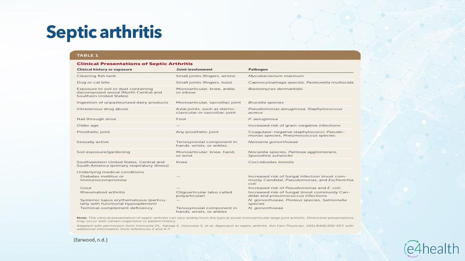
    https://www.hopkinsmedicine.org/health/conditions -and- diseases/ankylosing-spondylitis • Chauhan K, Jandu JS, Brent LH, et al. Rheumatoid Arthritis. [Updated 2023 Jan 10]. In: StatPearls [Internet]. Treasure Island (FL): StatPearls Publishing; 2023 Jan-. Available from: https://www.ncbi.nlm.nih.gov/books/NBK441999/ • Chen, Leon L. DNP, AGACNP-BC, FCCP, FAANP, FNYAM; Fasolka, Brian PhD, RN, CEN; Treacy, Caitlin MS, AGACNP-BC. Necrotizing fasciitis: A comprehensive review. Nursing 50(9):p 34-40, September 2020. | DOI: 10.1097/01.NURSE.0000694752.85118.62 • Earwood, J. S. (n.d.). Septic Arthritis: Diagnosis and Treatment. AAFP. https://www.aafp.org/pubs/afp/issues/2021/1200/p589.html#afp20211200p589-t1 • Facr, W. C. S. J. M. F. (2023, April 17). Aseptic Necrosis Causes, Symptoms, Treatment, Medications, Prevention. MedicineNet. https://www.medicinenet.com/aseptic_necrosis/article.htm • Gastrointestinal stromal tumor: MedlinePlus Genetics. (n.d.). https://medlineplus.gov/genetics/condition/gastrointestinal-stromal- tumor/#:~:text=A%20gastrointestinal%20stromal%20tumor%20%28GIST%29%20is%20a%20type,of%20Cajal%20%28ICCs%29%20or %20precursors%20to%20these%20cells. • Groner, L. K., Green, D. J., Weisman, S. V., Legasto, A. C., Toy, D., Gruden, J. F., & Escalon, J. G. (2021). Thoracic Manifestations of Rheumatoid Arthritis. Radiographics, 41(1), 32–55. https://doi.org/10.1148/rg.2021200091