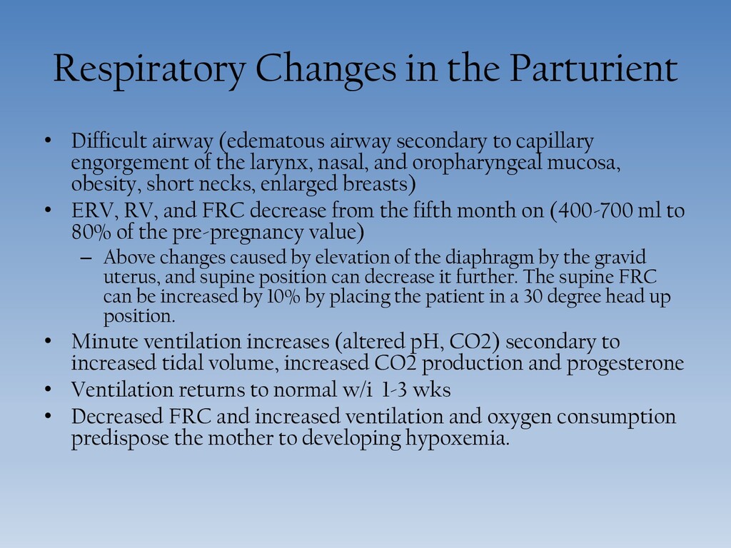
secondary to capillary engorgement of the larynx, nasal, and oropharyngeal mucosa, obesity, short necks, enlarged breasts) • ERV, RV, and FRC decrease from the fifth month on (400-700 ml to 80% of the pre-pregnancy value) – Above changes caused by elevation of the diaphragm by the gravid uterus, and supine position can decrease it further. The supine FRC can be increased by 10% by placing the patient in a 30 degree head up position. • Minute ventilation increases (altered pH, CO2) secondary to increased tidal volume, increased CO2 production and progesterone • Ventilation returns to normal w/i 1-3 wks • Decreased FRC and increased ventilation and oxygen consumption predispose the mother to developing hypoxemia.
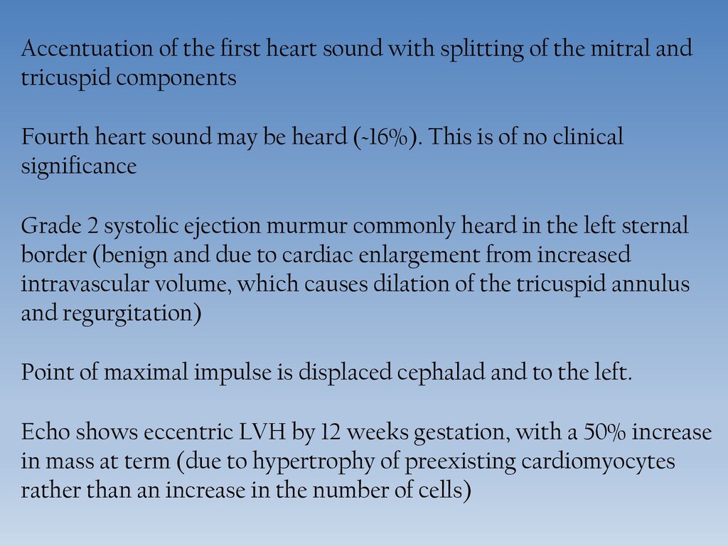
mitral and tricuspid components Fourth heart sound may be heard (~16%). This is of no clinical significance Grade 2 systolic ejection murmur commonly heard in the left sternal border (benign and due to cardiac enlargement from increased intravascular volume, which causes dilation of the tricuspid annulus and regurgitation) Point of maximal impulse is displaced cephalad and to the left. Echo shows eccentric LVH by 12 weeks gestation, with a 50% increase in mass at term (due to hypertrophy of preexisting cardiomyocytes rather than an increase in the number of cells)
the end of the first trimester, 50% in second trimester, largest increase immediately postpartum (75%) Increased heart rate (first trimester) Increased stroke volume (second trimester) Larger EF secondary to increased left ventricular end diastolic volume Increased myocardial contractility Cardiac output returns to pre-pregnancy values between 12-24 weeks postpartum Systemic Vascular Resistance decreases (~20%) from the development of a low resistance vascular bed (the intervillous space) as well as vasodilation caused by prostacyclin, estrogen and pregesterone.
system with perfusion that is largely pressure dependent. Pain and stress may reduce uteroplacental blood flow through sympathetic stimulation and the release of circulating catecholamines. This can cause abnormal FHR patterns. Hypotension that occurs during neuraxial anesthesia may decrease blood flow due to the reduction in perfusion pressure, reflex release of vasoconstrictors, steal of blood to the lower limbs. Spinal may be more pronounced than epidural cause of the rapid onset of a sympathetic block. IV preload is commonly administered before neuraxial block to prevent hypotension, although the efficacy of this approah has been questioned.
is evident at term. Can occur as early as 13-16 weeks increased femoral venous pressures decrease in right atrial pressures 10-20% decline in stroke volume and cardiac output which can decrease uterine blood flow by ~20% Higher SVR secondary to compression of the aorta Tachycardia/Bradycardia