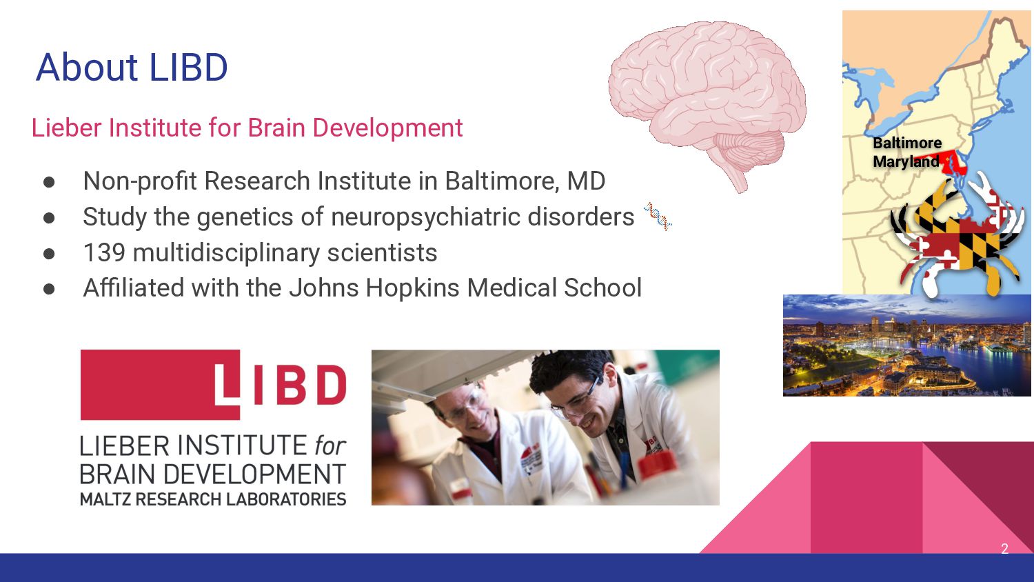
Institute in Baltimore, MD • Study the genetics of neuropsychiatric disorders 🧬 • 139 multidisciplinary scientists • Affiliated with the Johns Hopkins Medical School 2 Baltimore Maryland 🔸
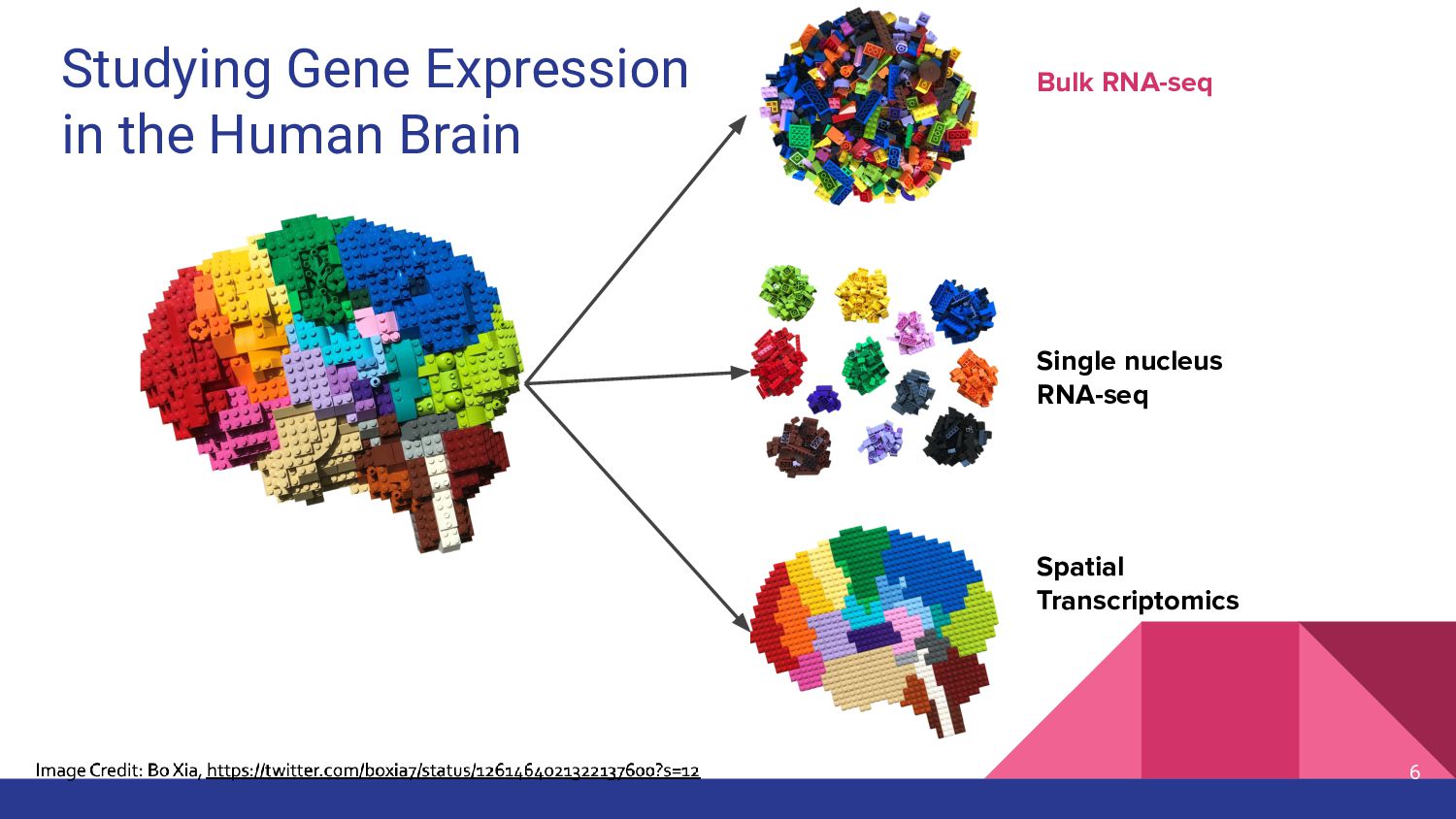
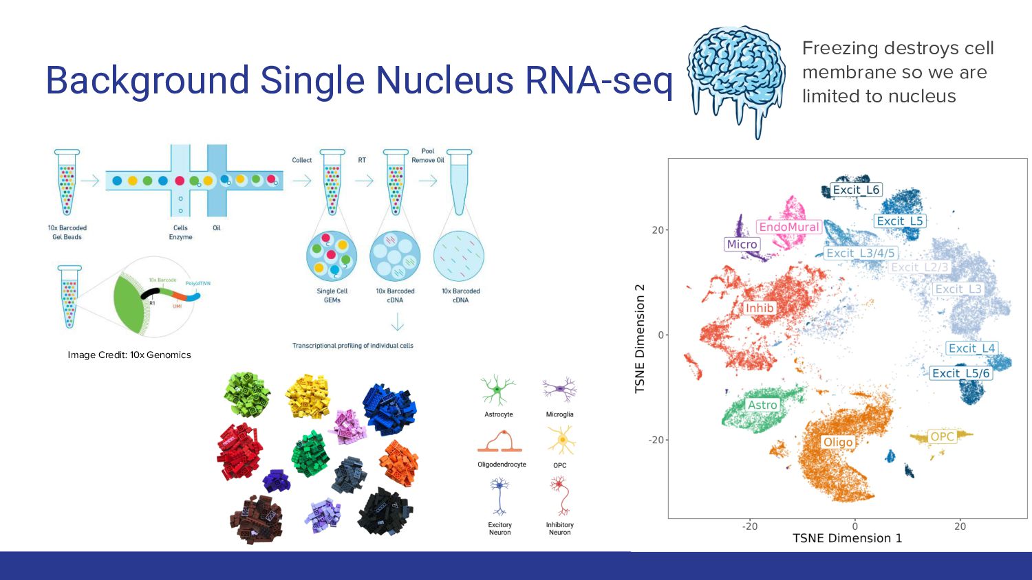
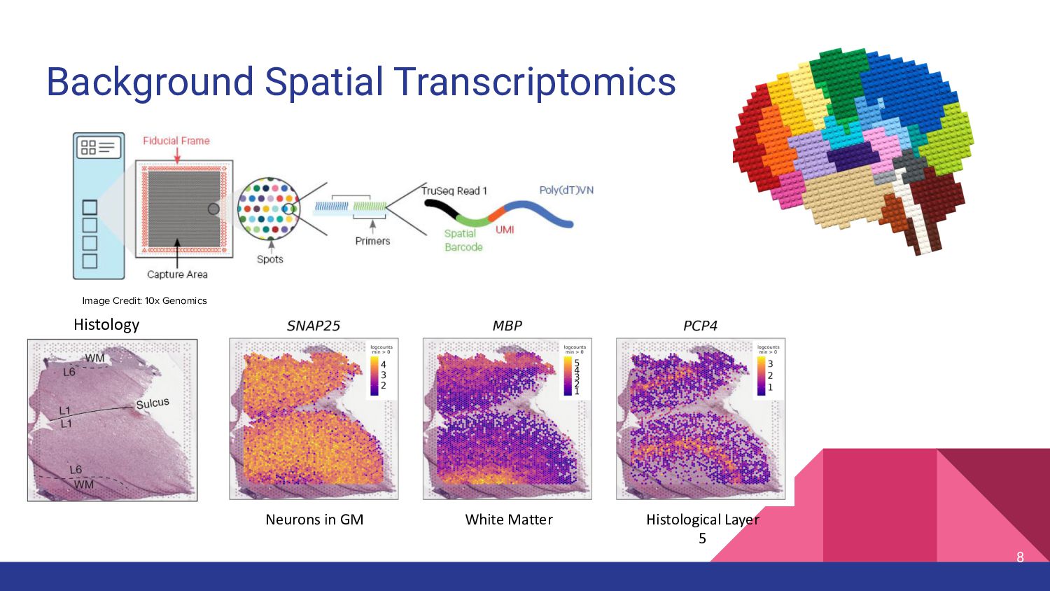
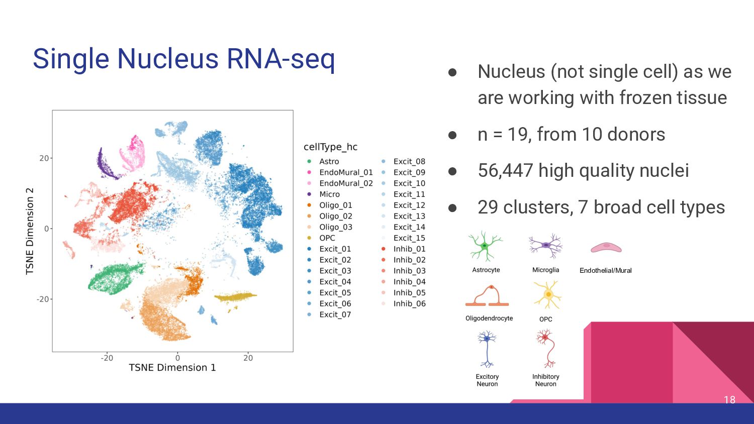
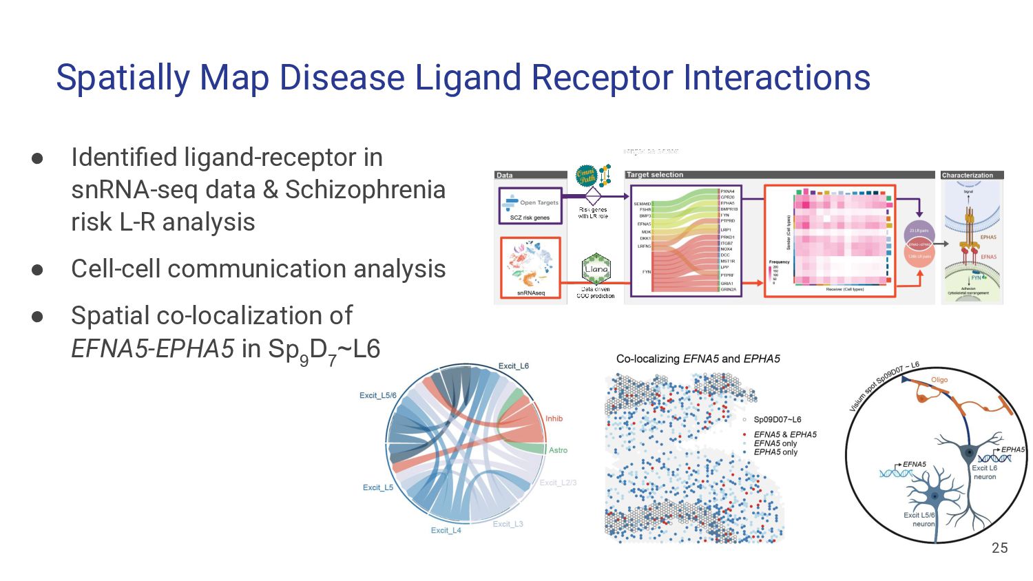
Apply paired spatial and snRNA-seq data to study cell type composition & cell-cell interactions 2. Identify cell type populations in Single Nucleus RNA-seq data
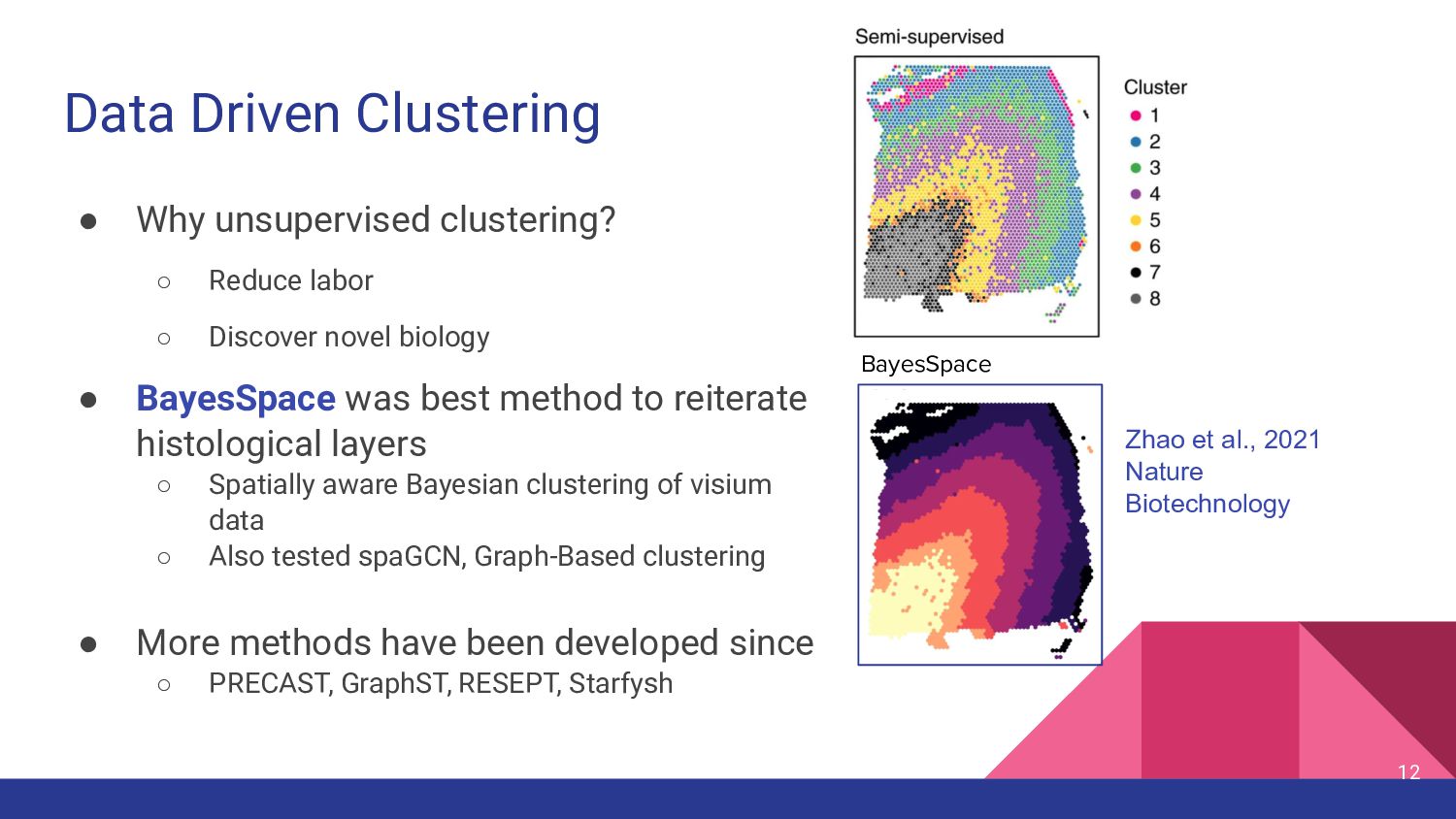
◦ Discover novel biology • BayesSpace was best method to reiterate histological layers ◦ Spatially aware Bayesian clustering of visium data ◦ Also tested spaGCN, Graph-Based clustering • More methods have been developed since ◦ PRECAST, GraphST, RESEPT, Starfysh 12 Zhao et al., 2021 Nature Biotechnology BayesSpace
• k=2: separate white vs. grey matter • k=9: best reiterated histological layers • k=16: data-driven optimal k based on fast H+ statistic 13 More Clusters = More Complexity
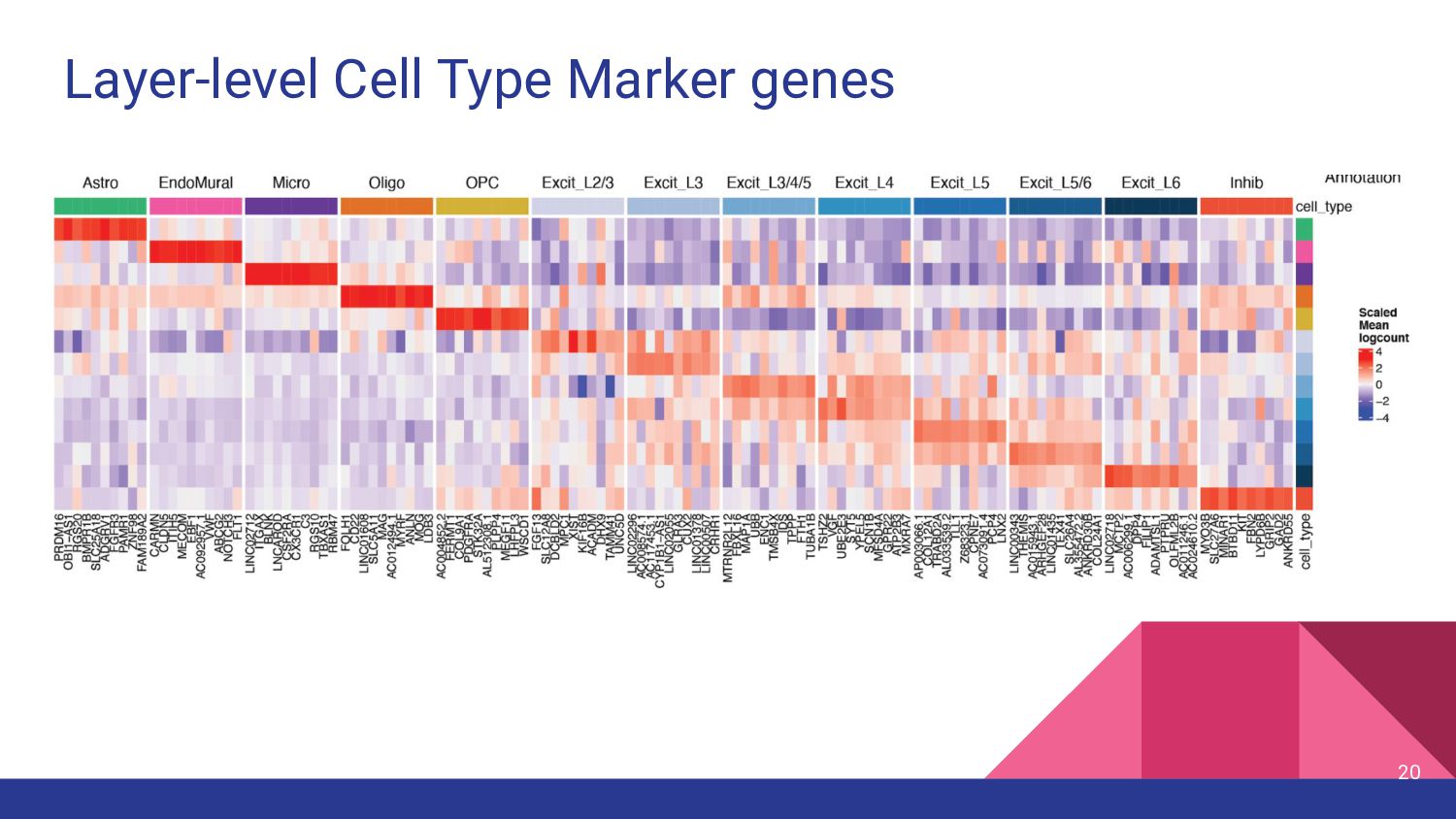
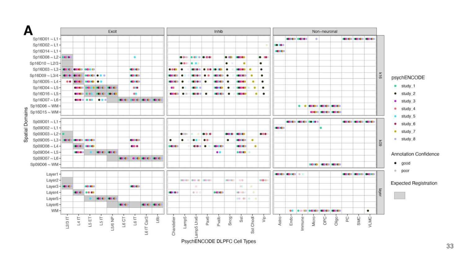
structure • Correlate enrichment t-statistics for top marker genes of reference ◦ Cluster vs. manual annotation • Annotate with strongly associated histological layer 14 Sp k D d ~L
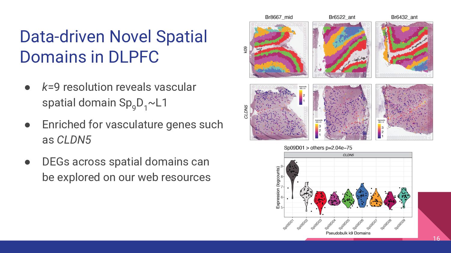
vascular spatial domain Sp 9 D 1 ~L1 • Enriched for vasculature genes such as CLDN5 • DEGs across spatial domains can be explored on our web resources 16
Apply paired spatial and snRNA-seq data to study cell type composition & cell-cell interactions 2. Identify cell type populations in Single Nucleus RNA-seq data
Apply paired spatial and snRNA-seq data to study cell type composition & cell-cell interactions 2. Identify cell type populations in Single Nucleus RNA-seq data
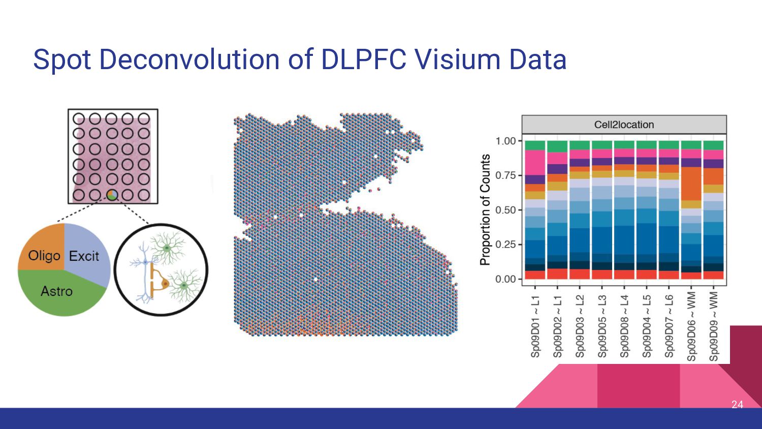
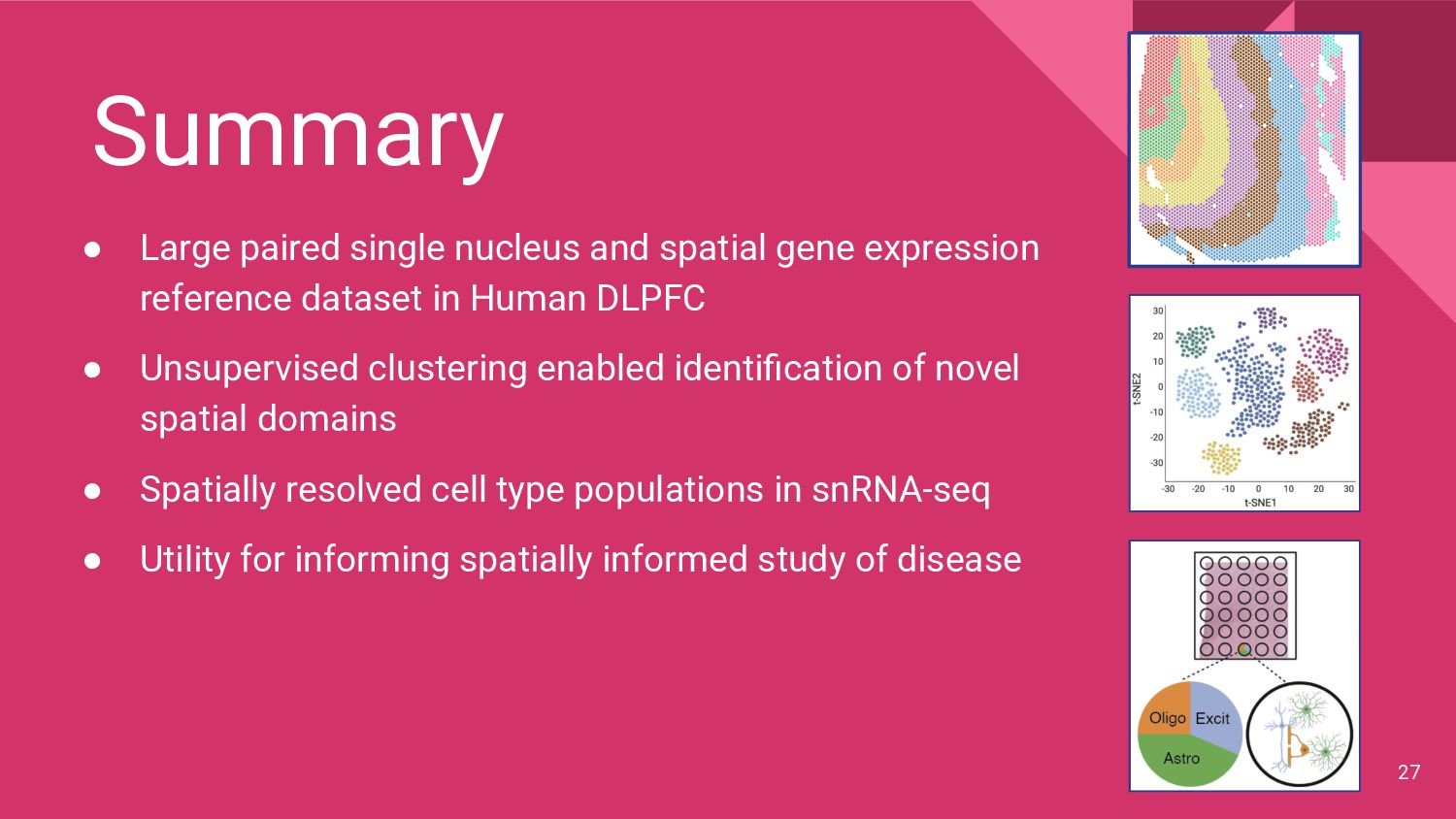
reference dataset in Human DLPFC • Unsupervised clustering enabled identification of novel spatial domains • Spatially resolved cell type populations in snRNA-seq • Utility for informing spatially informed study of disease 27
Montgomery Sang Ho Kwon Heena R. Divecha Madhavi Tippani Chaichontat Sriworarat Annie B. Nguyen Matthew N. Tran Arta Seyedian Thomas M. Hyde Joel E. Kleinman Stephanie C. Page Keri Martinowich Leonardo Collado-Torres Kristen R. Maynard JHU Biostatistics Dept Boyi Guo Stephanie C. Hicks JHU Biomed Engineering Prashanthi Ravichandran Alexis Battle University College London Genetics and Genomic Medicine Melissa Grant-Peters Mina Ryten PsychENCODE Consortium Get in touch! lahuuki.github.io @lahuuki Download these slides: speakerdeck.com/lahuuki Thank you! 🧠 Any Questions?