Upgrade to Pro — share decks privately, control downloads, hide ads and more …

JOINTS

 JOINTS

Introduction to JointsJoint (Articulation): Any point where two or more bones connect in the skeletal system. Arthrology: The scientific study of joints. Components of a Joint: Made of bones held and supported by connective tissues: Tendons: Tough connective tissues that attach bones to skeletal muscles. Ligaments: Strong fibrous bands that connect two bones together to stabilize the joint. Cartilage: Smooth, protective tissue covering bone ends that prevents wear and tear caused by friction.

Avatar for Pawan Kumar Sahu

Pawan Kumar Sahu

October 16, 2026

More Decks by Pawan Kumar Sahu

Other Decks in Education

Transcript

  1. Introduction to Joints Joint (Articulation): Any point where two or

    more bones connect in the skeletal system. Arthrology: The scientific study of joints. Components of a Joint: Made of bones held and supported by connective tissues: Tendons: Tough connective tissues that attach bones to skeletal muscles. Ligaments: Strong fibrous bands that connect two bones together to stabilize the joint. Cartilage: Smooth, protective tissue covering bone ends that prevents wear and tear caused by friction. Department of Pharmacy | Pawan Kumar Sahu, Lecturer, Rudauli College of Pharmacy, Ayodhya (U.P.)
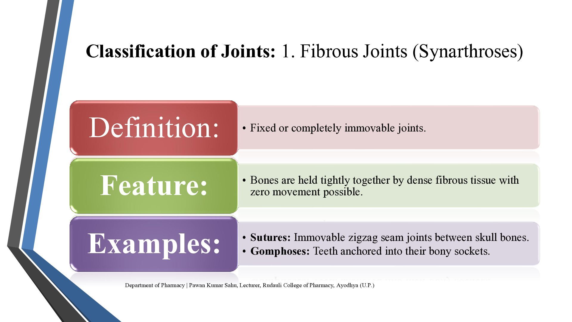
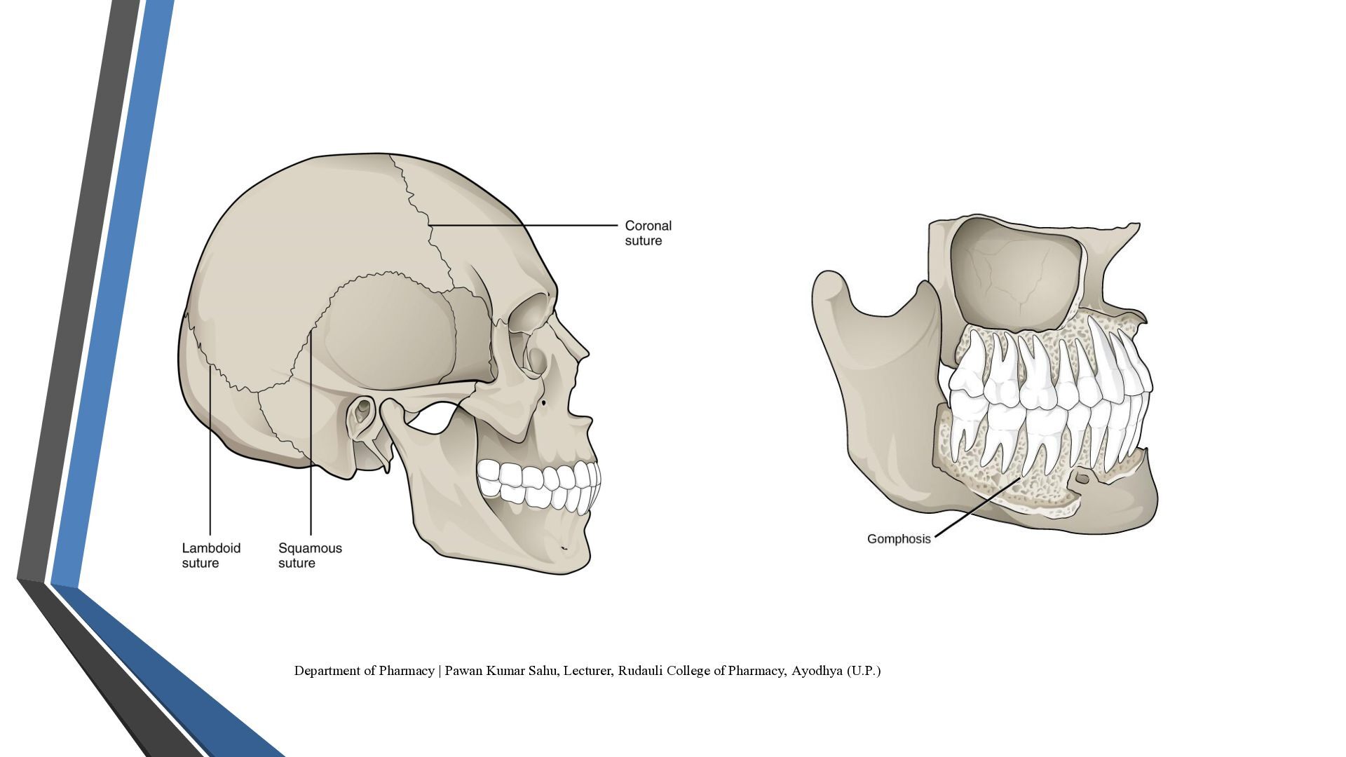
  2. Classification of Joints: 1. Fibrous Joints (Synarthroses) Definition: • Fixed

    or completely immovable joints. Feature: • Bones are held tightly together by dense fibrous tissue with zero movement possible. Examples: • Sutures: Immovable zigzag seam joints between skull bones. • Gomphoses: Teeth anchored into their bony sockets. Department of Pharmacy | Pawan Kumar Sahu, Lecturer, Rudauli College of Pharmacy, Ayodhya (U.P.)
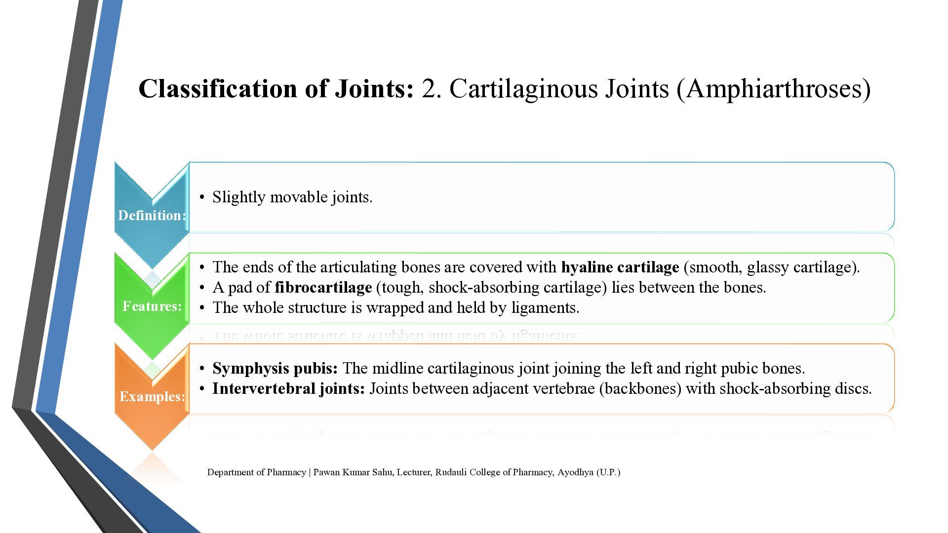
  3. Classification of Joints: 2. Cartilaginous Joints (Amphiarthroses) • Slightly movable

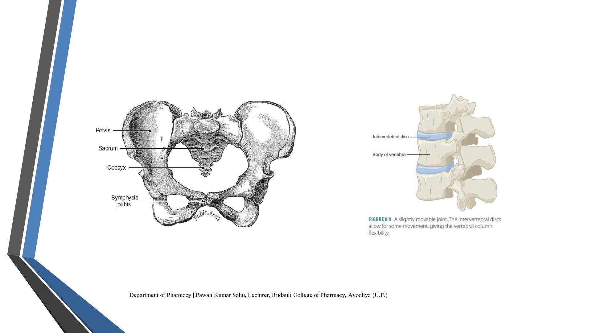
    joints. Definition: • The ends of the articulating bones are covered with hyaline cartilage (smooth, glassy cartilage). • A pad of fibrocartilage (tough, shock-absorbing cartilage) lies between the bones. Features: • The whole structure is wrapped and held by ligaments. • Symphysis pubis: The midline cartilaginous joint joining the left and right pubic bones. • Intervertebral joints: Joints between adjacent vertebrae (backbones) with shock-absorbing discs. Examples: Department of Pharmacy | Pawan Kumar Sahu, Lecturer, Rudauli College of Pharmacy, Ayodhya (U.P.)
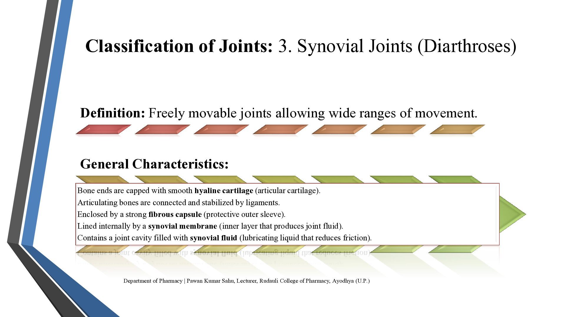
  4. Classification of Joints: 3. Synovial Joints (Diarthroses) Definition: Freely movable

    joints allowing wide ranges of movement. General Characteristics: Bone ends are capped with smooth hyaline cartilage (articular cartilage). Articulating bones are connected and stabilized by ligaments. Enclosed by a strong fibrous capsule (protective outer sleeve). Lined internally by a synovial membrane (inner layer that produces joint fluid). Contains a joint cavity filled with synovial fluid (lubricating liquid that reduces friction). Department of Pharmacy | Pawan Kumar Sahu, Lecturer, Rudauli College of Pharmacy, Ayodhya (U.P.)
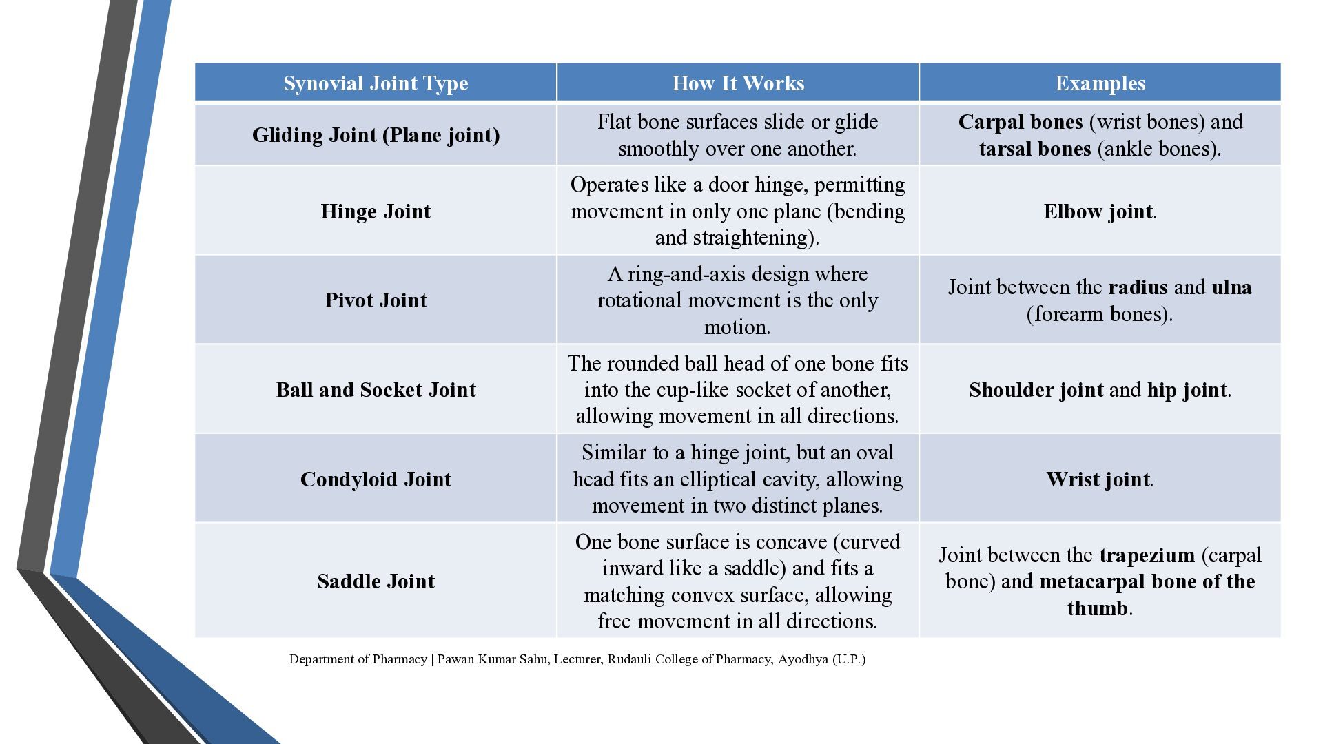
  5. Synovial Joint Type How It Works Examples Gliding Joint (Plane

    joint) Flat bone surfaces slide or glide smoothly over one another. Carpal bones (wrist bones) and tarsal bones (ankle bones). Hinge Joint Operates like a door hinge, permitting movement in only one plane (bending and straightening). Elbow joint. Pivot Joint A ring-and-axis design where rotational movement is the only motion. Joint between the radius and ulna (forearm bones). Ball and Socket Joint The rounded ball head of one bone fits into the cup-like socket of another, allowing movement in all directions. Shoulder joint and hip joint. Condyloid Joint Similar to a hinge joint, but an oval head fits an elliptical cavity, allowing movement in two distinct planes. Wrist joint. Saddle Joint One bone surface is concave (curved inward like a saddle) and fits a matching convex surface, allowing free movement in all directions. Joint between the trapezium (carpal bone) and metacarpal bone of the thumb. Department of Pharmacy | Pawan Kumar Sahu, Lecturer, Rudauli College of Pharmacy, Ayodhya (U.P.)
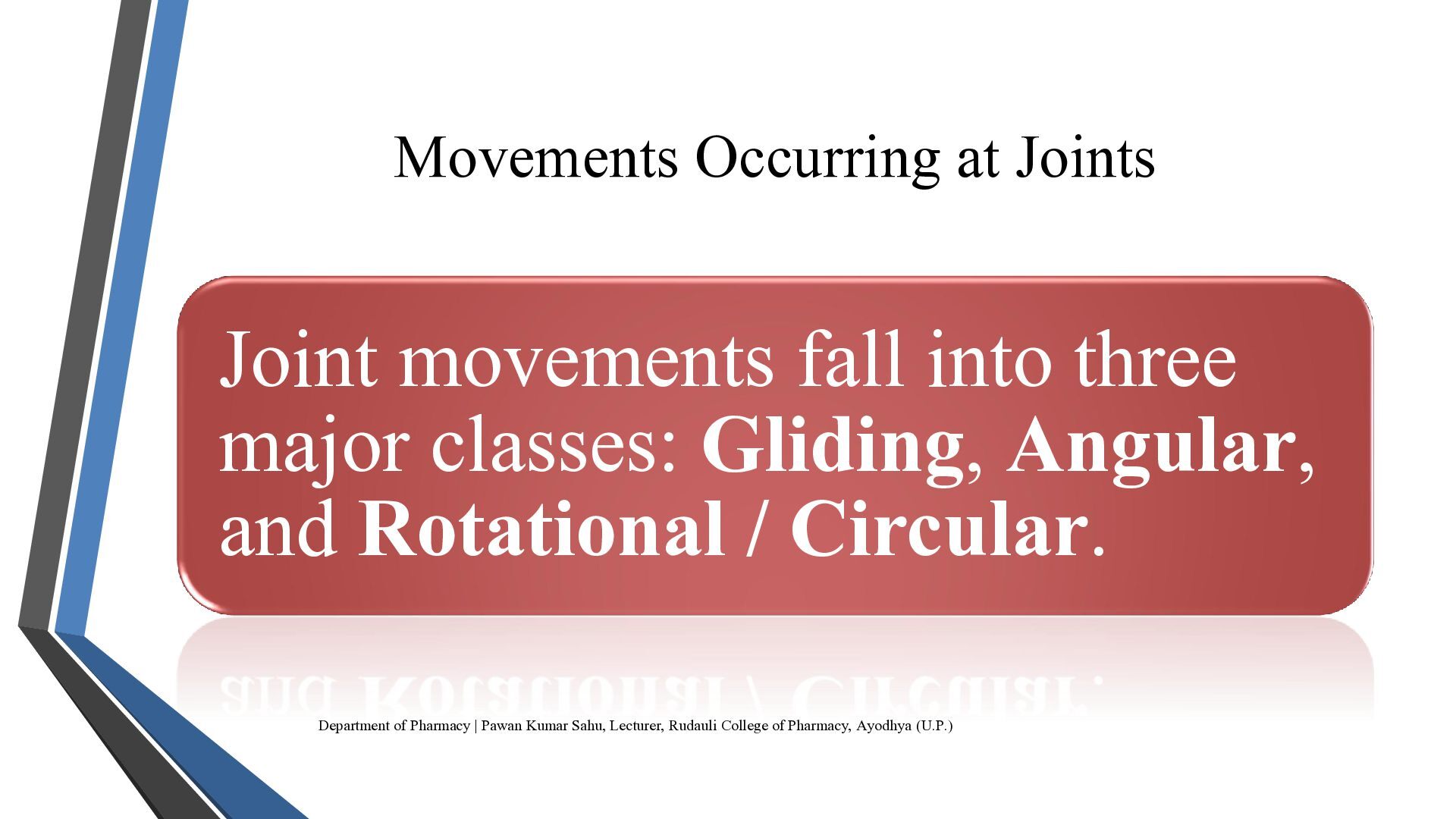
  6. Movements Occurring at Joints Joint movements fall into three major

    classes: Gliding, Angular, and Rotational / Circular. Department of Pharmacy | Pawan Kumar Sahu, Lecturer, Rudauli College of Pharmacy, Ayodhya (U.P.)
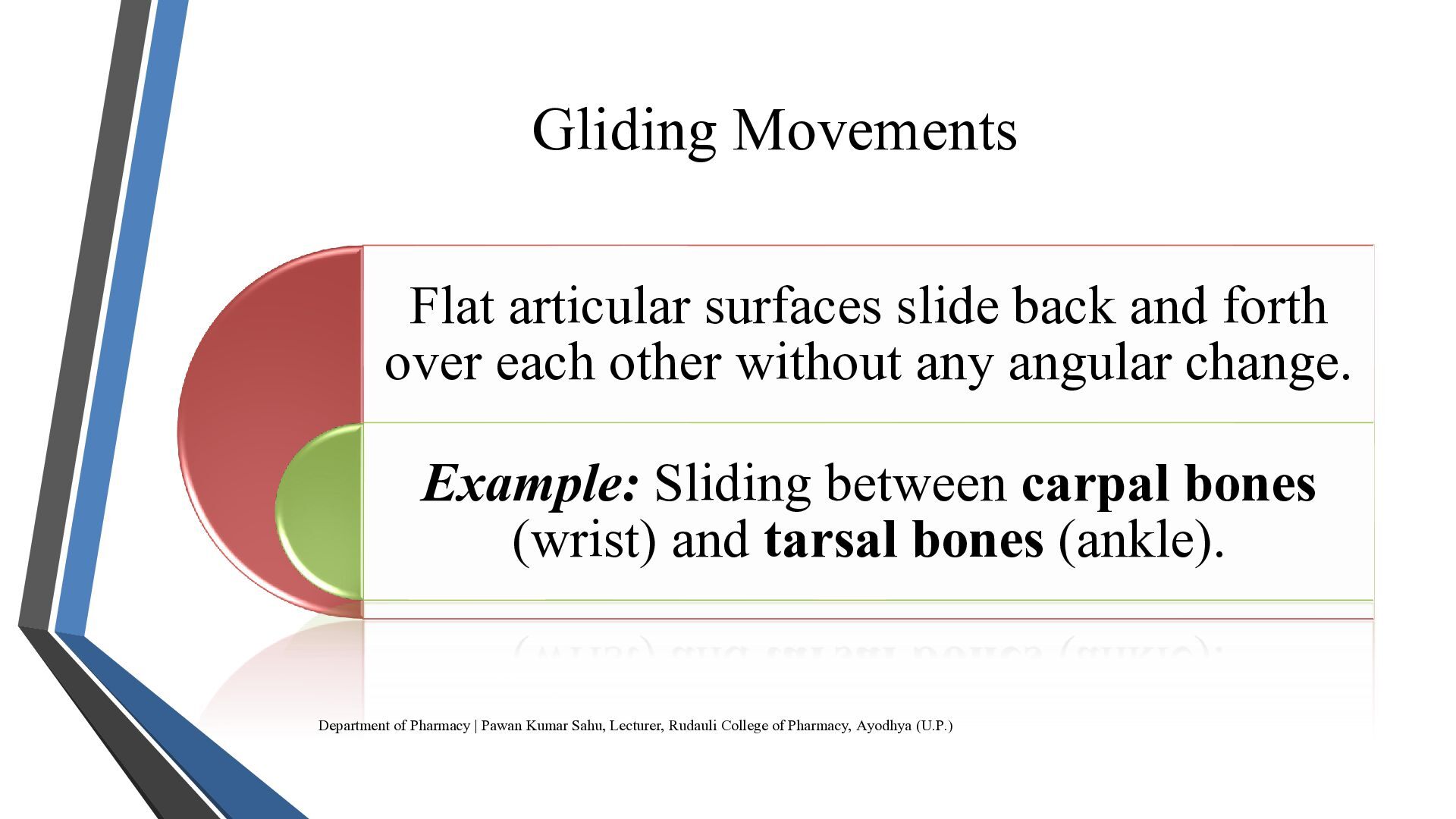
  7. Gliding Movements Flat articular surfaces slide back and forth over

    each other without any angular change. Example: Sliding between carpal bones (wrist) and tarsal bones (ankle). Department of Pharmacy | Pawan Kumar Sahu, Lecturer, Rudauli College of Pharmacy, Ayodhya (U.P.)
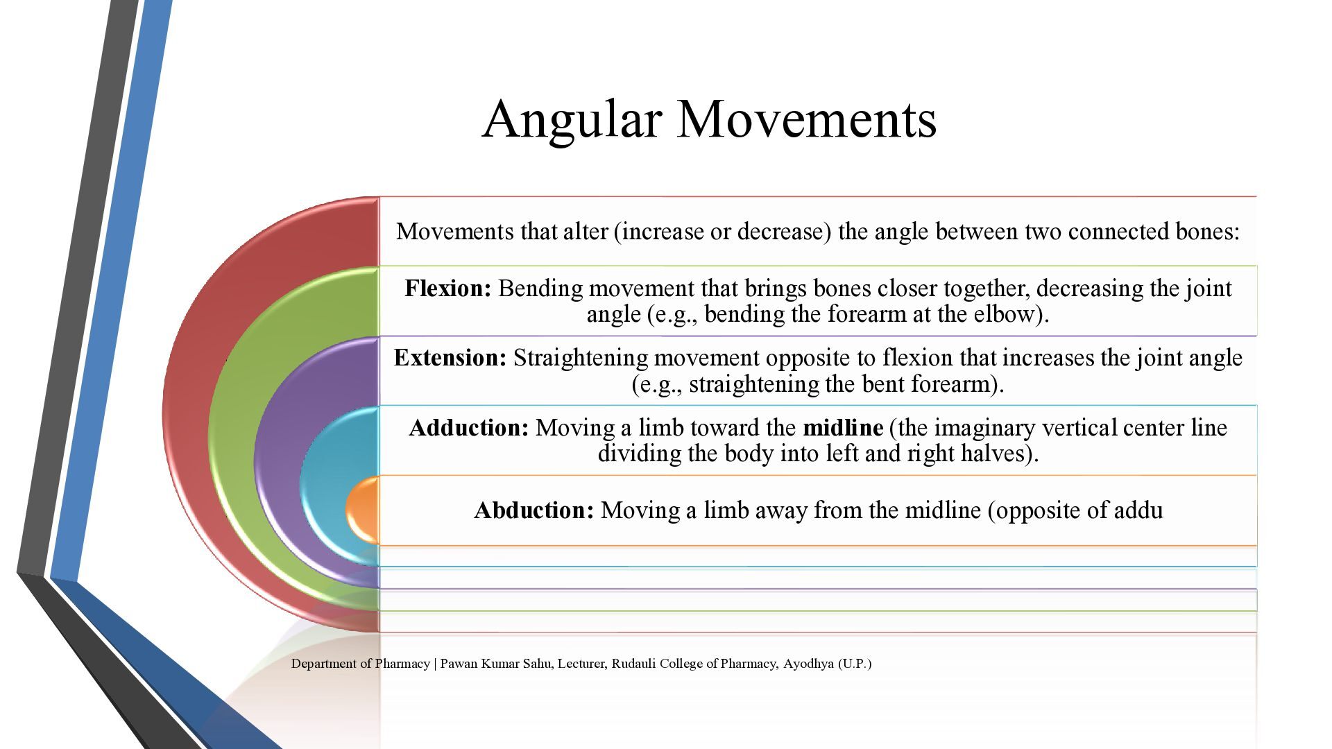
  8. Angular Movements Movements that alter (increase or decrease) the angle

    between two connected bones: Flexion: Bending movement that brings bones closer together, decreasing the joint angle (e.g., bending the forearm at the elbow). Extension: Straightening movement opposite to flexion that increases the joint angle (e.g., straightening the bent forearm). Adduction: Moving a limb toward the midline (the imaginary vertical center line dividing the body into left and right halves). Abduction: Moving a limb away from the midline (opposite of addu Department of Pharmacy | Pawan Kumar Sahu, Lecturer, Rudauli College of Pharmacy, Ayodhya (U.P.)
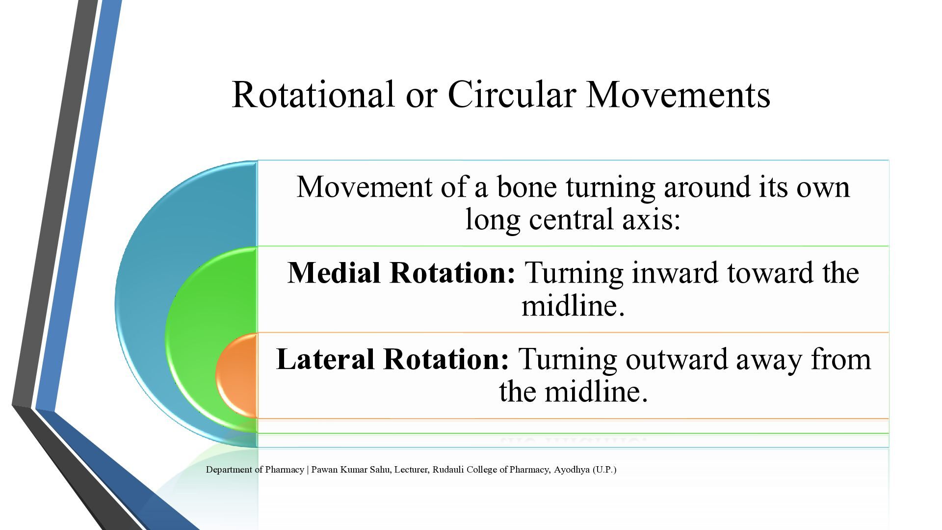
  9. Rotational or Circular Movements Movement of a bone turning around

    its own long central axis: Medial Rotation: Turning inward toward the midline. Lateral Rotation: Turning outward away from the midline. Department of Pharmacy | Pawan Kumar Sahu, Lecturer, Rudauli College of Pharmacy, Ayodhya (U.P.)
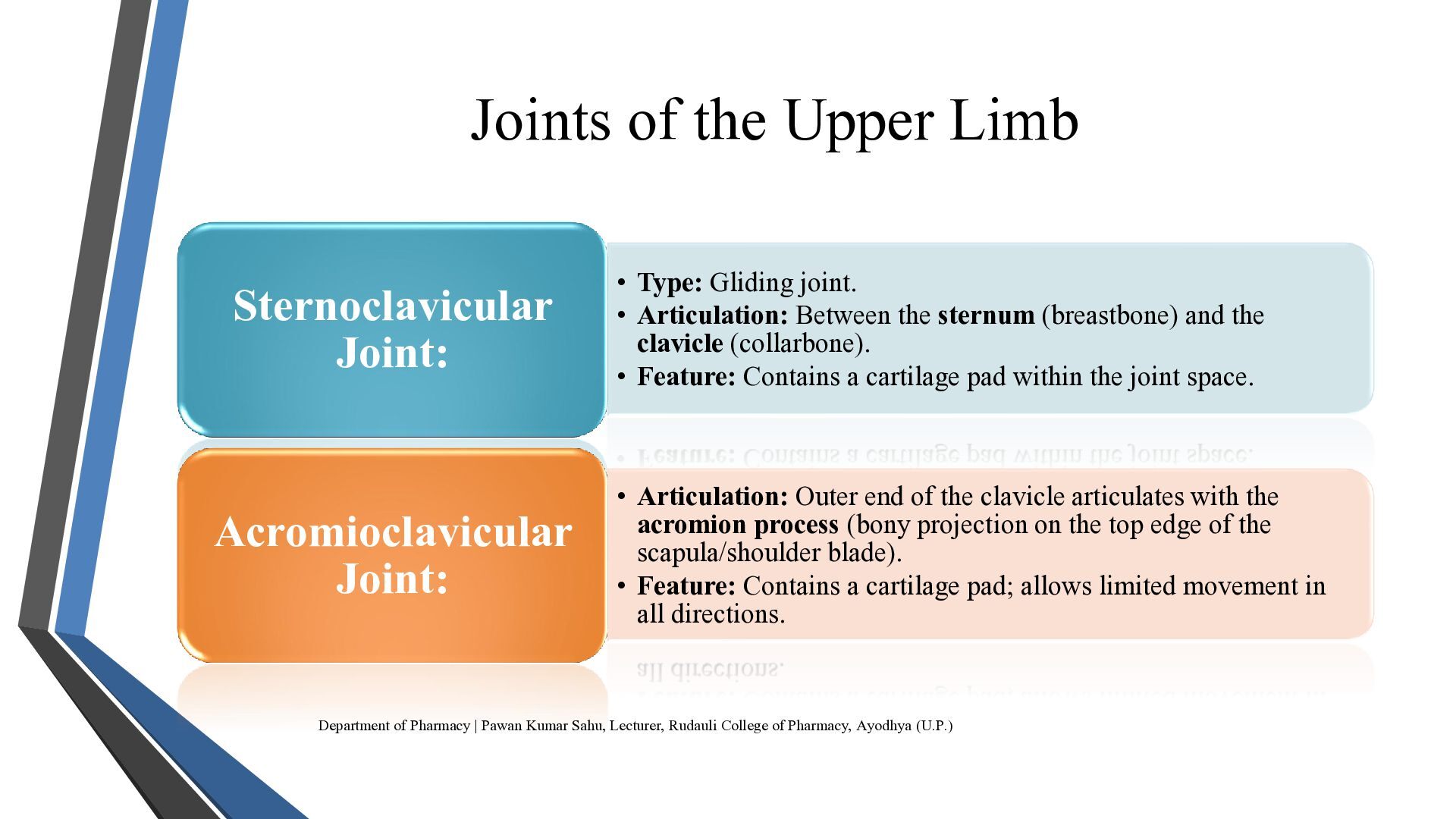
  10. Joints of the Upper Limb Sternoclavicular Joint: Acromioclavicular Joint: •

    Type: Gliding joint. • Articulation: Between the sternum (breastbone) and the clavicle (collarbone). • Feature: Contains a cartilage pad within the joint space. • Articulation: Outer end of the clavicle articulates with the acromion process (bony projection on the top edge of the scapula/shoulder blade). • Feature: Contains a cartilage pad; allows limited movement in all directions. Department of Pharmacy | Pawan Kumar Sahu, Lecturer, Rudauli College of Pharmacy, Ayodhya (U.P.)
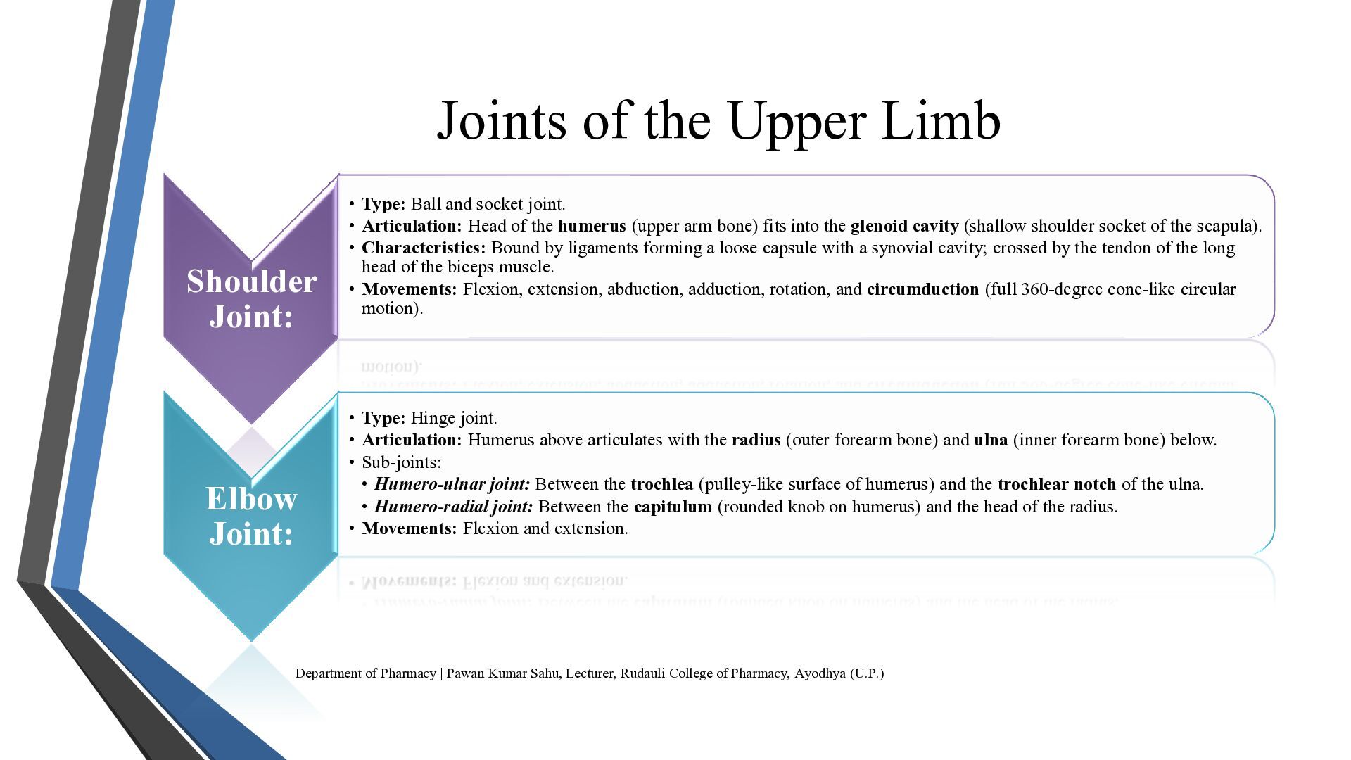
  11. Joints of the Upper Limb Shoulder Joint: Elbow Joint: •

    Type: Ball and socket joint. • Articulation: Head of the humerus (upper arm bone) fits into the glenoid cavity (shallow shoulder socket of the scapula). • Characteristics: Bound by ligaments forming a loose capsule with a synovial cavity; crossed by the tendon of the long head of the biceps muscle. • Movements: Flexion, extension, abduction, adduction, rotation, and circumduction (full 360-degree cone-like circular motion). • Type: Hinge joint. • Articulation: Humerus above articulates with the radius (outer forearm bone) and ulna (inner forearm bone) below. • Sub-joints: • Humero-ulnar joint: Between the trochlea (pulley-like surface of humerus) and the trochlear notch of the ulna. • Humero-radial joint: Between the capitulum (rounded knob on humerus) and the head of the radius. • Movements: Flexion and extension. Department of Pharmacy | Pawan Kumar Sahu, Lecturer, Rudauli College of Pharmacy, Ayodhya (U.P.)
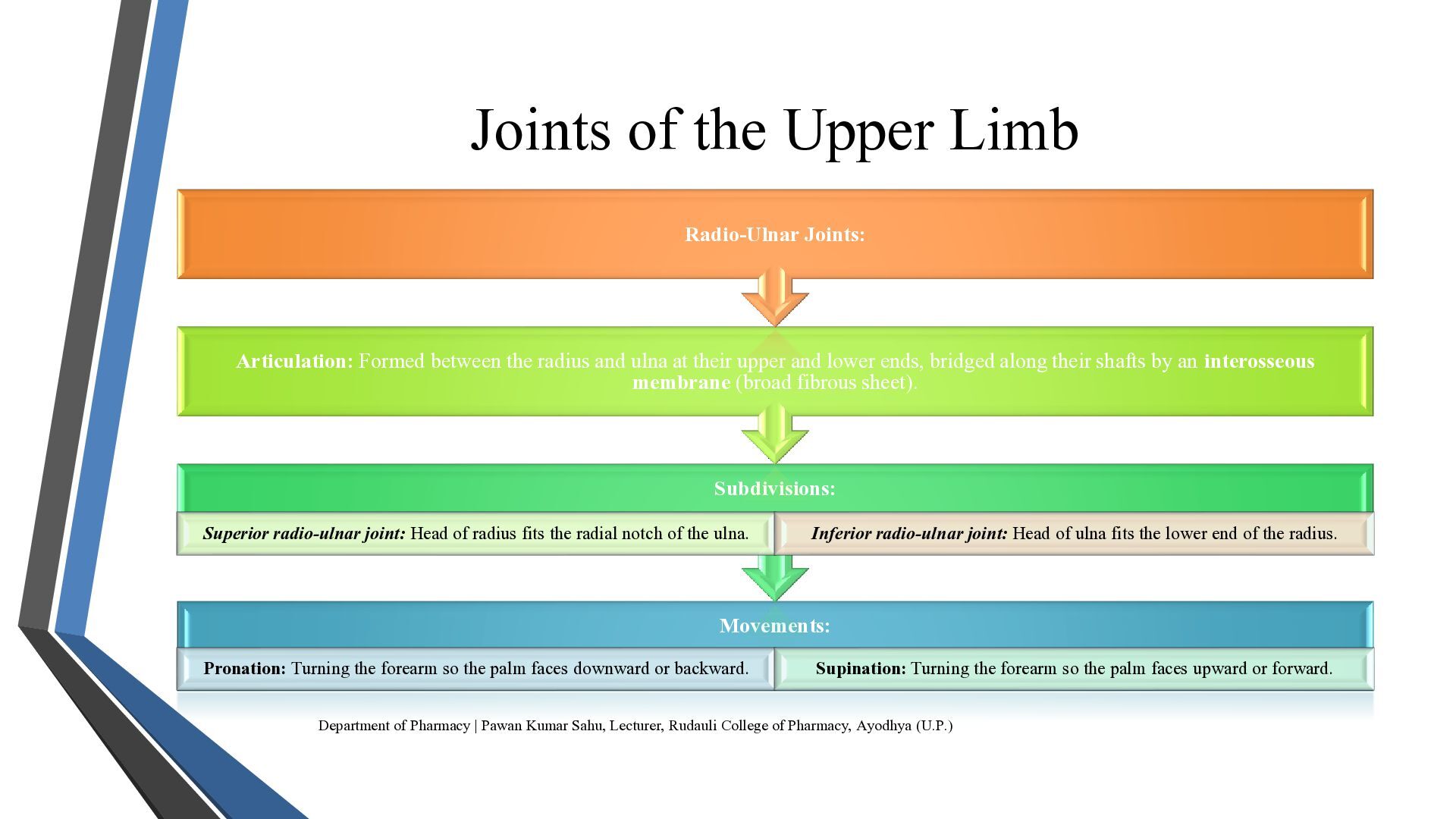
  12. Joints of the Upper Limb Radio-Ulnar Joints: Articulation: Formed between

    the radius and ulna at their upper and lower ends, bridged along their shafts by an interosseous membrane (broad fibrous sheet). Subdivisions: Superior radio-ulnar joint: Head of radius fits the radial notch of the ulna. Inferior radio-ulnar joint: Head of ulna fits the lower end of the radius. Movements: Pronation: Turning the forearm so the palm faces downward or backward. Supination: Turning the forearm so the palm faces upward or forward. Department of Pharmacy | Pawan Kumar Sahu, Lecturer, Rudauli College of Pharmacy, Ayodhya (U.P.)
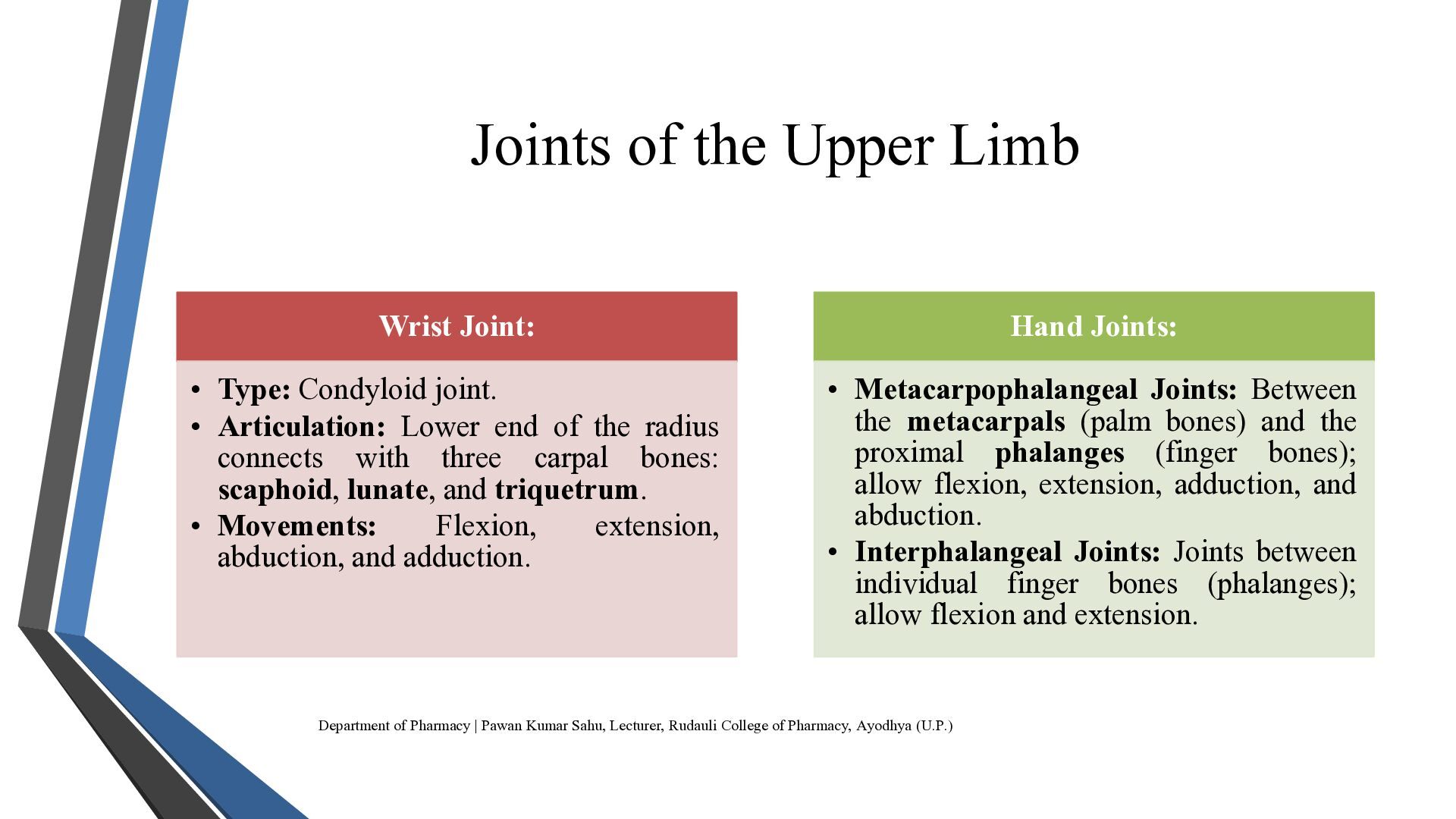
  13. Joints of the Upper Limb Wrist Joint: Hand Joints: •

    Type: Condyloid joint. • Articulation: Lower end of the radius connects with three carpal bones: scaphoid, lunate, and triquetrum. • Movements: Flexion, extension, abduction, and adduction. • Metacarpophalangeal Joints: Between the metacarpals (palm bones) and the proximal phalanges (finger bones); allow flexion, extension, adduction, and abduction. • Interphalangeal Joints: Joints between individual finger bones (phalanges); allow flexion and extension. Department of Pharmacy | Pawan Kumar Sahu, Lecturer, Rudauli College of Pharmacy, Ayodhya (U.P.)
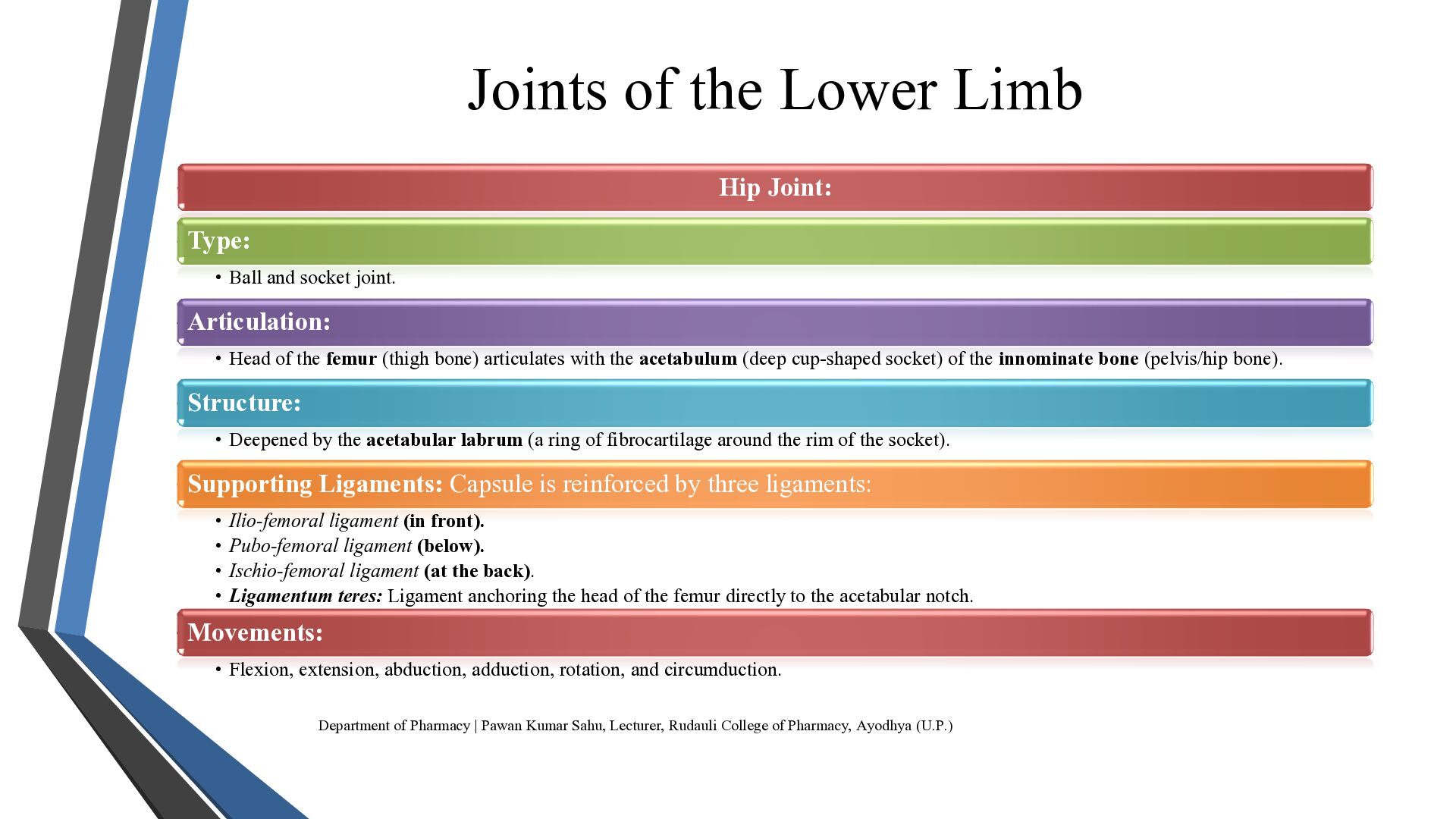
  14. Joints of the Lower Limb Hip Joint: Type: • Ball

    and socket joint. Articulation: • Head of the femur (thigh bone) articulates with the acetabulum (deep cup-shaped socket) of the innominate bone (pelvis/hip bone). Structure: • Deepened by the acetabular labrum (a ring of fibrocartilage around the rim of the socket). Supporting Ligaments: Capsule is reinforced by three ligaments: • Ilio-femoral ligament (in front). • Pubo-femoral ligament (below). • Ischio-femoral ligament (at the back). • Ligamentum teres: Ligament anchoring the head of the femur directly to the acetabular notch. Movements: • Flexion, extension, abduction, adduction, rotation, and circumduction. Department of Pharmacy | Pawan Kumar Sahu, Lecturer, Rudauli College of Pharmacy, Ayodhya (U.P.)
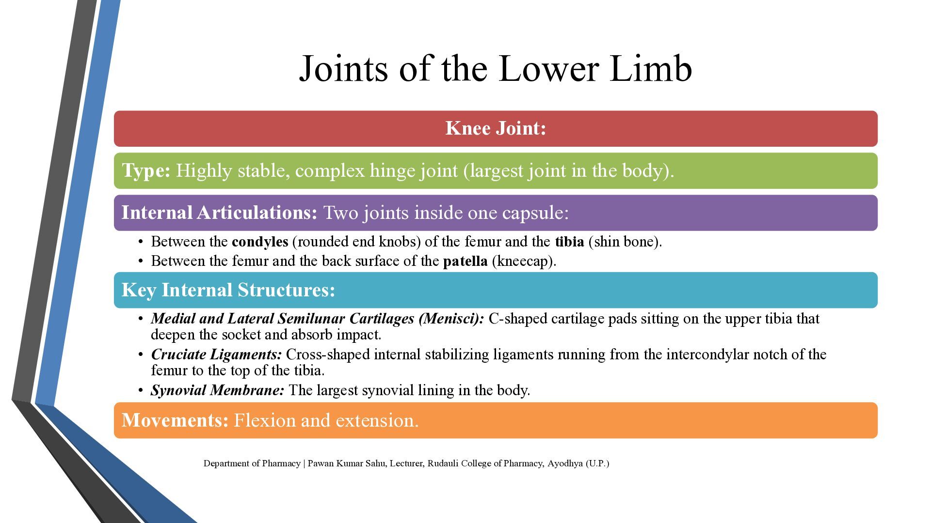
  15. Joints of the Lower Limb Knee Joint: Type: Highly stable,

    complex hinge joint (largest joint in the body). Internal Articulations: Two joints inside one capsule: • Between the condyles (rounded end knobs) of the femur and the tibia (shin bone). • Between the femur and the back surface of the patella (kneecap). Key Internal Structures: • Medial and Lateral Semilunar Cartilages (Menisci): C-shaped cartilage pads sitting on the upper tibia that deepen the socket and absorb impact. • Cruciate Ligaments: Cross-shaped internal stabilizing ligaments running from the intercondylar notch of the femur to the top of the tibia. • Synovial Membrane: The largest synovial lining in the body. Movements: Flexion and extension. Department of Pharmacy | Pawan Kumar Sahu, Lecturer, Rudauli College of Pharmacy, Ayodhya (U.P.)
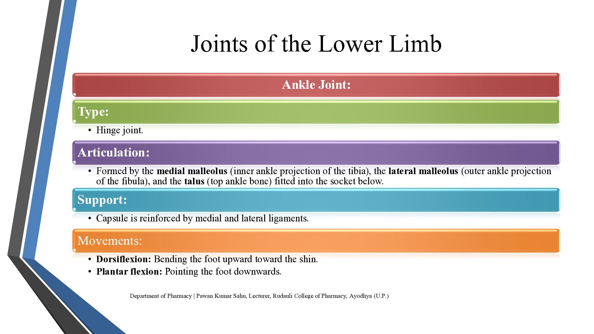
  16. Joints of the Lower Limb Ankle Joint: Type: • Hinge

    joint. Articulation: • Formed by the medial malleolus (inner ankle projection of the tibia), the lateral malleolus (outer ankle projection of the fibula), and the talus (top ankle bone) fitted into the socket below. Support: • Capsule is reinforced by medial and lateral ligaments. Movements: • Dorsiflexion: Bending the foot upward toward the shin. • Plantar flexion: Pointing the foot downwards. Department of Pharmacy | Pawan Kumar Sahu, Lecturer, Rudauli College of Pharmacy, Ayodhya (U.P.)
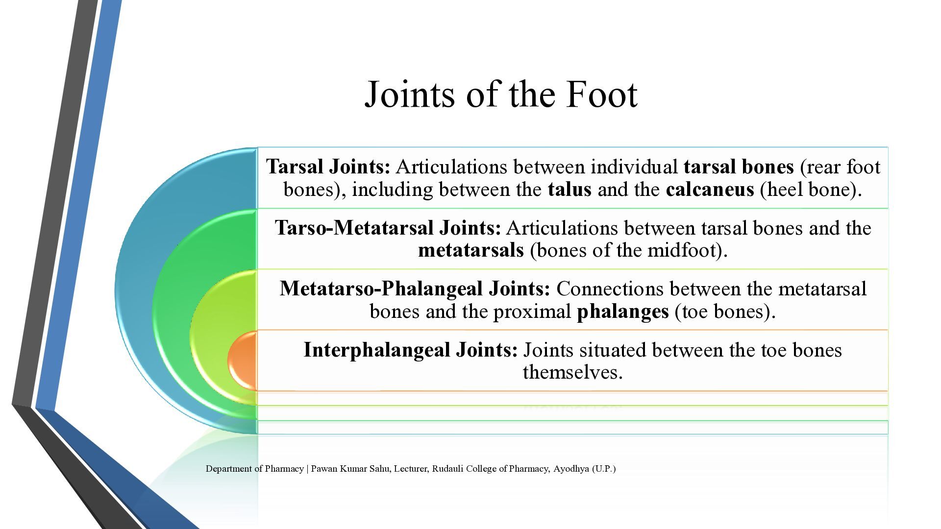
  17. Joints of the Foot Tarsal Joints: Articulations between individual tarsal

    bones (rear foot bones), including between the talus and the calcaneus (heel bone). Tarso-Metatarsal Joints: Articulations between tarsal bones and the metatarsals (bones of the midfoot). Metatarso-Phalangeal Joints: Connections between the metatarsal bones and the proximal phalanges (toe bones). Interphalangeal Joints: Joints situated between the toe bones themselves. Department of Pharmacy | Pawan Kumar Sahu, Lecturer, Rudauli College of Pharmacy, Ayodhya (U.P.)
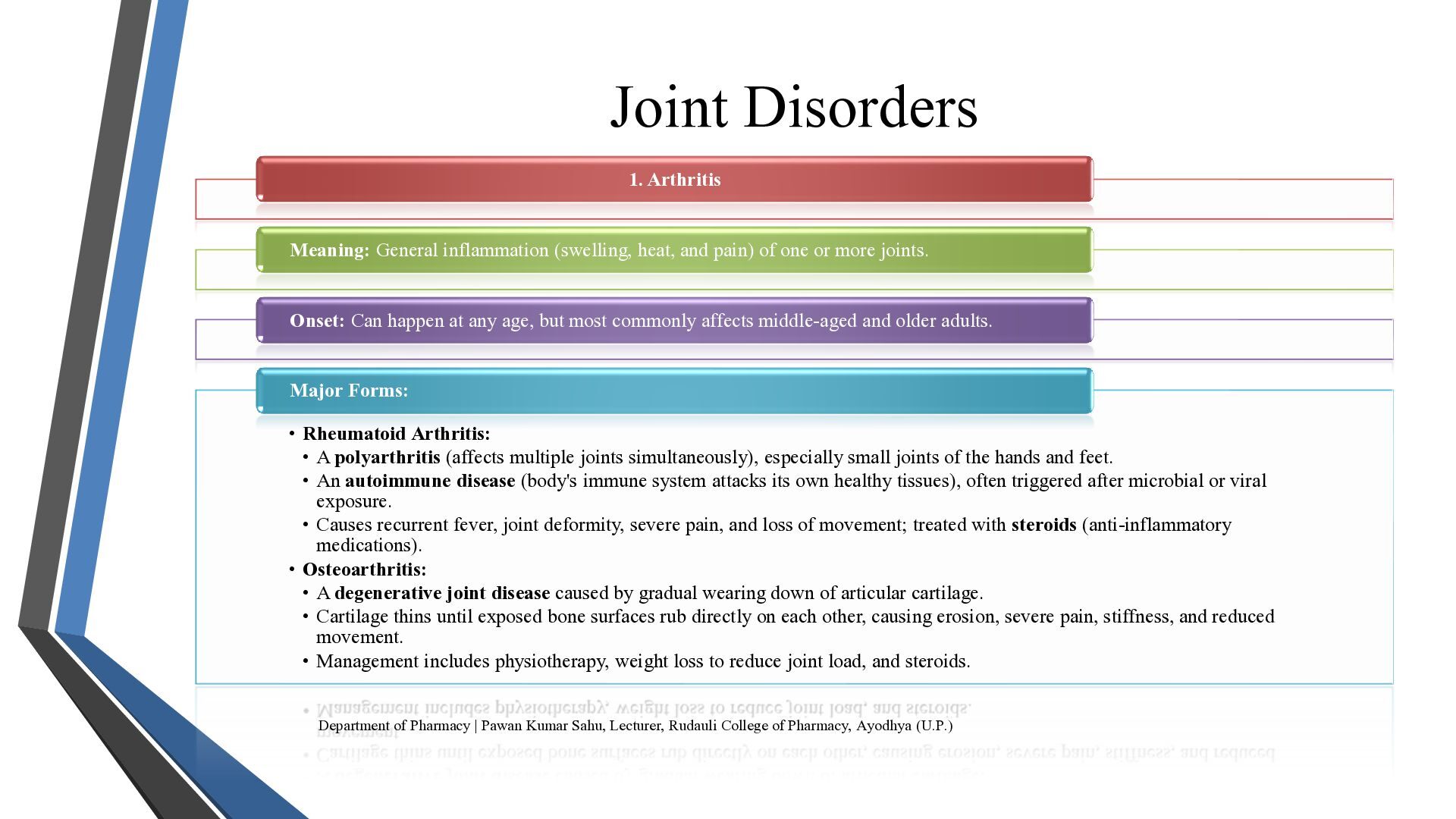
  18. Joint Disorders 1. Arthritis Meaning: General inflammation (swelling, heat, and

    pain) of one or more joints. Onset: Can happen at any age, but most commonly affects middle-aged and older adults. Major Forms: • Rheumatoid Arthritis: • A polyarthritis (affects multiple joints simultaneously), especially small joints of the hands and feet. • An autoimmune disease (body's immune system attacks its own healthy tissues), often triggered after microbial or viral exposure. • Causes recurrent fever, joint deformity, severe pain, and loss of movement; treated with steroids (anti-inflammatory medications). • Osteoarthritis: • A degenerative joint disease caused by gradual wearing down of articular cartilage. • Cartilage thins until exposed bone surfaces rub directly on each other, causing erosion, severe pain, stiffness, and reduced movement. • Management includes physiotherapy, weight loss to reduce joint load, and steroids. Department of Pharmacy | Pawan Kumar Sahu, Lecturer, Rudauli College of Pharmacy, Ayodhya (U.P.)
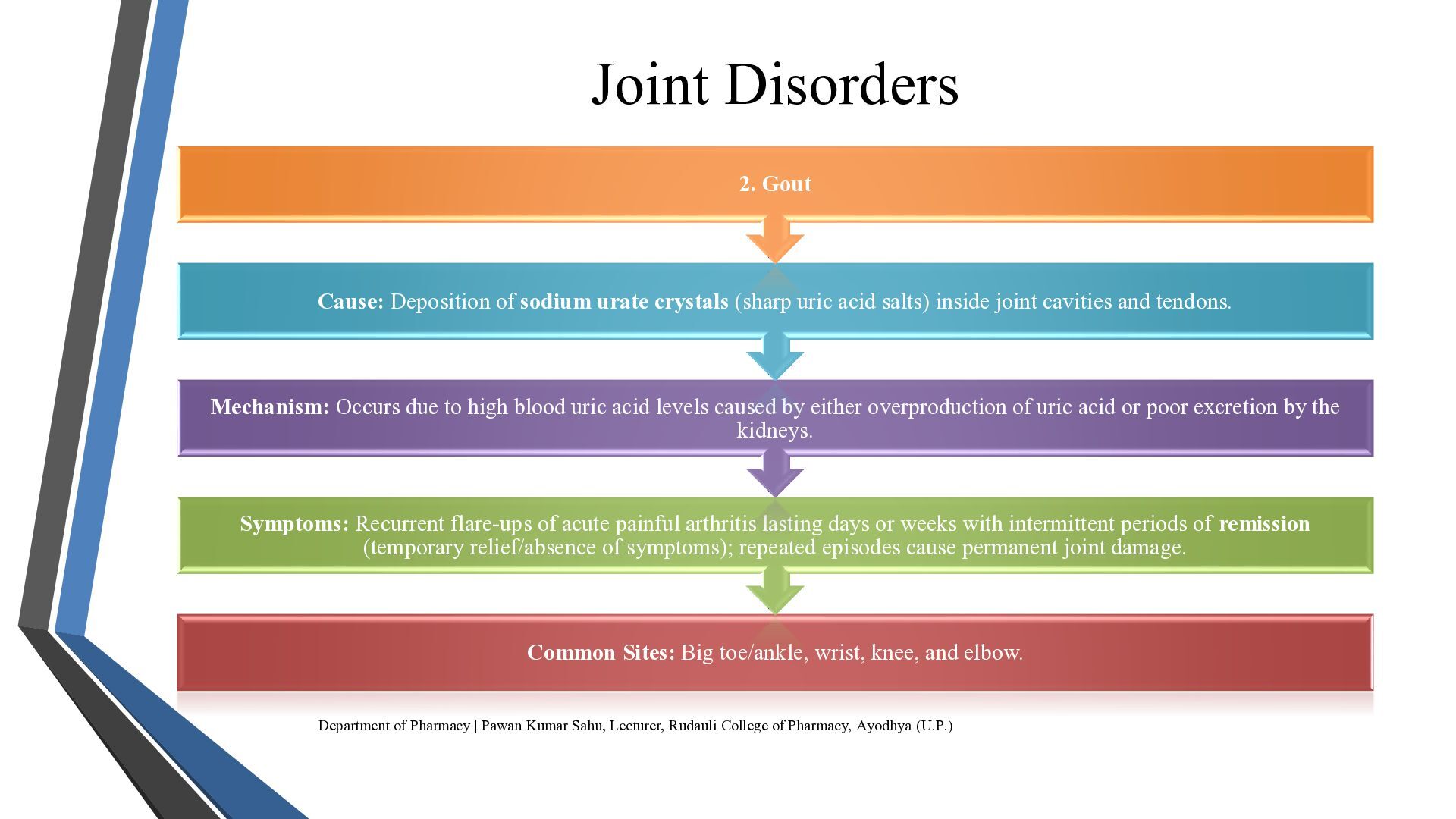
  19. Joint Disorders 2. Gout Cause: Deposition of sodium urate crystals

    (sharp uric acid salts) inside joint cavities and tendons. Mechanism: Occurs due to high blood uric acid levels caused by either overproduction of uric acid or poor excretion by the kidneys. Symptoms: Recurrent flare-ups of acute painful arthritis lasting days or weeks with intermittent periods of remission (temporary relief/absence of symptoms); repeated episodes cause permanent joint damage. Common Sites: Big toe/ankle, wrist, knee, and elbow. Department of Pharmacy | Pawan Kumar Sahu, Lecturer, Rudauli College of Pharmacy, Ayodhya (U.P.)
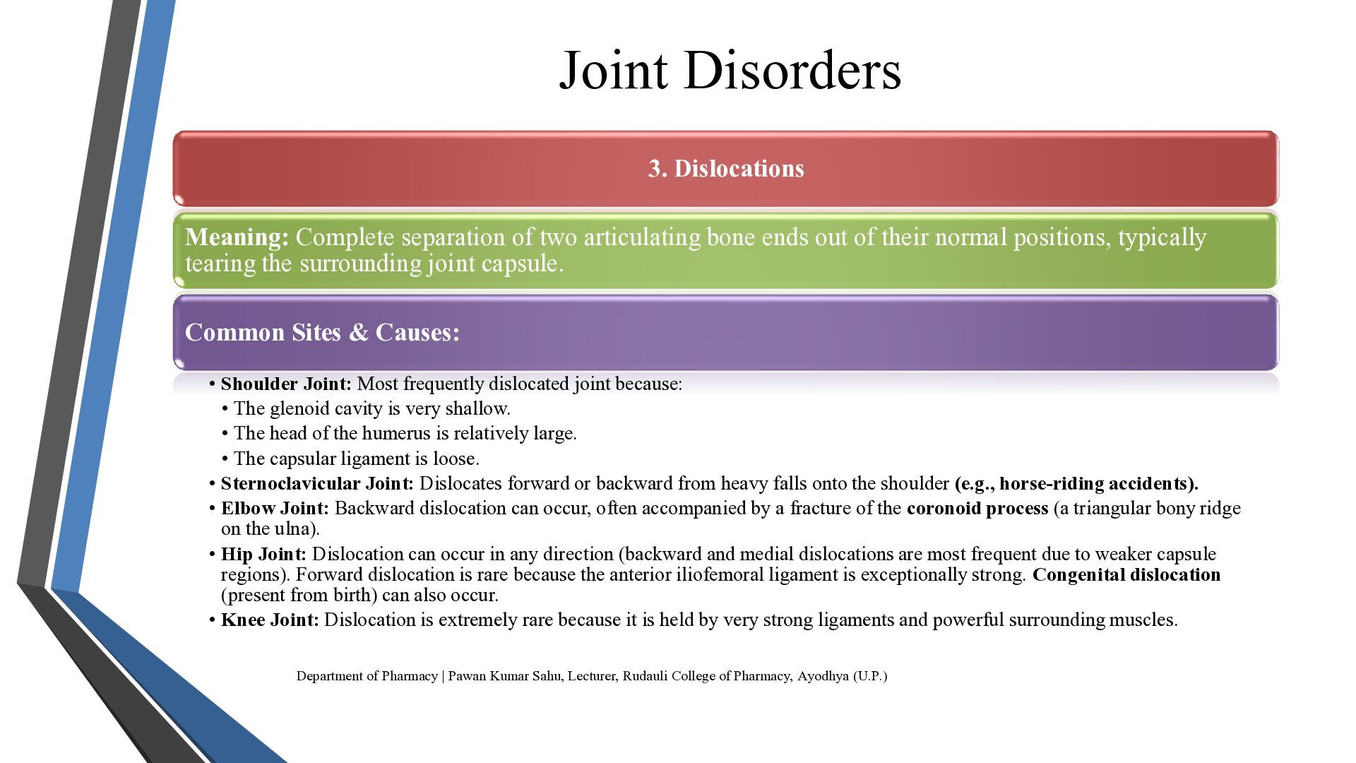
  20. Joint Disorders 3. Dislocations Meaning: Complete separation of two articulating

    bone ends out of their normal positions, typically tearing the surrounding joint capsule. Common Sites & Causes: • Shoulder Joint: Most frequently dislocated joint because: • The glenoid cavity is very shallow. • The head of the humerus is relatively large. • The capsular ligament is loose. • Sternoclavicular Joint: Dislocates forward or backward from heavy falls onto the shoulder (e.g., horse-riding accidents). • Elbow Joint: Backward dislocation can occur, often accompanied by a fracture of the coronoid process (a triangular bony ridge on the ulna). • Hip Joint: Dislocation can occur in any direction (backward and medial dislocations are most frequent due to weaker capsule regions). Forward dislocation is rare because the anterior iliofemoral ligament is exceptionally strong. Congenital dislocation (present from birth) can also occur. • Knee Joint: Dislocation is extremely rare because it is held by very strong ligaments and powerful surrounding muscles. Department of Pharmacy | Pawan Kumar Sahu, Lecturer, Rudauli College of Pharmacy, Ayodhya (U.P.)
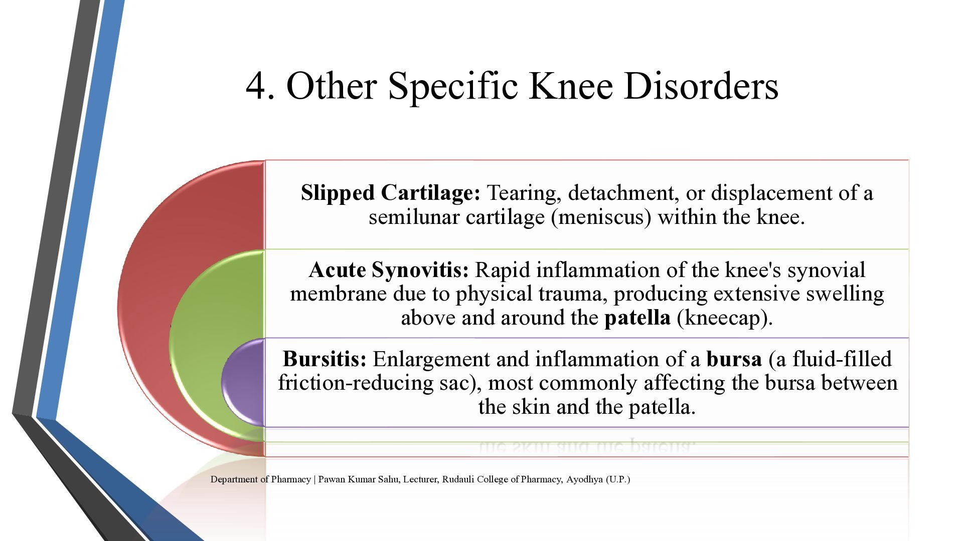
  21. 4. Other Specific Knee Disorders Slipped Cartilage: Tearing, detachment, or

    displacement of a semilunar cartilage (meniscus) within the knee. Acute Synovitis: Rapid inflammation of the knee's synovial membrane due to physical trauma, producing extensive swelling above and around the patella (kneecap). Bursitis: Enlargement and inflammation of a bursa (a fluid-filled friction-reducing sac), most commonly affecting the bursa between the skin and the patella. Department of Pharmacy | Pawan Kumar Sahu, Lecturer, Rudauli College of Pharmacy, Ayodhya (U.P.)
  22. THANK YOU Department of Pharmacy | Pawan Kumar Sahu, Lecturer,

    Rudauli College of Pharmacy, Ayodhya (U.P.)