Upgrade to Pro — share decks privately, control downloads, hide ads and more …

Team lcolladotor Journal Club August 2025

Team lcolladotor Journal Club August 2025

Team lcolladotor Journal Club Presentation: "Cell-type deconvolution methods for spatial transcriptomics"
doi: https://doi.org/10.1038/s41576-025-00845-y
Presented By: Manisha Barse
Date: August 13, 2025

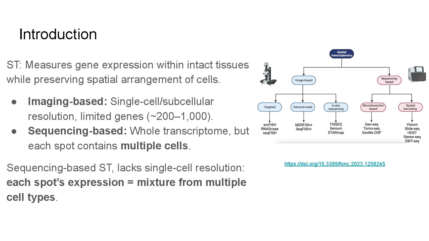
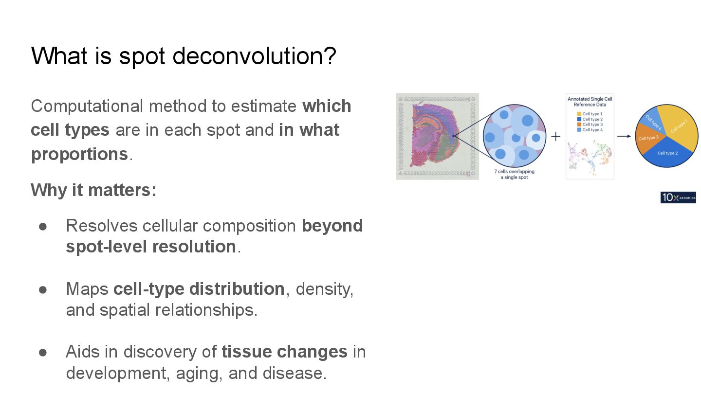
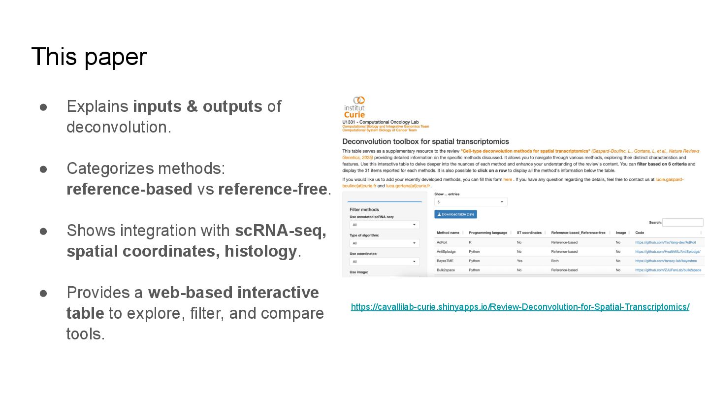
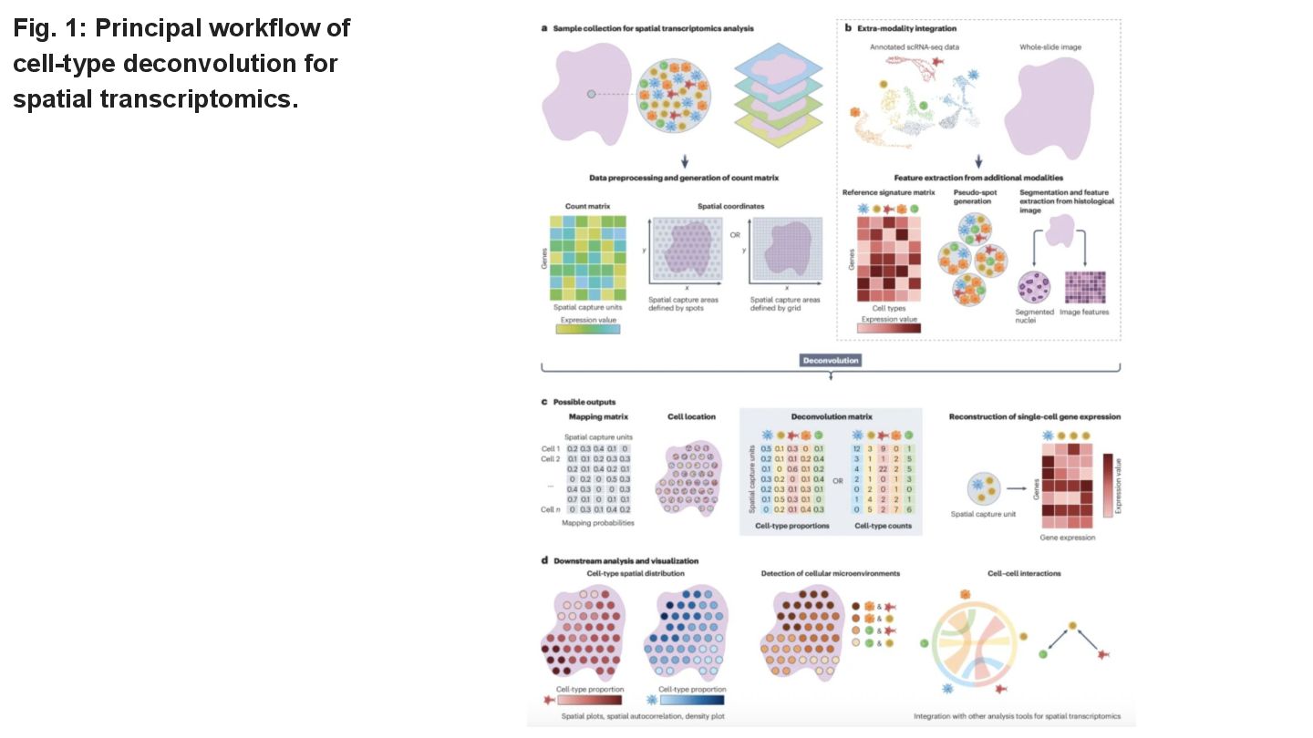
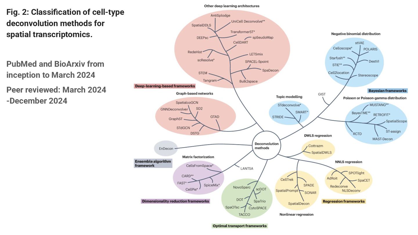
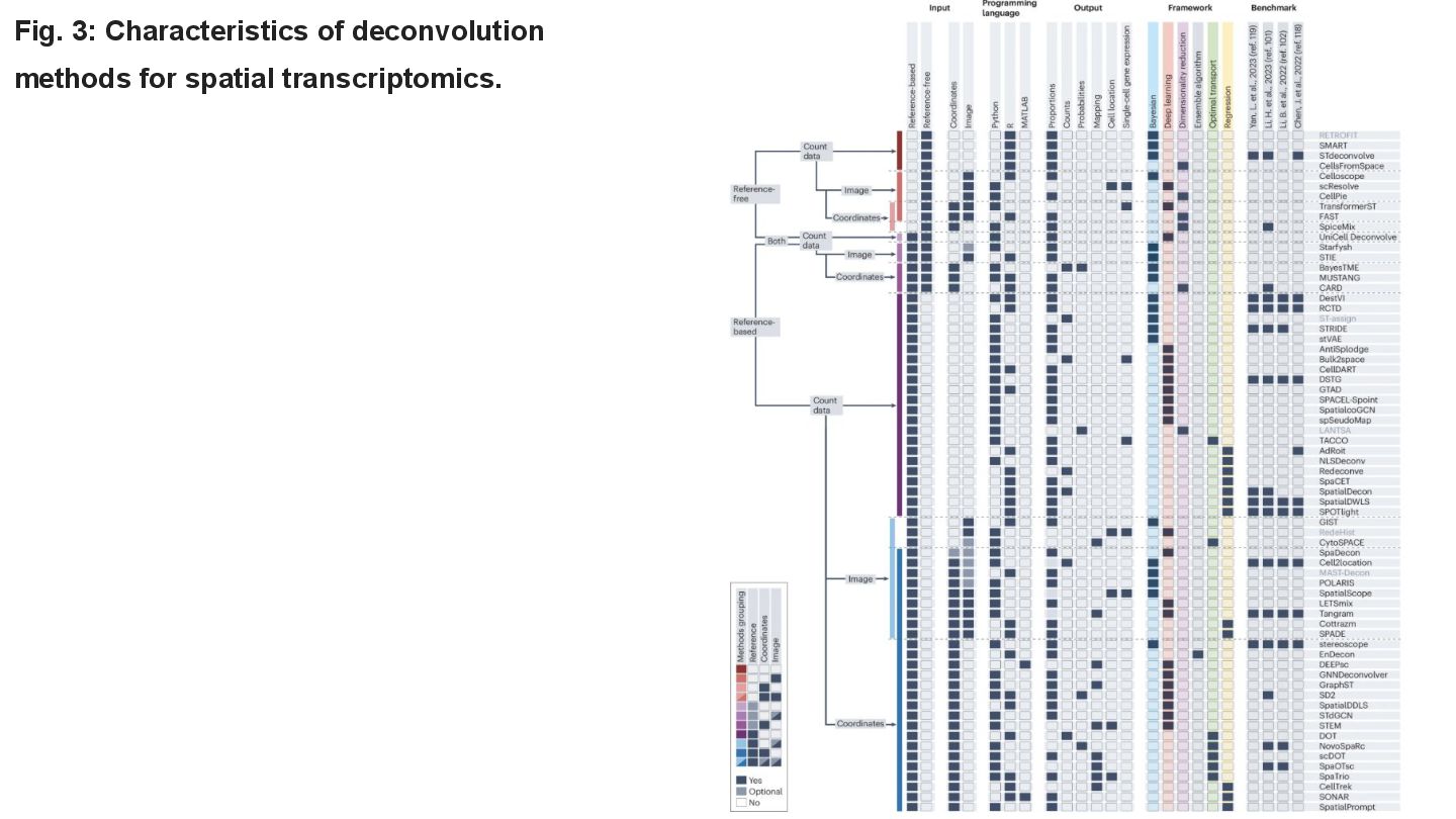
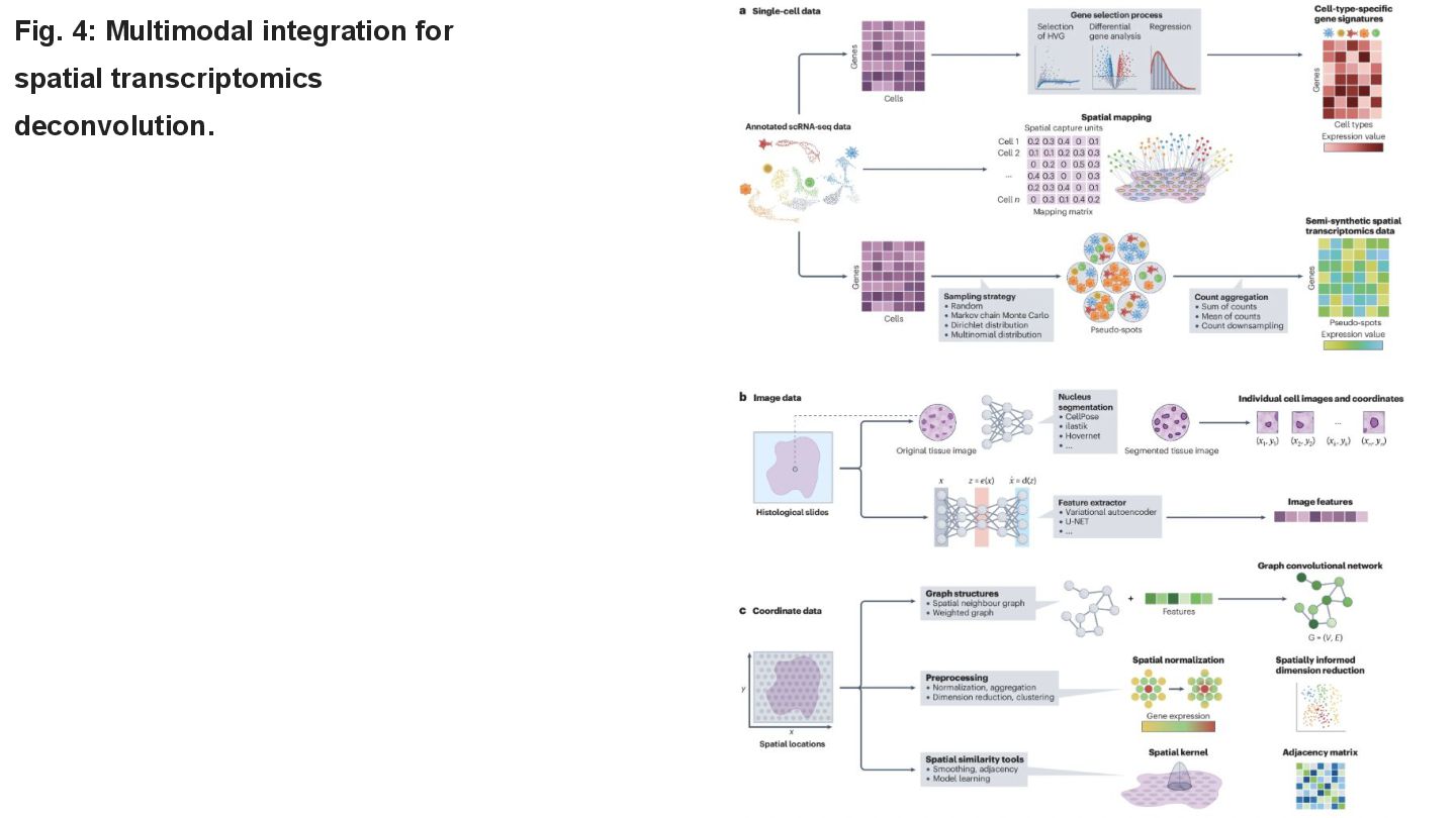
This paper reviews the latest computational approaches for cell-type deconvolution in spatial transcriptomics data. It categorizes existing tools, compares their features, and highlights how they integrate different data modalities, along with a useful interactive tool to compare them.
#transcriptomics #spot-deconvolution #benchmarks

Avatar for Manisha Barse

Manisha Barse

August 13, 2025
Tweet

More Decks by Manisha Barse

Other Decks in Research

Transcript

  1. Cell-type deconvolution methods for spatial transcriptomics Gaspard-Boulinc et.al (2025) doi:

    https://www.nature.com/articles/s41576-025-00845-y Team lcolladotor Journal Club Presented By: Manisha Barse August 13, 2025
  2. Introduction ST: Measures gene expression within intact tissues while preserving

    spatial arrangement of cells. • Imaging-based: Single-cell/subcellular resolution, limited genes (~200–1,000). • Sequencing-based: Whole transcriptome, but each spot contains multiple cells. Sequencing-based ST, lacks single-cell resolution: each spot’s expression = mixture from multiple cell types. https://doi.org/10.3389/fonc.2023.1258245
  3. What is spot deconvolution? Computational method to estimate which cell

    types are in each spot and in what proportions. Why it matters: • Resolves cellular composition beyond spot-level resolution. • Maps cell-type distribution, density, and spatial relationships. • Aids in discovery of tissue changes in development, aging, and disease.
  4. This paper • Explains inputs & outputs of deconvolution. •

    Categorizes methods: reference-based vs reference-free. • Shows integration with scRNA-seq, spatial coordinates, histology. • Provides a web-based interactive table to explore, filter, and compare tools. https://cavallilab-curie.shinyapps.io/Review-Deconvolution-for-Spatial-Transcriptomics/
  5. Pseudo-spot Generation for Model Training & Validation • Pseudo-spots simulate

    spatial transcriptomics spots from single-cell data. • Used for training models and benchmarking deconvolution methods. • Sampling strategies: random or guided to reflect realistic cell-type distributions. • Pseudo-spots help address data scarcity and imbalance, especially for rare cell types. • Limitations: do not fully capture spatial complexity or batch effects; quality depends on single-cell annotation.
  6. Fig. 2: Classification of cell-type deconvolution methods for spatial transcriptomics.

    PubMed and BioArxiv from inception to March 2024 Peer reviewed: March 2024 -December 2024
  7. Key Questions to Guide Method Selection • What are the

    tissue characteristics? (structured vs disordered) • How much prior knowledge is available? (reference profiles or unsupervised) • Which input datasets are available? (single-cell, images, spatial coordinates) • What type of output is desired? (proportions, deconvolved expression, single-cell maps) • Is model complexity or interpretability a priority? • Is computational efficiency important for large datasets? • How robust must the method be to noise and batch effects? • How was the method validated and benchmarked? • Has it been tested in similar tissue or disease contexts? • How did it perform in systematic benchmarking?
  8. Compositional Data Analysis (CoDA) in Deconvolution • Deconvolution outputs are

    proportions, a type of compositional data. • Compositional data sum to a constant and require special statistical treatment. • Traditional analyses may misinterpret relationships due to sum constraint. • CoDA uses log-ratio transformations (additive or centered) to properly analyze proportions. • Enables accurate modeling and interpretation of relative cell-type abundance changes.
  9. Future Perspectives • Development of reference-free deconvolution methods for rare

    or uncharacterized tissues. • Need for robust methods enabling multi-sample and comparative analyses. • Improved multimodal integration, especially image and spatial data. • Comprehensive benchmarking with diverse datasets and new metrics. • Advances in high-resolution spatial transcriptomics demand new aggregation and deconvolution methods.