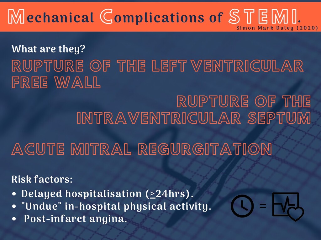
OF THE LEFT VENTRICULAR FREE WALL RUPTURE OF THE INTRAVENTRICULAR SEPTUM Risk factors: S T E M I S i m o n M a r k D a l e y ( 2 0 2 0 ) echanical omplications of . ACUTE MITRAL REGURGITATION What are they? M C =
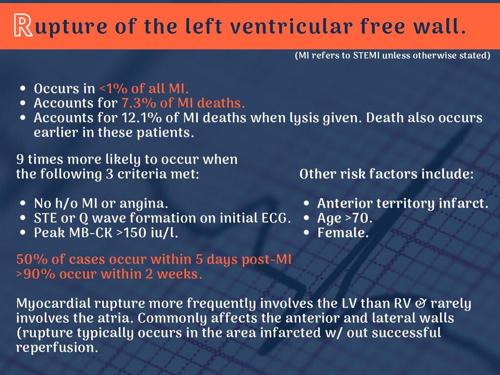
<1% of all MI. Accounts for 7.3% of MI deaths. Accounts for 12.1% of MI deaths when lysis given. Death also occurs earlier in these patients. No h/o MI or angina. STE or Q wave formation on initial ECG. Peak MB-CK >150 iu/l. 9 times more likely to occur when the following 3 criteria met: 50% of cases occur within 5 days post-MI >90% occur within 2 weeks. Myocardial rupture more frequently involves the LV than RV & rarely involves the atria. Commonly affects the anterior and lateral walls (rupture typically occurs in the area infarcted w/ out successful reperfusion. (MI refers to STEMI unless otherwise stated) Anterior territory infarct. Age >70. Female. Other risk factors include:
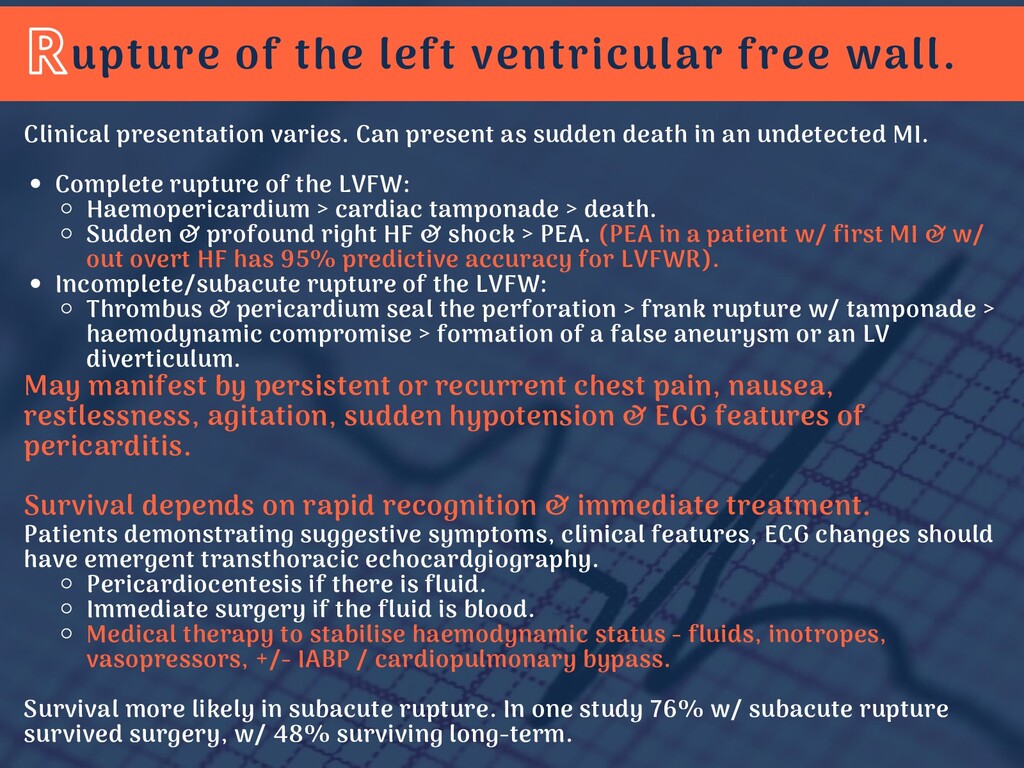
of the LVFW: Haemopericardium > cardiac tamponade > death. Sudden & profound right HF & shock > PEA. (PEA in a patient w/ first MI & w/ out overt HF has 95% predictive accuracy for LVFWR). Incomplete/subacute rupture of the LVFW: Thrombus & pericardium seal the perforation > frank rupture w/ tamponade > haemodynamic compromise > formation of a false aneurysm or an LV diverticulum. Pericardiocentesis if there is fluid. Immediate surgery if the fluid is blood. Medical therapy to stabilise haemodynamic status - fluids, inotropes, vasopressors, +/- IABP / cardiopulmonary bypass. Clinical presentation varies. Can present as sudden death in an undetected MI. May manifest by persistent or recurrent chest pain, nausea, restlessness, agitation, sudden hypotension & ECG features of pericarditis. Survival depends on rapid recognition & immediate treatment. Patients demonstrating suggestive symptoms, clinical features, ECG changes should have emergent transthoracic echocardgiography. Survival more likely in subacute rupture. In one study 76% w/ subacute rupture survived surgery, w/ 48% surviving long-term.
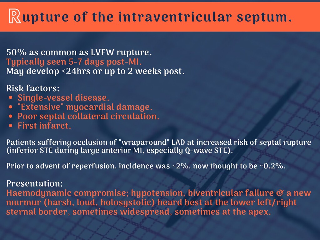
damage. Poor septal collateral circulation. First infarct. 50% as common as LVFW rupture. Typically seen 5-7 days post-MI. May develop <24hrs or up to 2 weeks post. Risk factors: Patients suffering occlusion of "wraparound" LAD at increased risk of septal rupture (inferior STE during large anterior MI, especially Q-wave STE). Prior to advent of reperfusion, incidence was ~2%, now thought to be ~0.2%. Presentation: Haemodynamic compromise; hypotension, biventricular failure & a new murmur (harsh, loud, holosystolic) heard best at the lower left/right sternal border, sometimes widespread, sometimes at the apex.
pulmonary artery balloon catheter to document L to R shunt. Transthoracic echocardiography. Timing of surgical repair is controversial. Survival rates lower in those repaired >6 weeks post-MI, however this reflected selection bias. Haemodynamic stabilisation should be attempted w/ vasodilators, inotropes, diuretics +/- IABP. Operative mortality is HIGH. In patients not effectively reperfused, septal rupture associated w/ persistent STE for >72hrs. Risk of septal rupture increases in RV infarction. Diagnosis: Management: In cardiogenic shock, death is inevitable w/ out surgery. Delayed surgical repair in the absence of shock is feasible, but there is potential for unpredictable & rapid deterioration. Adverse outcomes correlate w/ advanced age & delay between rupture & repair. Transcatheter closure of VSD is an option when surgery is not possible/appropriate.
or true aneurysm. Papillary muscle or chordal rupture. Moderate-severe MR in the context of AMI seen in 3% of patients. Mortality is ~24% at 30 days & 52% at 1 year. Includes: 1. 2. 3. In cardiogenic shock after MI, mod-sev MR seen in 39% of patients. These patients had significantly poorer survival at 1 year than those with mild or no MR (31% vs 58%). Some patients w/ acute MR are haemodynamically stable. Many of these improve w/ medical therapy & revascularisation. Silent MR: Intensity of murmur doesn't correlate w/ severity of the murmur. Some patients w/ acute MR attributable to rupture have early equalisation of LV & LA pressures, resulting in silent MR or a soft/indistinct murmur in as many as 50% of patients.
of total MI mortality. Usually 2-7 days post-MI. May be partial or complete. Papillary muscle rupture: Rupture of the posteriomedial (PM) papillary muscle occurs 6-12 times more frequently than anterolateral (AL) papillary muscle due to blood supply. The PM papillary muscle is supplied by the PDA, while the AL papillary muscle is dual supplied by the LAD & circumflex arteries. Papillary muscle rupture (PMR) occurs in both STEMI & NSTEMI. Most patients have relatively small areas of necrosis with poor collaterals; up to 50% have single vessel disease. >80% of PMRs occur with first MI. Recurrent &/or post-MI anginal pain before or during admission indicated higher likelihood of PMR suggesting that post-MI ischaemia or infarct expansion are contributing factors.
systolic murmur in the setting of AMI. Typically confirmed by transthoracic echocardiography. In some cases transoesophageal echocardiography is required. Diagnosis: Outcomes in PMR are poorer than for other causes of acute mitral regurgitation, w/ operative mortality rate ~50%. Outcomes are worse in medical therapy w/ mortality of 75% at 24hrs & 96% within 2 weeks of complete rupture. Risk factors for poor surgical outcomes include advanced age, female gender & poor LV function. In some patients the risk may be so high as to be considered futile.
therapy prior to emergent surgery are all necessary for a favourable outcome. Medical therapy focused on reducing afterload to reduce the regurgitant fraction - Nitrates, sodium nitroprusside, diuretics & IABP. Emergent surgery remains the treatment of choice despite high operative mortality (20-25%). Survival in medical only patients is very low. Mitral valve repair > replacement where possible. Treatment: 10-year survival 35%. 10-year survival w/ out HF only 23%. In partial PMR some surgeons prefer to stabilize & delay surgery to 6-8 weeks post-MI, but many patients cannot be stabilised.