Upgrade to Pro — share decks privately, control downloads, hide ads and more …

Pediatric Breathing - Dr. Stein

us414
February 28, 2021

Pediatric Breathing - Dr. Stein

Pediatric Breathing - Dr. Stein

us414

February 28, 2021
Tweet

More Decks by us414

Other Decks in Education

Transcript

  1. Pediatric Breathing  Circuits  Endotracheal tubes  Inhalation induction

    by Mark H. Stein, MD Pediatric Breathing  Circuits  Endotracheal tubes  Inhalation induction by Mark H. Stein, MD Pediatric Breathing  Circuits  Endotracheal tubes  Inhalation induction by Mark H. Stein, MD Pediatric Breathing  Circuits  Endotracheal tubes  Inhalation induction by Mark H. Stein, MD
  2. With any breathing circuit, you want to prevent re-breathing of

    exhaled carbon dioxide, and to reduce the work of breathing during spontaneous ventilation by minimizing resistance within the circuit. In order to meet those goals, the first breathing circuits (i.e. the Ayres T-piece and its modifications) did not have in-line unidirectional valves or CO2 absorbers, and a relatively high rate of fresh gas flow was used. Even with the best of these circuits, some of which are still in use today, a relatively high fresh gas flow is needed to prevent re-breathing, whether the patient is breathing spontaneously or controlled. This results in a lot of waste of oxygen, inhalational anesthesia agents, loss of body temperature due to breathing the cold fresh gas, inadequate humidification, and inadequate scavenging of exhaled gases from these circuits. Breathing circuits, back in the day…
  3. Mapleson Breathing Circuits In 1954, Dr. William Mapleson [1926-2018], a

    British physiologist, analyzed all the available anesthesia breathing circuits at the time. He classified 5 groups, naming them A-E. (The F circuit was a later modification of the E.) His work is based on several principles. These principles will become clearer as we go over each circuit: 1. There are 3 types of gas in any circuit- fresh, alveolar, and dead space gas. 2. The breathing circuits are open to atmospheric pressure during spontaneous breathing. 3. During spontaneous breathing, the reservoir bag fills up to atmospheric pressure; anything beyond that will blow out through the pop-off valve. 4. The pop-off valve is left maximally loosened during spontaneous breathing, but must be partially closed during controlled ventilation to allow positive pressure to be generated when the reservoir bag is squeezed. 5. All circuits are comprised of a fresh gas inlet, an expiratory outlet (most also have a pop-off valve at the expiratory outlet), a gas reservoir (a length of corrugated tubing and/or a reservoir bag), and a connection to the patient. 6. Circuits are classified by the relative positions of the items mentioned in #5.
  4. The Mapleson circuits were commonly used in the past for

    pediatric patients The circle breathing system with valves is the most commonly used system today, but you’ll still find valve-less Mapleson breathing circuits, or their modifications, in use (e.g. the Ambu bag). The Maplesons were recommended for many years for children weighing less than 10 kg, because of the decreased resistance to spontaneous breathing (years ago, most pediatric cases were done with spontaneous breathing), the better ‘feel’ of the rebreathing bag in the hand of the anesthesiologist, and the faster induction and emergence times using the higher fresh gas flows those circuits require for non- rebreathing.
  5. Arguments in favor of the Maplesons have gradually lost out

    to modern improvements and changes in practice. • Newer machines and circuits have low-resistance valves, reduced dead space at connections, and improved CO2 absorbent canister design. • Capnography and pulse oximetry are now the standard of care, guiding us during ventilation. • Controlled ventilation, rather than spontaneous, is favored for children having longer and more complex surgeries. • Nobody wants to manually ventilate a patient for hours and hours during a long complex surgical procedure, especially on a tiny and/or complicated patient. (Most of the Maplesons required manually ventilating when controlled ventilation was needed.) The Mapleson D could be connected to the machine ventilator, but you had to bypass some of the ventilator alarms and risked making a wrong connection. • Modern less-soluble inhalation agents allow faster induction and emergence, thus mitigating the influence of the breathing circuit.
  6. Mapleson Breathing Circuits (cont.) These are a group of 6

    different semiclosed breathing circuits, labeled A-F. • They have no unidirectional valves and no soda lime carbon dioxide absorption. • Analyzing how a patient breathes, either spontaneously or controlled, via each of the Mapleson circuits, is a good exercise in understanding the problems of ventilating a patient, and how our modern anesthesia breathing circuits evolved. CO2 rebreathing, during spontaneous or controlled ventilation, was realized as a serious problem when the modern practice of anesthesiology emerged. Since spontaneous ventilation was the most common modality in anesthesiology years ago, the ‘work of breathing’ was noted to be an an important issue, especially for small children.
  7. Mapleson Breathing Circuits (cont.) How much CO2 rebreathing occurs with

    each Mapleson circuit, either during spontaneous or controlled ventilation, is an important difference among the circuits. Imagine if you didn’t have a CO2 absorber, or unidirectional valves. How would you prevent rebreathing? • The amount of rebreathing depends on fresh gas inflow rate, minute ventilation, mode of ventilation (spontaneous or controlled), tidal volume, respiratory rate, inspiratory to expiratory time ratio, duration of the expiratory pause, peak inspiratory flow rate, volume of the reservoir tube, volume of the breathing bag, and dead space. • With enough fresh gas flow, you will minimize rebreathing, and the work of breathing will be very low because the patient doesn’t have to work against any unidirectional valves, a CO2 absorber, or any other mechanical impediments in the breathing circuit.
  8. Physiologic dead space of the respiratory system refers to the

    space in which oxygen (O2 ) and carbon dioxide (CO2 ) gases are not exchanged across the alveolar membrane in the respiratory tract. Physiologic dead space is the sum of the anatomic and alveolar dead space. • Anatomic dead space specifically refers to the volume of air located in the segments of the respiratory tract that are responsible for conducting air to the alveoli and respiratory bronchioles but do not take part in the process of gas exchange itself. These segments of the respiratory tract include the upper airways, trachea, bronchi, and terminal bronchioles. Anatomic dead space normally is estimated at 2mL/kg of body weight and comprises 1/3 of the tidal volume in a healthy adult patient; it is even higher in pediatric patients [3.3ml/kg in infants, then approaches adult values after age 6 yrs]. Effectively, 1/3 of a TV of inhaled air is rebreathed due to dead space. • Alveolar dead space refers to the volume of air in alveoli that are ventilated but not perfused, and thus gas exchange does not take place. Alveolar dead space typically is negligible in a healthy individual. https://www.ncbi.nlm.nih.gov/books/NBK442016/
  9. Dead space in a patient is the volume of a

    breath that does not participate in gas exchange. Physiologic or total dead space is the sum of anatomic dead space (where there are no alveoli) and alveolar dead space (alveoli that aren’t getting perfused). Dead space in an anesthesia breathing system, or mechanical dead space, exists where fresh and exhaled gases are mixed. You can try to minimize dead space in a Mapleson system by the arrangement of the circuit components, and the amount of fresh gas flow. Mechanical dead space should not exist when fresh and exhaled gases are completely separated.
  10. Dead Space in an anesthesia breathing system is the volume

    from the patient-end to the point up to which, to and fro movement of mixed expired and fresh gas takes place. Extent of dead space in various systems: 1: Fresh gas flow originates further from the patient than the expiratory valve. The apparatus dead space extends up to the expiratory valve positioned near the patient (eg. Mapleson types A, B, and C). 2: If the fresh gas enters the system near the patient-end, the dead space extends up to the point of fresh gas entry (Mapleson types D, E, and F). 3: In systems where inspiratory and expiratory limbs are separate, dead space extends up to the point of bifurcation (the circle system is an example of this). Some circuits have a septum at the Y bifurcation to enhance the separation of the inspiratory and expiratory limbs. 1 2 3
  11. Factors that increase physiologic or mechanical dead space: • General

    anesthesia – multifactorial, including loss of skeletal muscle tone and bronchoconstrictor tone • Anesthesia apparatus/circuit: Tubing from a ventilator or breathing circuit increases dead space volume- depending on the position of unidirectional valves and the Y connection by the patient- by adding volume to the effective space not participating in a gas exchange. • Artificial airway (mask, LMA, filters, long ET tube) • Neck extension and jaw protrusion • Positive pressure ventilation (i.e. increased airway pressure) • Upright posture as opposed to supine (because of decreased perfusion to the uppermost alveoli) • Pulmonary embolus, PA thrombosis, hemorrhage, hypotension, surgical manipulation of pulmonary artery tree – anything that decreases perfusion to well-ventilated alveoli • Emphysema (i.e. damaged alveoli- blebs, loss of alveolar septa and vasculature) • Age • Anticholinergic drugs
  12. • Each type of Mapleson circuit has a T connection

    to: a fresh gas source (FG), an exhalation limb with a reservoir (bag and/or corrugated tube), and a connection to the patient. ✱ The exhalation limb outlet may or may not have a pressure relief (‘pop- off’) valve. The Mapleson Circuits
  13. Maplesons A, B and C have the exhalation outlet closer

    to the patient than the fresh gas source is. Maplesons D, E and F have the fresh gas source closer to the patient than the exhalation outlet is. Keep in mind: Spontaneous ventilation is a negative inspiratory force, generated by the patient. The circuit will open to the atmosphere via the pop-off valve if any positive pressure starts to build up in the circuit. Controlled ventilation is a positive inspiratory force, generated by the anesthesiologist squeezing the reservoir bag. The pop-off valve has to be partially shut to allow positive pressure to build up so that the tidal volume can be delivered. The Mapleson Circuits (cont.)
  14. Let’s start with the simplest of the Maplesons, the E.

    (It is based on the historic original and simplest breathing circuit, the Ayer’s T-piece.) You’ll probably recognize this as the ‘T-piece’ you connect to a patient when they’re still intubated but breathing spontaneously, and you want to transport them to the PACU. When you connect it to the patient, you are told to put the flow on 15 LPM “to prevent rebreathing”. How did they come up with 15 LPM? Let’s do some arithmetic... The Mapleson Circuits (cont.)
  15. Let’s say the patient weighs 80 kg: • Normal tidal

    volume with spontaneous breathing is about 5cc/kg. • Normal respiratory rate is about 12 breaths per minute. • Therefore, 80 x 5 = 400 cc tidal volume, and 400 x 12 = 4800 cc minute ventilation. How did they decide that 3 times the fresh gas flow was needed to prevent rebreathing? There are 60 sec in a minute, and 12 breaths per minute, so the total time for each breath, inspiration and expiration together, takes 5 seconds: 60 ÷ 12 = 5 If the normal I:E ratio is 1:2, and total of inspiratory time plus expiratory time per breath has to equal 5 seconds, that means you’ve got an inspiratory time of about 1.67 sec and an expiratory time of 3.33 sec per breath. That means you’ve got to deliver the 400 cc of tidal volume in 1.67 sec, which is an inspiratory flow rate of . = 239.5 𝑐𝑐 𝑝𝑒𝑟 𝑠𝑒𝑐𝑜𝑛𝑑, 𝑜𝑟 14.4 𝐿𝑃𝑀. That is 3 times the minute ventilation. (4800 x 3 = 14,400 cc) The Mapleson Circuits (cont.)
  16. The Mapleson Circuits (cont.) We know that during the beginning

    of inspiration, a very rapid initial flow rate occurs, in the range of 60 LPM or more, for a brief second. Even if you set the fresh oxygen flow into a Mapleson E circuit at 3x the minute ventilation (approximately 15 LPM), the initial phase of inspiration needs more. That’s why you want the corrugated reservoir to contain enough volume of fresh oxygen to supply the extra volume needed for that initial rapid flow. It’s only for a second (or less) timewise, but unless you’ve got enough volume of fresh oxygen in your corrugated reservoir, the patient will entrain previously exhaled gas, or room air.
  17. The Mapleson E System (cont.) Therefore, to prevent rebreathing, you

    should have a length of corrugated tubing long enough to hold a tidal volume, in addition to a fresh oxygen flow of 3x minute ventilation. Unfortunately, we just use a short piece of corrugated tubing on the T-pieces in our PACU. That length of corrugated tubing added to the expiratory limb acts as a fresh gas reservoir for that rapid initial flow needed during the next inspiration. During expiration: the exhaled gas enters the corrugated tubing, and during the expiratory pause, it will be flushed out with fresh oxygen flow. During inspiration: the fresh oxygen stored in the expiratory part of the circuit, combined with fresh oxygen flow, will provide adequate tidal volume and prevent rebreathing.
  18. You can’t give a manual breath with a Mapleson E

    circuit. Dr. Gordon Jackson-Rees, a British anesthesiologist who worked at Liverpool Children’s Hospital, decided to add a reservoir bag at the end of the corrugated tubing, thus allowing manual ventilation. Now you’ve got a Mapleson F, also known as a Jackson-Rees circuit. There is an opening at the end of the bag, allowing you to vent excess pressure.
  19. The Type F Mapleson Circuit Jackson-Rees (Type F Mapleson) The

    Mapleson F system, a modification of the E system, is also known as the Jackson- Rees system. It adds a reservoir bag. Exhaled and fresh gas will collect and mix in the bag. The next inspiration will result in the patient inhaling fresh gas from both the fresh gas inlet and from that stored in the expiratory part of the circuit. The presence of the bag allows the anesthetist to observe ventilation during spontaneous breathing as well as control ventilation by squeezing the bag in the absence of spontaneous breathing. Rebreathing would be minimized by insuring adequate fresh gas flows, about 2.5 - 3 times the minute ventilation rate. The bag will also allow waste gas venting because it has an opening that can go to a scavenging system.
  20. The Mapleson D system Now let’s take the Mapleson F

    circuit, and move the pop-off valve to be in front of the bag. Now you’ve got the Mapleson D system. This setup tends to be somewhat more efficient for controlled ventilation than the other Mapleson systems.
  21. The Mapleson D System and spontaneous ventilation With exhalation, fresh

    gas as well as dead space and alveolar gas will enter the tubing, and with more fresh gas flow, more of the expired gas will be vented out of the circuit. • To eliminate rebreathing you need a fresh gas flow (FGF) of 3x times minute ventilation, because you need to wash out the expired gas that enters the corrugated tubing and bag, as explained earlier. • As expiratory time increases the circuit becomes MORE efficient - this is because more gas from the FGF flushes out the alveolar gas from the last expiration. • Length of the corrugated reservoir tubing volume should be ≥ tidal volume, as mentioned when we talked about the Mapleson E.
  22. The Mapleson D System and controlled ventilation During expiration the

    corrugated tubing will first fill with exhaled gas. The length of corrugated tubing should be 1 meter, and should be able to hold an exhaled tidal volume. Some exhaled gas may also enter the reservoir bag. Fresh gas will then enter the corrugated tube during end- expiration and wash out the exhaled gas through the pop-off valve. If you had partly closed the pop-off valve, any additional fresh gas flow will enter the reservoir bag. If you use an I:E ratio to allow more expiratory time, efficiency is improved as the FGF has more time to wash out the exhaled gas from the corrugated tube. A higher FGF rate will also have the same effect. Manually squeezing the reservoir bag (during inspiration) will cause fresh gas to enter the patient from the corrugated tubing, and then fresh gas flow enters the patient. Any excess pressure on the bag will cause pop-off valve activation and the release of any remaining contents of the bag. At least 1.5x the minute ventilation gas flow rate is required to minimize rebreathing during controlled ventilation with the Mapleson D. Therefore, it’s considered the most efficient of all the Maplesons for controlled ventilation. You can also detach the reservoir bag and connect the anesthesia ventilator hose to where the bag was, to allow mechanical ventilation.
  23. The Bain Circuit is a modification of the Mapleson D.

    BAIN CIRCUIT It was used for all pediatric anesthesiology cases in our department until 1991.
  24. Mapleson D system (coaxial modification, Bain circuit): A commonly used

    variant of the Mapleson D system, the fresh gas tube is within the corrugated expiratory tubing. The exhaled gas travels through the external corrugated tube to the reservoir bag and pop-off valve. The outer tube is transparent so that the inner tube can be seen. The system is used just like the Mapleson D. The reservoir bag can be dettached and the anesthesia ventilator hose connected to where the bag was, to allow mechanical ventilation. The Bain Circuit (cont.) BAIN CIRCUIT
  25. The Bain Circuit (cont.) System advantages: • There is some

    warming of the fresh gas by the exhaled gas present in the outer tubing. • The Bain circuit can be connected to an anesthesia machine ventilator. • Since the overflow valve is located far away from the patient, expiratory gases may be readily scavenged. BAIN CIRCUIT
  26. System disadvantages: unrecognized disconnection of the inner fresh gas tube

    at the patient end could cause mixing of fresh gas and exhaled gas. • Therefore, prior to use, the Bain system must be carefully checked. • The transparency of the outer tube is important because should the inner tube leak or if the inner tube is detached from the fresh gas port, a significant increase in system dead space could occur. • There are specific procedures to check for a leak. One such method requires high-flow oxygen filling the circuit while occluding the patient end until the reservoir bag has filled. Then, the patient end is opened allowing oxygen to flush the system. If the inner tube is structurally intact and properly aligned, then a decrease in pressure within the circuit will be observed and the reservoir bag will deflate (Venturi effect). If the inner tube is leaking, however, fresh gas will escape into the expiratory part of the circuit and the reservoir bag will remain inflated. The Bain Circuit (cont.)
  27. The Mapleson A compared to the Mapleson D system The

    fresh gas inlet and the expiratory outlet are reversed. As a result, Dr. Mapleson found that A is the most efficient for spontaneous breathing, and the D is more efficient for controlled ventilation. The corrugated tubing volume needs to be at least the size of the tidal volume, so it’s usually 3 feet long. Having the pop-off (APL) valve close to the patient adds dead space (100cc) and weight to the circuit. Unsuitable for very small children. The Mapleson A, known as the Magill Circuit
  28. Spontaneous ventilation: During exhalation, exhaled gas first move down the

    corrugated tube and towards the reservoir bag. Meanwhile, fresh gas flow is entering the reservoir bag. Once the reservoir bag is full, the pop-off valve will open, and the contents of the corrugated tubing (the gas that had just been exhaled, along with fresh gas entering the corrugated tube) will flow out of the pop-off valve. During the end-expiratory pause, the fresh gas flow will clear out more of the contents of the corrugated tubing via the pop-off valve. As long as fresh gas flow is ≥ minute ventilation, and the volume of the corrugated tubing is ≥ tidal volume, no rebreathing occurs. The Mapleson A circuit is considered the most efficient of the Maplesons for spontaneous ventilation. Dr. Mapleson determined that in order to limit or prevent rebreathing, the fresh gas flow rate should be equal to or slightly greater than minute ventilation. That way, you keep the corrugated tube and reservoir bag relatively free of previously exhaled gas. http://www.creaghbrown.co.uk/anae/bc.htm The Mapleson A, known as the Magill Circuit (cont.)
  29. Controlled ventilation: Squeezing the bag initiates inflation of the lungs.

    A mix of previously exhaled dead space gas, alveolar gas, and fresh gas goes to the patient. The pop-off valve has to be partially closed so that the lungs can be inflated. Once the lungs are inflated, the remaining gas mixture in the circuit can exit via the pop-off valve. Inadequate removal of previously exhaled alveolar gas results in re-breathing. To optimize CO2 removal, you want a short inspiratory:expiratory ratio [that is, allow more exp. time] along with a large tidal volume with high fresh gas flow; you’ll need a fresh gas flow rate about 3x minute ventilation. With this approach CO2 removal may be optimized but at the expense of wasting oxygen and anesthetic agent. Therefore, the Mapleson A is considered the least efficient of the Maplesons for controlled ventilation because of the high fresh gas flow rate required to prevent rebreathing. The Mapleson A, known as the Magill Circuit (cont.)
  30. If we add a nonrebreathing valve to a Mapleson A

    circuit at the site of the popoff valve in order to improve efficiency, you’d have an AMBU bag. • The nonrebreathing valve used in the AMBU is called a Rubens valve. We still use about 3x minute ventilation as our fresh gas flow when using an AMBU bag, which is why we set about 15 LPM of O2 flow. Fun fact about the Mapleson A circuit
  31. An AMBU bag, which is a self-reinflating bag with non-

    rebreathing valves (contained within the Rubens valve) to provide positive pressure ventilation, is a modification of a Mapleson circuit. The AMBU has an inspiratory valve in addition to the expiratory valve contained within the Rubens valve. Now you can understand why we set the flow rate to about 15-25 LPM when using the AMBU.
  32. The Mapleson B and C systems To improve the efficiency

    of the Mapleson A, the Mapleson B and C systems were created, with the fresh gas inflow port closer to the patient. Excess volume in the circuit- a mix of exhaled and fresh gas- collects in the reservoir bag and corrugated tubing. To prevent rebreathing, with either spontaneous or manual ventilation, you would still need fresh gas flows at least 2x minute ventilation. The Mapleson B and C systems were not a significant improvement on the Mapleson A, and so they never gained popularity.
  33. The Circle Breathing System • Uses CO2 absorbents to prevent

    rebreathing of CO2 • Allows partial or complete rebreathing of other gases (saves $$$) • Can be semi-open, semi-closed, or closed, depending on the fresh gas flow. – Semi-open has no rebreathing, a high fresh gas flow, and resembles a Mapleson Circuit. – Closed has complete rebreathing with CO2 absorption, and the pop- off stays closed. – Semi-closed has some re-breathing.
  34. The Circle Breathing System (cont.) • The Circle Breathing System

    has 7 primary components: Fresh gas inflow source, inspiratory and expiratory unidirectional valves, inspiratory and expiratory corrugated tubes, Y- piece connector, overflow (pop-off or adjustable pressure limiting [APL]) valve, reservoir bag, canister containing CO2 absorbent.  Main advantages of the Circle System-- maintenance of relatively stable inspired gas concentrations, conserves anesthesia gases, conserves respiratory moisture and heat, prevents OR pollution.  Main disadvantages of the Circle System-- complexity and multiple connections can result in potentially catastrophic misconnections, disconnections, obstructions, leaks.
  35. The Circle System (cont.) • Circle systems for pediatrics have

    the same advantages as for adults including low fresh gas flow requirements, conservation of heat, humidity, and anesthetic agents, and minimal environmental pollution. • Shorter, narrow-caliber tubing helps to minimize the compliance of pediatric circuits. • Dead space of a breathing system is an important issue, because of the potential of rebreathing that dead space mixture. • Dead space in a circle breathing system exists up to the point where fresh gas meets and mixes with exhaled gas (i.e. at the Y-piece). Dead space is minimized with a median septum (to try to prevent mixing) in the Y-piece of a circle system. Anything that increases the volume between the patient and the Y-piece will worsen dead space, such as goose neck extensions and filters.
  36. Limitations of the Circle System in Children The compliance of

    the breathing system (absorber canister, circle system, and hoses) allows a portion of the volume delivered by the ventilator to be compressed in the system during inspiration. The amount of desired delivered gas “lost” to the system depends on the inspiratory pressure and the total compliance of the breathing system. As the peak inspiratory pressure increases, less and less tidal volume ultimately is delivered to the patient.
  37. Limitations of the Circle System in Children (cont.) • The

    compression volume of the breathing circuit is an important influence on ventilation in children, because their tidal volumes are so small. – Breathing circuits with large compression volumes take up a significant portion of the delivered volume meant for the patient. – Pediatric circle systems should be shorter than for adults and have a smaller radius of the tubing, and are therefore less distensible (Young/LaPlace Equation- smaller radius makes them less distensible at a given pressure).
  38. Limitations of the Circle System in Children (cont.) • Delivered

    tidal volume in a circle system depends on the tidal volume, respiratory rate, inhalation to exhalation (I:E) ratio, and total fresh gas flow (FGF). Alterations in FGF can significantly affect minute ventilation unless compensated for by alterations in bellows volume, ventilatory rate, or drawing more FGF from a reservoir. • New machines can automatically perform such compensation by drawing more FGF from the reservoir bag.
  39. Limitations of the Circle System in Children (cont.) • The

    use of uncuffed endotracheal tubes in pediatric patients can cause variable amounts of leak, with resulting volume loss to the circuit, especially at low flows. Therefore, the volume set point may not accurately reflect the volume delivered to the patient. Likewise, volume returned from the patient may be inadequate to fill the bellows (or the rebreathing bag) during low flow anesthesia. • The reduction in exhaled gas volume because of an endotracheal tube leak will also result in inadequate spirometry through the exhalation limb of the breathing circuit, even if the delivered tidal volume is adequate.
  40. Pediatric Masks Requirements for pediatric masks include a minimum of

    dead space between the face and the mask. The inside of the mask should almost completely be filled by the child’s face. Mask anesthesia maybe used for short operations where there are no additional risks for aspiration, and the anesthesiologist has access to the airway at all times.
  41. Endotracheal Tubes Polyvinylchloride is still the most common material used

    in the production of endotracheal tubes, but polyurethane is now being used. Uncuffed ET tubes have been used in pediatric anesthesia since the 1950’s, since it was felt that the cricoid ring was the narrowest part of the airway, and sealing beyond that point with a cuffed ETT was unnecessary. However, nowadays we perform more complex surgeries, and in patients with poor lung compliance, so a cuffed tube is sometimes necessary. An air leak (>20-25) around an ETT has been advocated for the prevention of postintubation croup, [and to prevent subglottis stenosis after prolonged intubation], based upon a 1977 landmark study. Thanks to a better understanding of pediatric laryngeal anatomy and pathophysiology, and improved materials in the manufacture of ET tubes, the use of cuffed tubes is becoming more commonplace. Checking for the presence of a leak beyond 20-25 (whether cuffed or not), and checking the inflation pressure of the cuff, are still very important.
  42. Medical Considerations Most children and infants scheduled for elective surgical

    procedures are in good health, but common disorders such as upper respiratory tract infection (URI), reactive airways disease, gastroesophageal reflux, obesity, and hemodynamically stable congenital heart lesions can pose diagnostic and management challenges for the pediatric anesthesiologist.
  43. Upper Respiratory Tract Infections • URI is by far the

    most common problem the pediatric anesthesiologist encounters, especially in the ambulatory surgery setting. URI and the accompanying inflammation increase upper and lower airway irritability and secretions, and they may increase the incidence of laryngospasm, bronchospasm, and perioperative hypoxemia. • Risk factors for associated complications include nasal congestion, copious secretions, reactive airways disease, history of prematurity, passive smoking, airway surgery, endotracheal intubation, and laryngeal mask airway insertion. • Even in patients without a history of asthma, airway reactivity can develop with URI or lower respiratory tract infection and last as long as 6 to 8 weeks. • The decision to postpone the procedure depends on the urgency of the surgery, the severity of symptoms, and the need to instrument the airway. • In these patients, prophylactic bronchodilator treatment should be considered before the induction of anesthesia and before the emergence from anesthesia and extubation.
  44. Reactive Airways Disease • In children with reactive airways disease

    (RAD), a detailed past medical history must be obtained to determine the severity of the disease and the effectiveness of current medical treatment. Recurrent emergency visits and hospital admissions, especially those to critical care units and/or involving the use of steroids, are red flags for poor control of symptoms. • In a patient with active or recent bronchospasms, elective surgery should be postponed for 4 to 6 weeks so that the primary care physician can optimize the treatment regimen. • If surgery is required, preinduction treatment with a β2 - agonist is recommended to minimize respiratory complications.
  45. Congenital Heart Disease In the past, patients with nearly every

    type of congenital heart defect needed to receive antibiotics one hour before dental procedures or operations on the mouth, throat, gastrointestinal, genital, or urinary tract. However, in 2007 the American Heart Association simplified its recommendations. Today, antibiotics before dental procedures and upper respiratory tract surgery (eg. tonsillectomy) are only recommended for patients with the highest risk of IE: 1. A prosthetic heart valve or who have had a heart valve repaired with prosthetic material 2. A history of endocarditis 3. A heart transplant with abnormal heart valve function 4. Certain congenital heart defects including: – Cyanotic congenital heart disease (birth defects with oxygen levels lower than normal), that has not been fully repaired, including children who have had a surgical shunts and conduits. – A congenital heart defect that's been completely repaired with prosthetic material or a device for the first six months after the repair procedure. – Repaired congenital heart disease with residual defects, such as persisting leaks or abnormal flow at or adjacent to a prosthetic patch or prosthetic device.
  46. Procedures for which prophylaxis is reasonable in patients with cardiac

    conditions listed above: Prophylaxis is only recommended for “dental procedures that involve manipulation of gingival tissue or the periapical region of teeth or perforation of the oral mucosa” and “invasive procedure of the respiratory tract that involves incision or biopsy of the respiratory mucosa, such as tonsillectomy and adenoidectomy”. • Antibiotic prophylaxis is NOT recommended for the following dental procedures or events: routine anesthetic injections through noninfected tissue; taking dental radiographs; placement of removable prosthodontic or orthodontic appliances; adjustment of orthodontic appliances; placement of orthodontic brackets; and shedding of deciduous teeth and bleeding from trauma to the lips or oral mucosa. Taking antibiotics just to prevent endocarditis is not recommended for patients who have procedures involving the reproductive, urinary or gastrointestinal tracts.
  47. Whether sedated or awake, accompanied or alone, lying down or

    sitting up, a child breathing sevoflurane with nitrous oxide through a mask and circuit will become anesthetized. This has made inhalation induction the most commonly used technique in pediatric anesthesia in the United States. The induction of anesthesia is achieved with relative ease, speed, and safety, while avoiding the fear and pain of intravenous catheter insertion. Successfully selecting the approach, guiding the child and family through the process, and smoothly adapting to their needs is a challenge. Inhalation Induction
  48. Preanesthetic Preparations The preoperative interview, which is essential for obtaining

    pertinent positive and negative findings in the patient’s history and physical condition, may be the only opportunity for the anesthesiologist to assess child-family anxiety and establish a relationship of trust and confidence. Successful interaction with children requires skill and experience, but more importantly it requires honesty and professionalism. Most centers have presurgical tours and/or short movies to prepare children and families for the perioperative experience. Children are typically encouraged to bring a familiar object or favorite toy, and most pediatric waiting areas are designed as playrooms that are supplied with everything from busy boxes to video games.
  49. Psychological Considerations During the preoperative period, it is important to

    identify the children and families who are likely to develop pronounced fear and anxiety before and during the induction of anesthesia. Because the level of stress and underlying temperament that predispose individuals to extreme anxiety may not be overtly apparent, the pediatric anesthesiologist should evaluate each patient. Indicative behaviors, beyond the obvious crying and uncooperative child, include the absence of social interaction, vocalization, emotional expression, and age-appropriate independence from parents. It is also helpful to assess family members’ levels of anxiety and coping styles. Premedication with anxiolytics has been shown to be the most consistently effective intervention for facilitating induction and reducing postoperative complications in anxious patients.
  50. The anesthesiologist should sit at the child’s eye level and

    initiate communication based on the child’s present activity. The anesthesiologist should express genuine interest or play with the child. This may help gain the child’s confidence and divert or reduce fear and anxiety. Tone of voice and facial expression should be calm and friendly for all age groups. Good humor and empathy will go a long way in alleviating patient and family stress and anxiety. Most children can be well managed in this friendly environment. An anesthesia mask may be given to the child to play with in the waiting area before induction.
  51. Family responses, questions, and interactions with the child are important

    to note. It is essential for the pediatric anesthesiologist to include patients and families in decisions regarding anesthetic induction and care. Together they should agree on the needs for premedication, parental presence, and the type of induction technique (inhalation vs. intravenous) to ensure the smoothest and safest induction possible. Unfortunately, even the best of plans can fail, and back-up measures such as another pre-medication, using an induction room, or a different means of induction, must be readily at hand. For older, combative children scheduled for elective procedure, postponing the procedure until the child is properly prepared should be considered. Inhalation induction by force should be a measure of last resort.
  52. Premedication The use of premedication is the most reliably effective

    intervention for reducing preinduction anxiety and stress for young patients and their parents. Sedatives, typically by mouth, are administered before induction to facilitate child-parent separation, anesthetic mask acceptance, and/or patient cooperation. IM administration is almost now solely reserved for extremely agitated, uncontrollable children.
  53. Parental Presence during Induction Parental presence during induction of anesthesia

    (PPIA), in the operating room or a separate area such as an induction room, is now commonplace. Although the practice avoids separating children from their parents, it does not necessarily decrease patient anxiety or increase cooperation during induction. One benefit, which is important in terms of family-centered care, is an increase in parental satisfaction scores. Studies have shown that PPIA had a measurable benefit when a calm parent accompanied an anxious child, and a worsening effect when an anxious parent accompanied a calm child. There was no measurable benefit when both parent and child were calm. Parental preparation to reduce anxiety can significantly improve the outcome of PPIA.
  54. Once the patient has accepted the mask and is breathing

    the inhalation agent, we follow the progress of the induction. The first sign is usually nystagmus, especially if nitrous oxide is being used. The eyes then usually close; the limbs and head may relax; and respiration becomes slower, regular, and deeper— then more shallow and rapid. Often children will have uncoordinated movements during the hyperreflexic phase of anesthesia and develop “snoring” or noisy breathing as the anesthetic depth deepens and as the patient develops partial upper airway obstruction. Although prepared for this preoperatively, parents should be reminded and reassured that this is normal during the induction of anesthesia. Until the eyelash reflex is gone, nothing should be done to move or stimulate the patient, unless airway obstruction or a similar need arises. Nurses and surgeons should not touch the patient without getting permission from the anesthesiologist. As soon as general anesthesia is induced and the patient can tolerate moderately painful stimuli, an IV catheter can be started.
  55. Non-pharmacological interventionsfor assisting the induction of anaesthesia in children Peggy

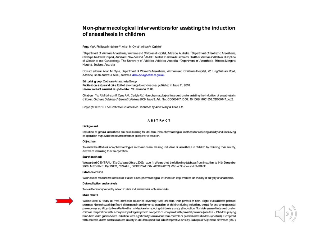
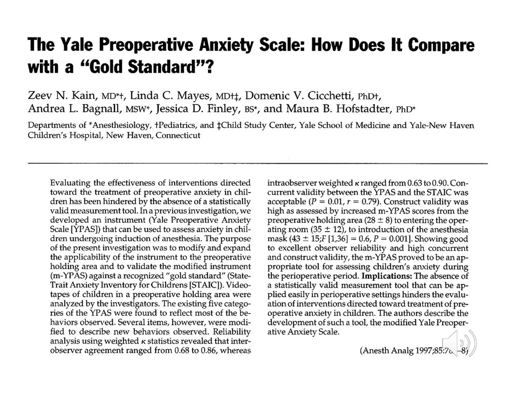
    Yip2, PhilippaMiddleton3, Allan M Cyna1, Alison V Carlyle4 1Department of Women’sAnaesthesia, Women’sand Children’sHospital, Adelaide, Australia. 2Department of Paediatric Anaesthesia, StarshipChildren’sHospital,Auckland,NewZealand.3ARCH:AustralianResearchCentrefor Healthof WomenandBabies,Discipline of Obstetrics and Gynaecology, The University of Adelaide, Adelaide, Australia. 4Department of Anaesthesia, Princess Margaret Hospital, Subiaco, Australia Contact address: Allan M Cyna, Department of Women’s Anaesthesia, Women’s and Children’s Hospital, 72 King William Road, Adelaide, South Australia, 5006, Australia. allan.cyna@ health.sa.gov.au. Editorial group: CochraneAnaesthesiaGroup. Publication statusand date: Edited (no changeto conclusions), published in Issue11, 2010. Review content assessed asup-to-date: 13 December 2008. Citation: Yip P , Middleton P , CynaAM, CarlyleAV. Non-pharmacological interventionsfor ass isting theinduction of anaesthesiain children. CochraneDatabas eof S ys te maticReviews2009, Issue3. Art. No.: CD006447. DOI: 10.1002/14651858.CD006447.pub2. Copyright © 2010 TheCochraneCollaboration. Published by John Wiley & Sons, Ltd. A B S T R A C T Background Induction of general anaesthesia can be distressing for children. Non-pharmacological methodsfor reducing anxiety and improving co-operation may avoid theadverseeffectsof preoperativesedation. Objectives To assesstheeffectsof non-pharmacological interventionsin assisting induction of anaesthesiain children by reducing their anxiety, distressor increasing their co-operation. Search methods Wesearched CENTRAL (TheCochraneLibrary2009, Issue1). Wesearched thefollowingdatabasesfrom inception to 14th December 2008: MEDLINE, PsycINFO, CINAHL, DISSERTATION ABSTRACTS, Web of Scienceand EMBASE. Selection criteria Weincluded randomized controlled trialsof anon-pharmacological intervention implemented on theday of surgery or anaesthesia. Data collection and analysis Two authorsindependently extracted dataand assessed risk of biasin trials. Main results We included 17 trials, all from developed countries, involving 1796 children, their parents or both. Eight trials assessed parental presence. Noneshowed significant differencesin anxiety or co-operation of children during induction, except for onewhereparental presencewassignificantly lesseffectivethan midazolam in reducing children’sanxiety at induction. Six trialsassessed interventionsfor children. Preparation with acomputer packageimproved co-operation compared with parental presence(onetrial). Children playing hand-held video gamesbeforeinduction weresignificantly lessanxiousthan controlsor premedicated children (onetrial). Compared with controls, clown doctorsreduced anxiety in children (modified YalePreoperativeAnxiety Scale(mYPAS): mean difference(MD)
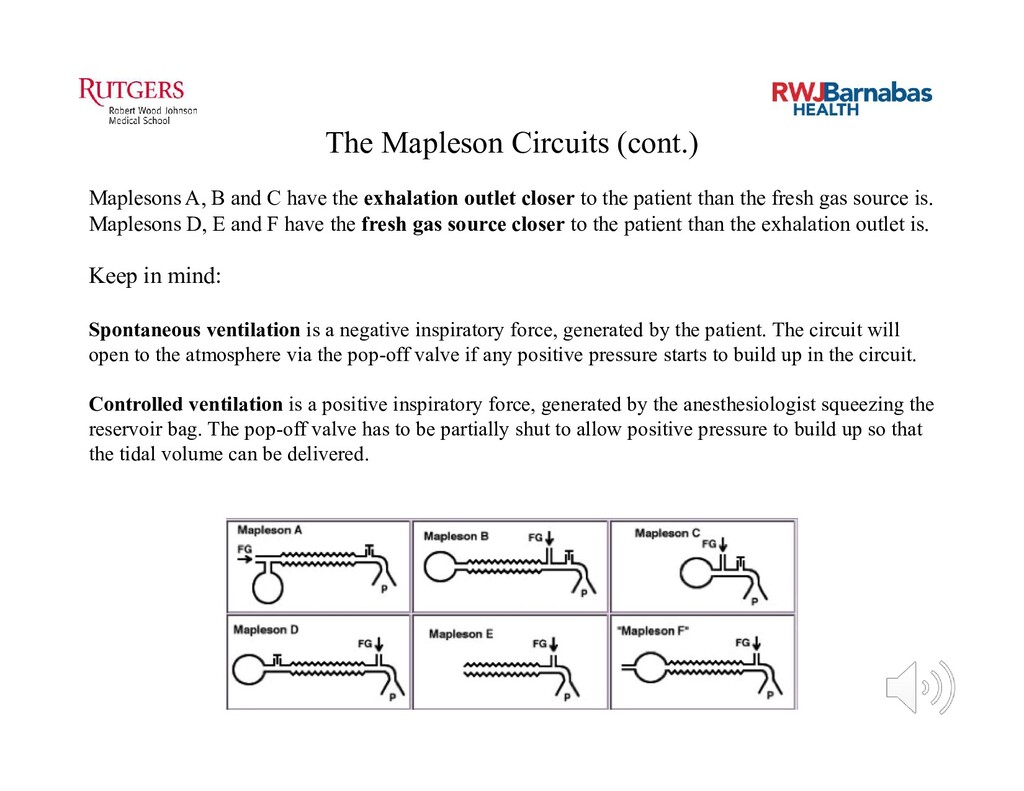
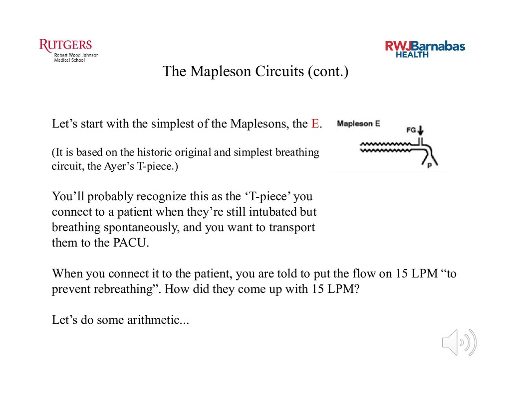
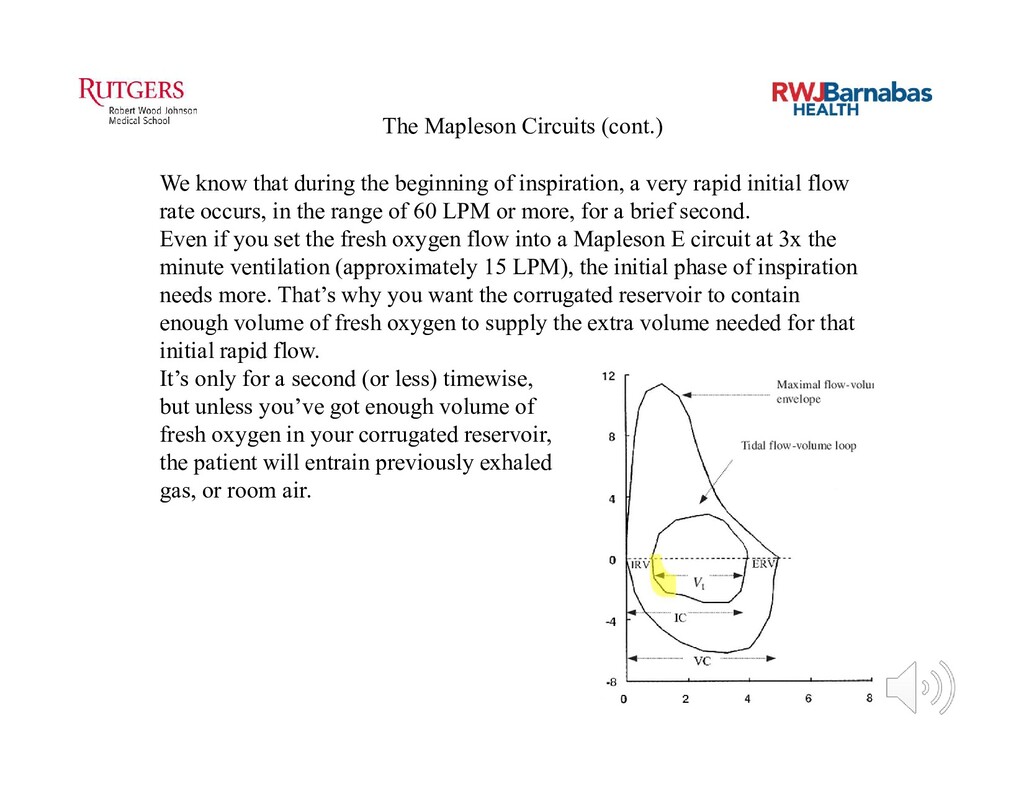
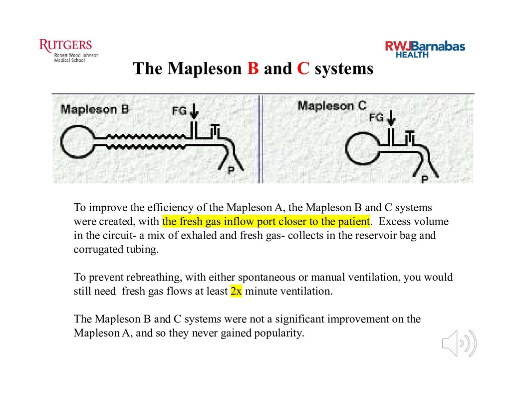
  56. News Articles, Anesthesiology/Pain Medicine, Surgery AAP responds to FDA warning

    on anesthesia use in children by Raeford E. Brown Jr. M.D., FAAP; Rita Agarwal M.D., FAAP The Academy has coordinated a response to a recent Food and Drug Administration (FDA) warning that cautions health care practitioners about the possibility of developmental problems associated with repeated or prolonged use of anesthetics in children younger than 3 years of age. The agency is requiring warning labels on all anesthetic agents and sedatives, including propofol, midazolam and all volatile anesthetic agents. An FDA Drug Safety Communication highlights the abundant animal data from more than a decade concerning suspected toxicities when these agents are used during surgeries or procedures lasting longer than three hours or when administered multiple times to children younger than 3 and pregnant women in their third trimester. Laboratory studies of multiple species, including primates, demonstrate that prolonged use and multiple anesthetics or sedations have been associated with developmental anomalies of cognition and memory and cell death in the developing brain. The findings cited in the warning are not new. They have been discussed by three FDA advisory committees since 2007. However, concerns have arisen recently that not all practitioners using these medications for sedation or surgical anesthesia in children are aware of these findings, reducing their ability to make informed decisions concerning the risks and benefits of procedures requiring sedation or anesthesia. In addition, lack of awareness reduces the clinician' s ability to educate families and get informed consent. The Academy, led by the Section on Anesthesiology and Pain Medicine and the Committee on Drugs, coordinated a response that aimed to place this warning in the perspective of recent controlled trials in humans and multiple epidemiological studies of large homogeneous populations. These studies demonstrate no developmental problems in children exposed to a single, short anesthetic or sedation. The response cautions parents and clinicians of the risks of delaying needed surgery and diagnostic procedures. Until additional information is available from the many ongoing studies in animals and humans, parents and providers should weigh the risks and benefits of each contemplated procedure prior to proceeding. In addition to the Academy, numerous other professional organizations endorsed the response, including the American Society of Anesthesiologists, the International Anesthesia Research Society, Society for Obstetric Anesthesia and Perinatology, Society for Pediatric Anesthesia, Congenital Cardiac Anesthesia Society, Pediatric Anesthesia Leadership Council and the Society for Pediatric Pain Medicine. Dr. Brown is chair-elect and Dr. Agarwal is chair of the AAP Section on Anesthesiology and Pain Medicine Executive Committee. Resources AAP response to the FDA Drug Safety Communication • FDA Drug Safety Communication •
  57. Funding Research To Ensure Pediatric Anesthesia Safety Each year, m

    illions of infants and toddlers require anesthesia and/or sedation for surgery, procedures, and tests. Concern has been raised about the safety of the medicines used for anesthesia and sedation in young children. This concern is based on research in anim als demonstrating long-term, possibly perm anent, injury to the developing brain caused by exposure to these medicines.This injury results in abnormalities in behavior, learning, and m emory in animals. The effect of exposure to anesthetic drugs in young children is unknown; however, som e but not all studies have suggested that problem s similar to those seen in animals could also occur in infants and toddlers. It is im portant to recognize that the studies in children suggest that similar def cits may occur. These studies in children have limitations that prevent experts from understanding whether the harmful effects were due to the anesthetic drugs or to other factors such as the surgery or related illness. Better research is required to understand whether children are harmed and if so, what alternative medicines might be used to minim ize risk from anesthesia. Because there is not enough information about the effects of anesthetic drugs on the brains of young children, it is not yet possible to know whether use of these medicines poses a risk, and if so, whether the risk is large enough to outweigh the benef t of the planned surgery, procedure, or test. Until further research clarif es the importance of these f ndings we recommend: F or healthcare providers Answers to questions from parents and caregivers related to these risks should highlight the differences between research f ndings in anim als and children and the uncertainty of any effect in children. It m ay also be em phasized that because most anesthetic drugs have been shown to cause injury in animal experiments, no specif c medications or technique can be chosen that are safer than any other. Clearly, anesthetic drugs are a necessary part of the care of children needing any surgery, procedure, or test that cannot be delayed. Decisions regarding the tim ing of a procedure requiring anesthesia should be discussed with all mem bers of the care team as well as the family or caregiver before proceeding. The benef ts of an elective procedure should always be weighed against all of the risks associated with anesthesia and surgery. F or parents and caregivers Discuss the tim ing of planned procedures with your child’s prim ary care physician, surgeon/proceduralist and anesthesiologist. Concerns regarding the unknown risk of anesthetic exposure to your child’s brain development must be weighed against the potential harm associated with cancelling or delaying a needed procedure. Each child’s care must be evaluated individually based on age, type and urgency of the procedure and other health factors. Your child’s doctors are best able to provide this advice. If you desire additional inform ation and updates on current research, please go to smarttots.org. Consensus Statement on the Use of Anesthetic and Sedative Drugs in Infants and Toddlers 44 Montgomery Street, S uite 1605 • San F rancisco, CA 94104 • t 415.296.6905 • f 415.296.6901 • SmartT ots.org • S martTots@ iars.org SPS sa f e a n d so u n d SO CI ET Y FO R P ED IATRI C SED ATI O N October 2015
  58. ‘TRUTH’ Hey ASA, bias much? These studies involved healthy kids

    coming for a single short elective procedure. No adverse effects were found on follow-up cognitive and intelligence testing.
  59. New research emerges every month. It’s mostly lab studies on

    animals. Damage is found. The questions remain: How ‘safe’ is anesthesia, especially on the very young, especially those who need longer procedures, repeated procedures, and have comorbidities? Are we knocking off a few neurons every time they come to the OR?