Upgrade to Pro — share decks privately, control downloads, hide ads and more …

Extensor Retinaculum, Anatomical snuff box and muscles of hand

Extensor Retinaculum, Anatomical snuff box and muscles of hand

This presentation describes Extensor Retinaculum, Anatomical snuff box and muscles of hand

Dr.Sherif Fahmy

August 01, 2019
Tweet

More Decks by Dr.Sherif Fahmy

Other Decks in Science

Transcript

  1. Attachments of Extensor Retinaculum Lateral: -Lower part of anterior border

    of radius. Medial: -Triquetral and pisiform bones.
  2. Extensor retinaculum Lateral attachment: -Lower part of anterior border of

    radius. Medial attachment -Triquetral and pisiform bone
  3. Superficial Relations Laterally: -Superficial radial nerve. -Beginning of cephalic vein.

    Medially: -Dorsal cutaneous branch of ulnar nerve. -Beginning of basilic vein.
  4. Deep Relations (Compartments) 1st compartment: -Tendon of abductor pollicis longus.

    -Tendon of extensor pollicis brevis. 2nd compartment: -Tendons of extensor carpi radialis longus and brevis. 3rd compartment: -Tendon of extensor pollicis longus. 4th compartment: -Tendons of extensor digitorum & extensor indicis. -Posterior interosseous nerve. -Anterior interosseous artery. 5th compartment: -Tendon of extensor digiti minimi. (Behind inferior radioulnar joint) 6th compartment: Behind lower end of radius
  5. Abductor pollicis longus Extensor pollicis brevis 1 Extensor carpi radialis

    longus Extensor carpi radialis brevis 2 Extensor pollicis longus 3 -Extensor digitorum and ext. indicis -Posterior interosseous nerve. -Anterior interosseous artery 4 Extensor digiti minimi 5 Extensor carpi ulnaris 6
  6. 1-Abductor pollicis longus & extensor pollicis brevis 2-Tendons of extensor

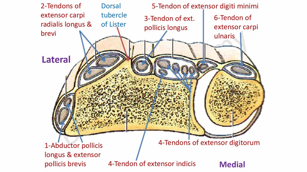
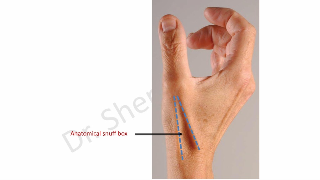
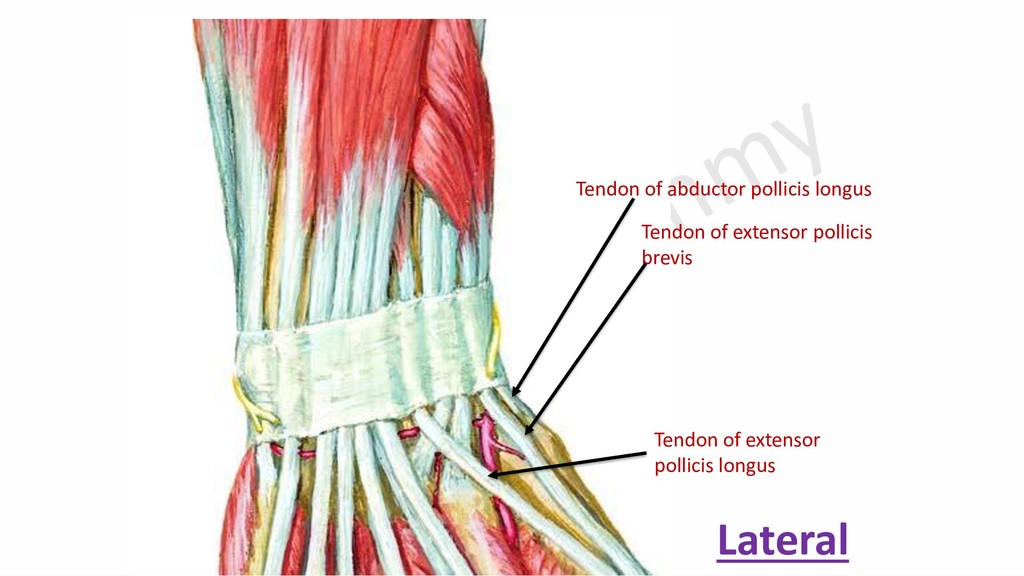
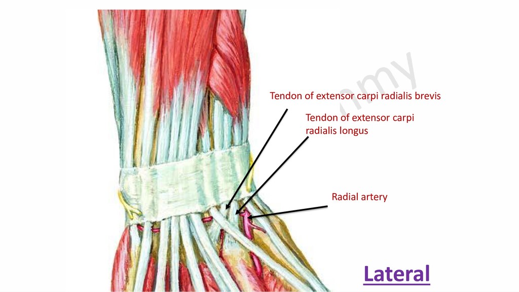
    carpi radialis longus & brevi 3-Tendon of ext. pollicis longus 4-Tendons of extensor digitorum 4-Tendon of extensor indicis 5-Tendon of extensor digiti minimi 6-Tendon of extensor carpi ulnaris Dorsal tubercle of Lister Lateral Medial
  7. Lateral Muscles of Palm 1- Thenar eminence muscles. a- Abductor

    pollicis brevis. b- Flexor pollicis brevis. c- Opponens pollicis. 2- Adductor Pollicis muscle.
  8. Abductor pollicis longus (cut) Adductor pollicis Opponens pollicis Flexor pollicis

    brevis (cut) Abductor digiti minimi Flexor digiti minimi Opponens digiti minimi
  9. Medial Muscles of the Palm (Hypothenar Muscles) 1- Abductor digiti

    minimi. 2- Flexor digiti minimi. 3- Opponens digiti minimi.
  10. Lat. Med. Palmar aponeurosis Medial palmar septum Lateral palmar septum

    Intermediate palmar septum 1st metacarpal bone 3rd metacarpal bone 5th metacarpal bone
  11. Palmar Compartments 1- lateral compartment (Thenar). 2- Medial compartment (Hypothenar).

    3- Intermediate compartment: A- Mid-palmar space. B- Thenar space.
  12. Lateral compartment Medial Compartment Adductor pollicis m. A line that

    divides middle compartment into mid-palmar (Medial) & thenar and adductor (Lateral)
  13. Lat. Med. Palmar aponeurosis Medial intermuscular septum Lateral intermuscular septum

    Intermediate intermuscular septum Adductor space containing adductor pollicis muscle Thenar space Mid-palmar space
  14. Lat. Med. Superficial palmar arch & Branches of median and

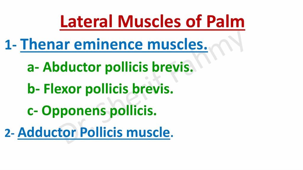
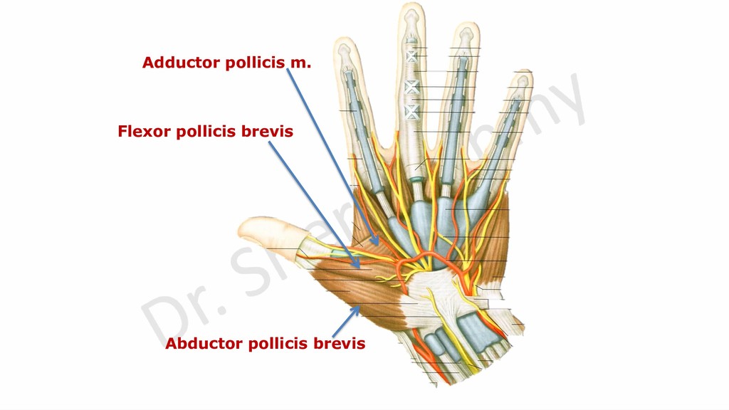
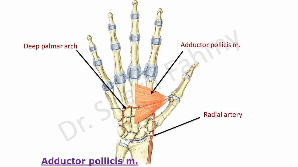
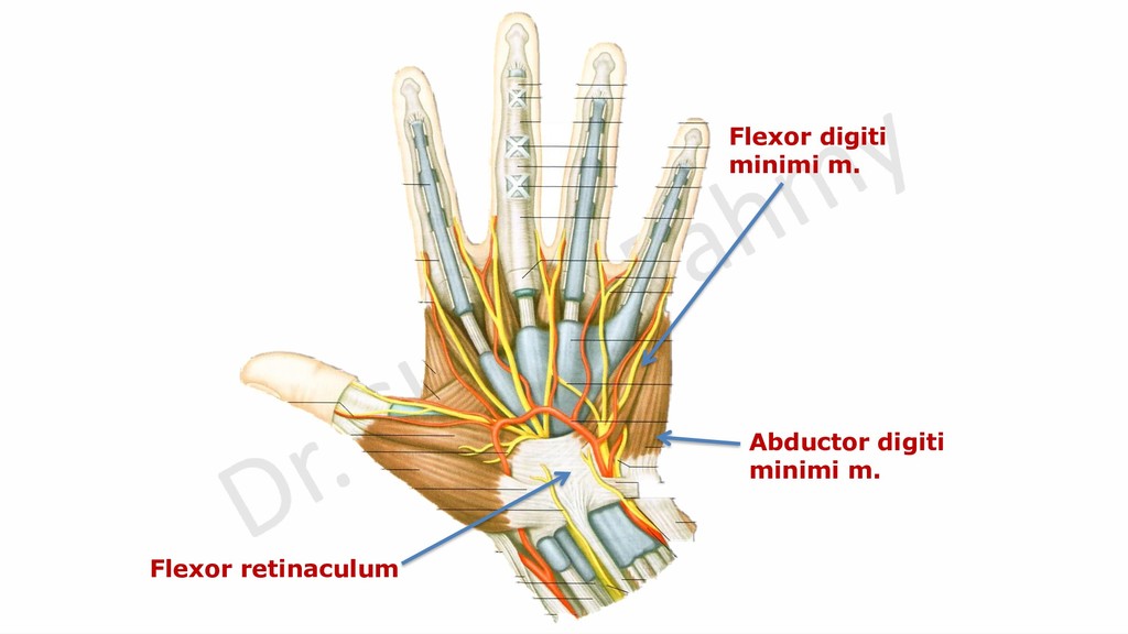
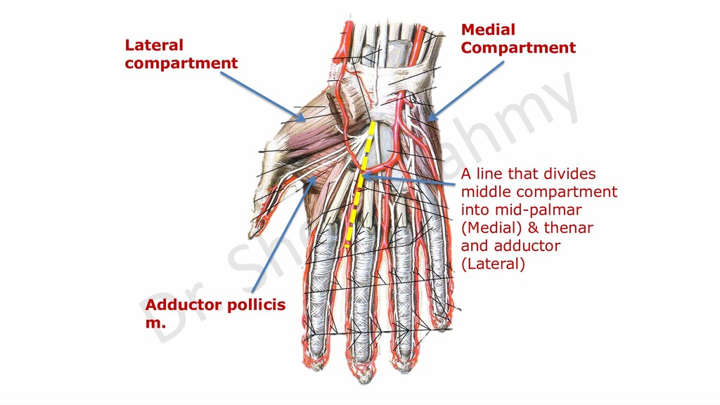
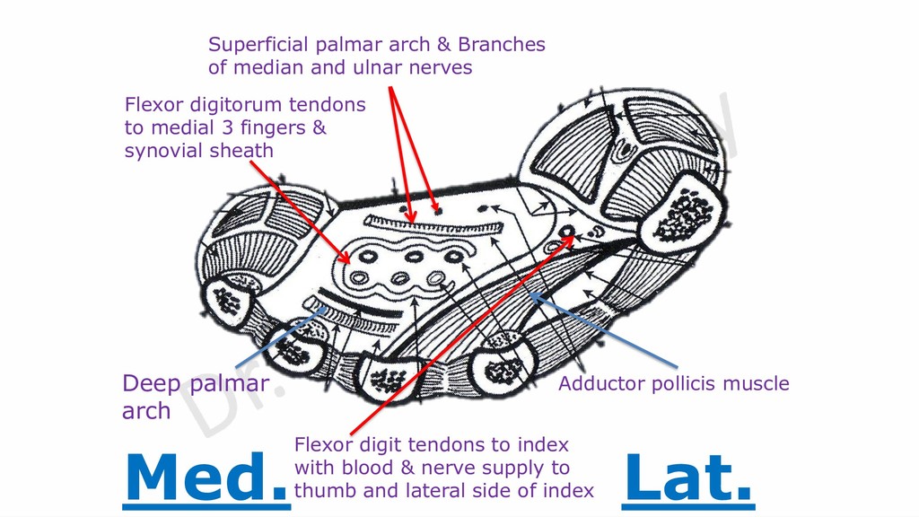
    ulnar nerves Flexor digitorum tendons to medial 3 fingers & synovial sheath Adductor pollicis muscle Flexor digit tendons to index with blood & nerve supply to thumb and lateral side of index Deep palmar arch