Upgrade to Pro — share decks privately, control downloads, hide ads and more …

9/30/20 - Dr. Stein - Perioperative Considerations Obesity

us414
September 29, 2020

9/30/20 - Dr. Stein - Perioperative Considerations Obesity

9/30/20 - Dr. Stein - Perioperative Considerations Obesity

us414

September 29, 2020
Tweet

More Decks by us414

Other Decks in Education

Transcript

  1. Perioperative Considerations for the Obese Patient Mark H. Stein, M.D.

    Professor, Department of Anesthesiology and Perioperative Medicine Rutgers, the State University of New Jersey Robert Wood Johnson Medical School and Robert Wood Johnson University Hospital New Brunswick, New Jersey
  2. As a CME provider accredited by the ACCME, Rutgers, The

    State University of New Jersey must insure balance, independence, objectivity, and scientific rigor in all its sponsored educational activities. As such, Rutgers requires all individuals in a position to control the content of an educational activity to provide a signed disclosure form to the Center for Continuing and Outreach Education (CCOE) prior to the initiation of the activity. In order to ensure its CME activities promote improvements or quality in healthcare and not a specific proprietary business interest of a commercial interest, CCOE will identify and resolve all conflicts of interest (e.g., peer review) prior to the delivery of the activity to the learner. Based on this disclosure information, CCOE may disqualify any individual from planning and implementation if a conflict of interest that may contribute to commercial bias is determined to exist and cannot be resolved. Individuals are required to disclose all relevant financial relationships with commercial interests (entities producing, marketing, re-selling, or distributing health care goods or services consumed by, or used on, patients) in any amount as well as the nature of the relationship within the past 12 months. In addition, an individual developing a presentation that provides information, in whole or in part, related to non-FDA approved uses for drug products or devices, must indicate his/her intention to CCOE by way of this form. The individual must also clearly identify the unlabeled indications or the investigational nature of the proposed uses to the learner. In accordance with the Essential Elements and Standards of Commercial Support set forth by ACCME, the undersigned understands and accepts the policies and standards as set forth in this document. All disclosure declarations must be communicated to the learner by means of a notation in the program or syllabus, or verbally by the activity director or moderator prior to the beginning of the activity. Individuals who do not provide the requested disclosure information will be disqualified from participating in the development and delivery of a CME activity. 1. Do you or any member of your immediate family have any relevant financial relationships w ith commercial interests (entities producing, marketing, re-selling, or distributing health care goods or services consumed by, or used on, patients) in any amount within the past 12 months? Yes No a) If Yes, please list (attach separate page if necessary) the commercial entities w ith the type of relationship listed below. Grant/Research Support Consultant Speakers Bureau Patent Holder Member, Scientific Advisory Board Member, Board of Directors Stock Shareholder (directly purchased) Other Financial Support (specify) Other Relationship/Affiliation (specify) b) If Yes, will your presentation include discussion of specific products/services of the commercial entities you’ve listed above? Yes No   Faculty Disclosure Declaration Form      
  3. Classification of Obesity Obesity is classified by Body Mass Index.

    BMI = relative weight for a given height = weight in kg/height in meters2  that formula was first proposed by Adolphe Quetelet in 1835 BMI has its limitations  deceptively high BMI- edema, high muscularity  deceptively low BMI- muscle wasting, dehydration  relationship between BMI and body fat also varies with age and gender  BMI doesn’t take your fat distribution into account, which has implications for associated morbidity (gynecoid vs. android distribution) Other methods of measuring total body fat aren’t very practical or necessarily more accurate.  accuracy varies: skin fold thickness, bio-electrical impedance, near infrared interactance  more accurate, but not very practical: hydrodensitometry (underwater weighing), dual- energy x-ray absorptiometry (DEXA), air displacement, total body electrical conductivity Hill JO, Howard BV, Klesges RC, et al. Clinical guidelines on the identification, evaluation, and treatment of overweight and obese adults. National Heart, Lung, and Blood Institute 1998; 98-4083: 1-228. http://www.nhlbi.nih.gov/guidelines/obesity/ob_gdlns.pdf Esco MR, Olson MS, Williford HN, et al. The accuracy of hand-to-hand bioelectrical impedance analysis in predicting body composition in college-age female athletes. Journal of Strength and Conditioning Research 2011; 25: 1040-1-45.
  4. Keys A, Fidanza F, Karvonen MJ, Kimuru N, Taylor HL.

    Indices of relative weight and obesity. J Chron Dis 1972. Vol. 25. pp. 329–343. http://www.nytimes.com/2004/11/23/obituaries/dr-ancel-keys-100-promoter-of-mediterranean-diet-dies.html?_r=0 “...it is assumed that a major reason for the use of a relative weight index is to remove the dependency of weight on height. Second, it is assumed that in the selection of an index attention should be given to the degree to which the index may indicate relative obesity or body fatness... it is proposed that this ratio, W/H2, be termed the body mass index.” “... the use of ideal or recommended weight confounds age and weight because on the average weight increases with age until the fifties while increase in height is over by the early twenties at the latest. The general trend to continue growth in weight may be undesirable but it has no relevance to the question of providing an objective description of relative body mass; it is scientifically indefensible to include a value judgment in that description. The characterization of persons in terms of desirable weight percentage has resulted in attributing to ‘overweight’ some tendencies to ill health and death that are actually only related to age.” • 12 cohorts of men from 5 different countries, 7426 total • Data gathered during 1947-67 • Body density measured by underwater weighing • Body “fatness” determined by skinfold measurement
  5. Who’s healthier? Young slim Elton or older heavier Elton? Young

    slim Billy or older heavier Billy? We tend to get heavier as we get older, even though height remains the same. While too much weight gain is undesirable, are older people less healthy because of some inevitable weight gain, or because of increased age? Should desirable BMI take age and gender into account?
  6. BMJ 2020;370:m3324 OBJECTIVE To quantify the association of indices of

    central obesity, including waist circumference, hip circumference, thigh circumference, waist-to-hip ratio, waist-to-height ratio, waist-to-thigh ratio, body adiposity index, and A body shape index, with the risk of all cause mortality in the general population, and to clarify the shape of the dose-response relations. WHAT IS ALREADY KNOWN ON THIS TOPIC Existing evidence suggests that central fatness might be more strongly associated with the risk of mortality than overall obesity, however the results have not been quantitatively gathered. Several large scale prospective cohort studies have suggested a positive linear or J shaped association between indices of central fatness with all cause mortality risk. Systematic reviews and meta-analyses assessing the dose-response association between indices of central fatness and all cause mortality risk are lacking. WHAT THIS STUDY ADDS Central fatness, reflected by large waist circumference, waist-to-hip ratio, and waist-to-height ratio, independent of overall adiposity, was associated with a higher risk of all cause mortality. The results suggest a nearly J shaped association between waist circumference and risk of all cause mortality. Larger hip circumference and thigh circumference were associated with a lower risk of all cause mortality. Considering the limitations of the current measures of general and abdominal adiposity, including body mass index (inability to distinguish lean mass from fat mass), waist circumference (strong correlation with body mass index), and waist-to-hip ratio (difficulty in measuring hip circumference), a new anthropometric measure entitled A body shape index has been developed. This measure is calculated by dividing waist circumference by (body mass index2/3 x height1/2). The A body shape index has a slight correlation with body mass index, height, and weight, and so could be independent of other anthropometric variables in predicting mortality. In the present review, we found a positive monotonic association between A body shape index and the risk of all cause mortality; the risk of premature death increased proportionally with increasing A body shape index values. The results are similar to those of the National Health and Nutrition Examination Survey, which indicated a positive linear association between A body shape index and risk of premature death.
  7. Scientific Reports (2020) 10:14541 https://doi.org/10.1038/s41598-020-71302-5 Abdominal and general adiposity are

    independently associated with mortality, but there is no consensus on how best to assess abdominal adiposity. We compared the ability of alternative waist indices to complement body mass index (BMI) when assessing all-cause mortality. We used data from 352,985 participants in the European Prospective Investigation into Cancer and Nutrition (EPIC) and Cox proportional hazards models adjusted for other risk factors. During a mean follow-up of 16.1 years, 38,178 participants died. Combining in one model BMI and a strongly correlated waist index altered the association patterns with mortality, to a predominantly negative association for BMI and a stronger positive association for the waist index, while combining BMI with the uncorrelated A Body Shape Index (ABSI) preserved the association patterns. Sex-specific cohort-wide quartiles of waist indices correlated with BMI could not separate high-risk from low-risk individuals within underweight (BMI < 18.5 kg/m2) or obese (BMI ≥ 30 kg/m2) categories, while the highest quartile of ABSI separated 18–39% of the individuals within each BMI category, which had 22–55% higher risk of death. In conclusion, only a waist index independent of BMI by design, such as ABSI, complements BMI and enables efficient risk stratification, which could facilitate personalization of screening, treatment and monitoring.
  8. The A Body Shape Index (ABSI) Waist Circumference ABSI =

    _____________________________________________________________ BMI2/3 x Height1/2 Units are in meters and kilograms. Height1 /2 is the square root of the height. BMI2/3 is the square of the BMI, then do the cube root of that. When you have a fractional exponent, the numerator is the power and the denominator is the root. https://journals.plos.org/plosone/article?id=10.1371/journal.pone.0039504 https://www.ncbi.nlm.nih.gov/pmc/articles/PMC5612697/
  9. Defining Childhood Obesity- BMI for Children and Teens For children

    and teens, BMI is age- and gender-specific and is often referred to as BMI-for-age. A child's weight status is determined using an age- and gender-specific percentile for BMI rather than the BMI categories used for adults. This is because children's body composition varies as they age and varies between boys and girls. Therefore, BMI levels among children and teens need to be expressed relative to other children of the same age and gender. http://www.cdc.gov/healthyweight/assessing/bmi/childrens_bmi/about_childrens_bmi.html
  10. Defining Childhood Obesity- BMI for Children and Teens (cont.) http://www.who.int/childgrowth/standards/en/

    http://apps.who.int/iris/bitstream/10665/44129/1/9789241598163_eng.pdf?ua=1 For children and teens, BMI is not a diagnostic tool and is used to screen for potential weight and health-related issues. For example, a child may have a high BMI for their age and gender, but to determine if excess fat is truly the reason, a health care provider would need to perform further assessments. These assessments might include skinfold thickness measurements, evaluations of diet, physical activity, family history, and other appropriate health screenings. The American Academy of Pediatrics recommends the use of BMI to screen for overweight and obesity in children beginning at 2 years old. For children under the age of 2 years old, to screen if the kids are alright, consult the WHO standards.
  11. Defining Childhood Obesity- BMI for Children and Teens (cont.) Remember-

    it’s a screening tool. There can be exceptions.
  12. Since 1912, the Metropolitan Life Insurance Company has published tables

    of ‘desirable, or ideal’ body weight for a given height.  “Desirable” weight for a given height is based upon which patients made the fewest life insurance claims. Ideal Body Weight is the weight associated with maximum life expectancy for a given height. Robinett-Weiss N, Hixson ML, Sieberg J. The Metropolitan Height-Weight Tables: perspectives for use. J Am Diet Assoc. 1984 Dec; 84(12): 1480-1.
  13. Classification of Overweight and Obesity by BMI Category Obesity Class

    BMI (kg/m2) Underweight <18.5 Normal 18.5-24.9 Overweight 25.0-29.9 Obesity 1 30.0-34.9 Obesity 2 35.0-39.9 Obesity 3 40.0-49.9 Super-obesity 4 50.0-59.9 Super-superobesity 5 60.0- Mechanick, JI, Kushner, RF, Sugerman, HJ, et al. American Association of Clinical Endocrinologists, The Obesity Society, and American Society for Metabolic and Bariatric Surgery Medical Guidelines for Clinical Practice for the Perioperative Nutritional, Metabolic, and Nonsurgical Support of the Bariatric Surgery Patient. Surgery for Obesity and Related Diseases 2008; 4: S109-S184. Newer descriptive terms have replaced Morbid Obesity. Severe OSA occurs in 10– 20% of patients with BMI > 35 kg/m2
  14. Measure the height, weight and neck circumference  Accuracy is

    important for drug dosing and BMI calculations don’t just ask the patient “what is your height and weight?”  The scale at RWJUH  goes up to 1000 lbs.  minimal step up  large standing platform  railing to hold on to  does height measurement  two such scales, located in Same Day Surgery preoperative area and in the Pre- admission Testing Suite  Measure the neck circumference (just above the cricoid) with a tape measure
  15. Mechanick, JI, Kushner, RF, Sugerman, HJ, et al. American Association

    of Clinical Endocrinologists, The Obesity Society, and American Society for Metabolic and Bariatric Surgery Medical Guidelines for Clinical Practice for the Perioperative Nutritional, Metabolic, and Nonsurgical Support of the Bariatric Surgery Patient. Surgery for Obesity and Related Diseases 2008; 4: S109-S184. Obesity-related Review of Organ Systems
  16.  Obstructive Sleep Apnea (OSA) syndrome of periodic episodes of

    partial or complete obstruction of the upper airway during sleep  episodes last more than 10 sec, could be as long as 60 sec.  O2 desaturation (at least 4% drop) during the episode loud snoring during sleep repetitive arousal from sleep to restore airway patency daytime hypersomnolence or other manifestations of disturbed sleep, such as poor concentration and fatigue associated with neck circumference: men ≥ 43 cm, women ≥ 41 cm associated with refractory HTN, CAD, CHF, CVA, GERD, ED higher incidence of adverse postoperative events and outcomes Obstructive Sleep Apnea and Obesity Hypoventilation Syndrome Sleep Disordered Breathing is a spectrum of sleep disorders produced by abnormal breathing patterns- they can be of obstructive, central, or mixed origin. • Each of these disorders has a spectrum of severity, described according to the number and severity of oxygen desaturations occurring every hour.
  17.  Daytime Symptoms daytime sleepiness, memory and concentration dysfunction, sexual

    dysfunction, GERD, behavioral irritability (depression, chronic fatigue, delirium, irritability) road traffic accident Signs and Symptoms of Obstructive Sleep Apnea  Nighttime Symptoms heavy persistent snoring (worse in supine position or after alcohol or sedatives) apnea witnessed by bed partner, sudden awakening with noisy breathing, accidents related to sleepiness, nocturnal sweating, wake up with dry mouth, nocturnal epilepsy, nocturia  Signs on examination a ‘crowded oropharynx’ edematous soft palate or uvula long soft palate and uvula decreased oropharyngeal dimensions nasal obstruction, maxillary hypoplasia, retrognathia central adiposity, increased neck circumference HTN, other cardiovascular consequences Eckert DJ and Malhotra A. Pathophysiology of adult obstructive sleep apnea. The Proceedings of the American Thoracic Society 2008; 5: 144-53. September 1 2, 201 9 | 1 2:05am NEWS 1 1 06 6 6 0 1 2 6 10 1 The NJ Transit engineer who caused the deadly Hoboken train crash of 201 6 will soon be back at the helm — on a “one-time , last chance basis” — after winning an appeal in court, a report says. Thomas Gallagher, who had been suspended and fired in 201 8 following a federal investigation, will be allowed to return to work in the train yards, but not passenger service , according to NJ.com. He was steering a Pascack Valley Line train into the Hoboken Terminal — at twice the posted speed limit — when it slammed into a bumper wall and killed one woman waiting on the platform, while injuring 1 08 others. The September 201 6 crash was investigated by the National Transportation Safety Board, which determined that Gallagher was suffering from obstructive sleep apnea and had passed out. A similar incident in January 201 7 — involving a Long Island Rail Road train that crashed By Chris Perez Getty Images
  18. Josh James’ Sleep Apnea Diagnosis and Treatment Ultimately Improves His

    Game Reggie White Dies Sunday Former Howard H.S., Tenn Vol & NFL Star Was 43 Years-Old Sunday, December 26, 2004 - by Tim Evearitt Former NFL star Reggie White died Sunday morning at his home near Huntersville, N.C.. White turned 43 on Dec. 19. A public viewing service for White will be held from 3 p.m. to 8 p.m. Wednesday at A.L. Jinwright Funeral Service in Charlotte. Sara White confirmed her husband's death saying that she believes White died of respiratory failure related to his sleep apnea. White was diagnosed with obstructive sleep apnea (OSA) as well as an inflammatory condition called sarcoidosis that affected his heart and lungs. He lived with these conditions for many years. The Knoxville News Sentinel reported that White died of a massive heart attack. Reggie White, nicknamed the "Minister of Defense" (a dual reference to his football prowess and to his Evangelical Christian ordination) was one of the American football's most prolific sackers in college, the USFL and the NFL. Reggie White was drafted by the Philadelphia Eagles. He played with the Eagles for eight seasons, picking up 124 sacks and becoming the Eagles' all-time sack leader. He also set a then-record season-best with 21 sacks in 1987. In 1993, White went to the Green Bay Packers, where he played for six more seasons. While not as prolific as his previous years, White still notched another 68.5 sacks, becoming the Packers' all-time leader in that category. He also helped the Packers to two Super Bowls, including a victory in Super Bowl XXXI. Following the 1998 season, Reggie White announced his retirement, but in 1999, he got the urge to play again and signed with the Carolina Panthers Following the 1999 season, White again retired. At the time of his retirement, White was the NFL's all-time sacks leader, with 198.
  19. Cardiovascular Consequences of Obstructive Sleep Apnea  Repetitive apneas expose

    the cardiovascular system to cycles of hypoxemia, exaggerated negative intrathoracic pressure, and arousals.  These noxious stimuli depress myocardial contractility, activate the sympathetic nervous system, raise blood pressure, heart rate, and myocardial wall stress, depress parasympathetic activity, provoke oxidative stress and systemic inflammation, activate platelets, and impair vascular endothelial function.  OSA increases the risk of developing cardiovascular diseases.  Studies have shown significant independent associations between OSA and HTN, CAD, arrhythmias, heart failure, and stroke.  Treating OSA with Positive Airway Pressure (PAP) may lower BP, attenuate signs of early atherosclerosis, diminish the risk of developing cardiovascular diseases, and improve cardiac function in patients with heart failure. Bradley TD, Floras JS. Obstructive sleep apnoea and its cardiovascular consequences. Lancet 2009; 373: 82-93. Yaggi HK, Concato J, Kernan WN, Lichtman JH, Brass LM, MohseninV. Obstructive sleep apnea as a risk factor for stroke and death.N Engl J Med 2005;353:2034–2041. Redline, Yenokyan, Gottlieb, et al.: Sleep Apnea and Incident Stroke. Am J Respir Crit Care Med Vol 182. pp 269–277, 2010. Campos-Rodriguez, Martinez-Garcia, Reyes-Nun˜ ez, et al.: Incident Stroke or Coronary Disease in Women with OSA. Am J Respir Crit Care Med Vol 189, 1544–1550, 2014.
  20. Interrelationship of Cardiovascular and Pulmonary Sequelae of Obesity Ogunnaike BO,

    Whitten CW. Anesthesia and obesity. In Barash PG, Cullen BF, Stoelting RK, eds. Clinical Anesthesia 5th ed.Philadelphia: Lippincott, Williams, and Wilkins 2006: 1040.
  21. Schematic representation of the typical pathophysiological sequence that occurs in

    obstructive sleep apnea (OSA) (shown in gray) and the associated physiological processes that occur throughout the cycle that are either protective/restorative (outside the circle) or perpetuating (inside the circle). UA = upper airway. Eckert DJ and Malhotra A. Pathophysiology of adult obstructive sleep apnea. The Proceedings of the American Thoracic Society 2008; 5: 144-53.
  22. CO2 works primarily through central receptors- brainstem- important for acute

    apneic episodes O2 works through peripheral receptors- carotid bodies and aortic bodies- important when the CO2 receptors become acclimated
  23. Loop Gain in Obstructive Sleep Apnea  The concept of

    loop gain is used to quantify the internal amplification of a system. It is the propensity of a system governed by feedback loops to develop unstable behavior. Respiration is such a system.  The gain can be influenced by control of the variables relating to hypercapnic and hypoxic ventilatory responses (‘controller’ gain, i.e. central and peripheral chemoreceptors ), or the ability to eliminate CO2 and the size of oxygen stores (‘plant’ gain; i.e. the respiratory tree and the circulation to and from the lungs).  Circulation time has an effect on the interaction between ventilation and controller gain. Upper airway factors (e.g. resistance, obstruction) have effects on the interaction between controller gain and ventilation.  The higher the loop gain, the potentially more unstable the respiratory control system becomes. A high loop gain promotes recurrent apneas because of an overcompensating response to a disturbance. A lowering of loop gain dampens those subsequent oscillations in breathing, in an effort to restore a normal pattern.  Loop gain principles apply to both central and obstructive sleep apneas. J Physiol. 2012 Apr 15; 590(Pt 8): 1781–1782 J Physiol 592.14 (2014) pp 2899–2901
  24. Simplified diagram of the control of breathing during sleep, showing

    the relationship between ventilation and the two gains in the feedback system. Loop Gain = response/disturbance It’s like a factory. Upstairs is the boss’s office, i.e. the controller. The boss’s office orders the factory downstairs (i.e. the plant) what to do. The boss finds out what the factory’s response is, then gives further orders.
  25. Loop Gain in Obstructive Sleep Apnea (cont.)  In a

    closed loop system, such as the breathing system, a disturbance in the controlled loop component (the respiratory apparatus, likened to a ‘factory’ i.e. ‘plant’) elicits changes in feedback (blood gas tensions) received by the ‘controller’ (respiratory centers in the brain), which in turn effects a compensatory response in the ‘plant’.  The initial response may partially correct the blood gas changes, with the residual changes being gradually corrected later. This is a stable response.  However, the initial response may result in over-correction of the blood gases (overshoot). The ventilatory apparatus is then inhibited, and a second hypopnea results. If that second hypopnea overshoots, the cycle can perpetuate. This happens with certain types of Central Sleep Apnea (CSA). The system eventually stabilizes, but repetitions will occur.  When an Obstructive Sleep Apnea (OSA) episode occurs, both the ‘plant’ and ‘controller’ struggle to restore stability to blood gas tensions, and the gain is increased in an effort to reopen the airway.  When multiple sites in the loop are effected, such as in patients with CHF and COPD in addition to OSA, there can be mixed picture of CSA/OSA.
  26. Schematic representation of the possible sites where each of the

    various pathophysiological traits would either predispose or tend to worsen OSA (inside the main circle). Some of the potential factors that may alleviate OSA at various points throughout the typical physiological cycle (dashed arrows and ovals) are located outside the main circle. Note that some factors are theoretical and largely untested whereas others are more proven therapies (i.e., continuous positive airway pressure [CPAP]). Eckert DJ and Malhotra A. Pathophysiology of adult obstructive sleep apnea. The Proceedings of the American Thoracic Society 2008; 5: 144-53. https://erj.ersjournals.com/content/erj/20/5/1130.full.pdf Surgery (e.g. UPPP), Inspire® device Medroxyprogesterone (respiratory stimulant), or acetazolamide (makes the blood more acidic) can be used to stimulate breathing.
  27.  affects as much as 24% of men, and about

    9% of women, in the general population. Most cases go undiagnosed.  found in 50% of obese men and 40% of obese women  found in 70% of patients undergoing bariatric surgery  average life span for untreated severe OSA is 58 years More about OSA… Ahmad S, Nagle A, McCarthy RJ, et al. Postoperative hypoxemia in morbidly obese patients with and without obstructive sleep apnea undergoing laparoscopic bariatric surgery. Anesthesia & Analgesia 2008; 107: 138-43.
  28. Polysomnography- the Sleep Study Polysomnography is the standard for diagnosing

    OSA.  Patient may need to spend the night in the sleep lab  Home testing options are available, but some patients will need the full sleep-lab study.  Includes having simultaneous EEG, electrooculogram, submental electromyogram, ECG, respiratory effort (thoracoabdominal excursion), respiratory inductive plethysmography, oronasal airflow (by nasal airflow pressure), snoring microphone, leg muscle activity, O2 saturation by pulse oximeter.  Apnea-Hypopnea Index (AHI) is the number of apnea or hypopnea episodes per hour during sleep.  Episodes are defined by degree of airflow reduction (hypopnea is at least a 30% reduction, lasts at least 10 sec), with O2 desaturation (at least 4%), with associated EEG arousal  OSA diagnosis based upon AHI index combined with findings of fragmented sleep and daytime sleepiness 5-15 mild OSA >15-30 moderate >30 severe
  29. Polysomnography and Obstructive Sleep Apnea Complete airway collapse causing cessation

    of airflow at nose and mouth for at least 10 seconds, with drop in oxygen saturation, and arousal from sleep cessation of airflow drop in oxygen saturation arousal from sleep
  30. Reasons most patients go to a sleep specialist:  Snoring,

    gasping/choking, stop breathing at night  Daytime sleepiness or tiredness  Drowsy driving  Morning headaches, dry mouth, sore throat  Decreased sex drive, waking up often to urinate  Mood, memory, attention problems, drowsy driving  Twitching and jerking in limbs at night  Difficulty falling or staying sleep
  31. Home Testing Options for Obstructive Sleep Apnea Diagnosis • For

    patients with no other serious comorbidities that could affect sleep or breathing • No other potential sleep disorders that could interfere with results
  32. Different OSA screening questionnaires have been developed:  ASA checklist

    3 categories of questions, physical exam findings, and measurements; need at least 2 categories to score 2 or more positive answers  Berlin questionnaire 10 questions about symptoms, then ask about history of HTN, age, gender, height, weight, and neck circumference  Other questionnaires include the Wisconsin, the Sleep Disorder Questionnaire (SDQ), the Hawaii, the Sleep Apnea Clinical Score (SACS)  STOP combined with BANG data (http://stopbang.ca/osa/screening.php)
  33. STOP-BANG Screening for Obstructive Sleep Apnea  A condensed and

    modified version of the lengthier Berlin and ASA questionnaires and checklists S Do you snore loudly? yes  no  T Do you often feel tired, fatigued, or sleepy during the daytime? yes  no  O Has anyone observed you stop breathing during your sleep? yes  no  P Do you have, or are you being treated for, high blood pressure? yes  no  B Body Mass Index (weight/height2) in [kg/meters2] More than 35? yes  no  A Age Over 50? yes  no  N Neck circumference Men ≥ 43 cm, women ≥ 41 cm? yes  no  G Gender Is patient male? yes  no  If 5 or more ‘yes’ answers on STOP-BANG, patient is at high risk for Obstructive Sleep Apnea. http://stopbang.ca/osa/screening.php Chung F, Yegneswaran B, Liao P, et al. STOP Questionnaire. Anesthesiology 2008; 108: 812-21. Chung F, Elsaid H. Screening for obstructive sleep apnea before surgery: why is it important? Current Opinion in Anesthesiology 2009; 22: 405-11. Chung F, Yegneswaran B, Liao P, et al. Validation of the Berlin Questionnaire and American Society of Anesthesiologists Checklist as screening tools for obstructive sleep apnea in surgical patients. Anesthesiology 2008; 108: 822-30.
  34. Validation of the STOP Questionnaire patients who completed the STOP

    questions and underwent polysomnography Validation of the BANG characteristics patients who had the BANG measurements and underwent polysomnography Positive predictive value of STOP with BANG
  35. Drug-Induced Sleep Endoscopy (DISE)  DISE is an upper airway

    evaluation technique in which fiberoptic examination is performed under conditions of unconscious sedation.  Due to the difficulty in establishing the site of obstruction in the conscious patient who carries a diagnosis of OSA, the diagnosis and treatment of OSA is a complex and multidimensional issue.  DISE allows evaluation of the collapsibility of the upper airway. This 3-dimensional examination of the airway may help to guide treatment selection.  To what extent can DISE can be compared to natural sleep? Correlation has been shown in terms of the tendency of the airway to collapse in sedation and during natural sleep. According to expert opinion, it is possible to imitate nocturnal collapsibility and perform a realistic investigation of the site of obstruction and vibration using DISE.
  36. Drug-Induced Sleep Endoscopy (DISE) cont.  Medications administered to the

    patient usually include the following:  Glycopyrrolate 0.2 mg IV x 1 - This must be given in the preoperative suite at least 15 minutes prior to DISE; it will decrease the salivary secretions, allowing optimal viewing during DISE  Oxymetazoline nasal spray - Two sprays into both nostrils 15 minutes prior to the procedure; it is best to avoid any topical lidocaine, as this could potentially remove and blunt any natural airway reflexes  Propofol infusion - Start 100 μ g/kg/min and titrate to patient’s snoring and OSA.  Try to avoid propofol boluses. Too much propofol will lead to a central apneic episode, making it difficult to distinguish between OSA and central apnea in the patient. This concern must be communicated with the anesthesiologist.  The anesthesiologist must carefully titrate the propofol infusion in order to cause obstructive apnea, but no central apnea.  When the patient can no longer be aroused by voice, introduce the flexible fiberoptic laryngoscope into the nasal cavity. Fully examine the nasal cavity for any airway obstructions and then advance into the nasopharynx. Wait in the nasopharynx until the patient begins snoring.
  37. Drug-Induced Sleep Endoscopy (DISE) cont.  Objective grading schemes provide

    a reliable and reproducible method of evaluating the upper airway during sleep endoscopy.  VOTE is a proposed system of classification that may be used in order to help establish a universal way for otolaryngologists to classify and communicate objective findings of airway obstruction in OSA patients.  Four common locations of obstruction are recognized. These locations can either contribute to OSA individually or in combination. The levels are velum, oropharynx, tongue base, and epiglottis (VOTE). A grade of 0-2 is assigned based on degree of obstruction: 0 = no obstruction; 1 = partial obstruction; 2 = total obstruction. It is also helpful to describe the type of collapse: lateral, anterior-posterior, or concentric.  In pediatric patients, the evaluated locations are adenoids, velum, lateral pharyngeal wall, tongue base, and supraglottis. Obstructions are scored on a specific 4-point grading system.
  38. Vanderveken, OM, Maurer JT, Hohenhorst W, et al. Evaluation of

    drug- induced sleep endoscopy as a patient selection tool for implanted upper airway stimulation for obstructive sleep apnea. J Clin Sleep Med. 2013;9(5):433-438. During an OSA episode, the upper airway can collapse at 4 different levels (‘VOTE’). • Velo-palate (most frequent) • Oropharynx • Tongue base (second most frequent) • Epiglottis These 4 areas are evaluated during Drug-Induced Sleep Endoscopy (DISE). DISE findings using propofol
  39. Society of Anesthesia and Sleep Medicine Guideline on Intraoperative Management

    of Adult Patients With Obstructive Sleep Apnea Stavros G. Memtsoudis, MD, PhD,*† Crispiana Cozowicz, MD,*† Mahesh Nagappa, MD,‡ Jean Wong, MD, FRCPC,§ Girish P. Joshi, MBBS, MD, FFARCSI,║ David T. Wong, MD, FRCPC,§ Anthony G. Doufas, MD, PhD,¶ Meltem Yilmaz, MD,# Mark H. Stein, MD,** Megan L. Krajewski, MD,†† Mandeep Singh, MBBS, MD, MSc, FRCPC,‡‡§§¶¶## Lukas Pichler, MD,*† Satya Krishna Ramachandran, MD,*** and Frances Chung, MBBS, FRCPC§ Anesthesia & Analgesia: October 2018 - Volume 127 - Issue 4 - p 967-987 2.3 Propofol 2.3.1 Question: Are patients with OSA at increased risk for adverse events from the use of propofol for procedural sedation? 2.3.1 Recommendation: Patients with OSA may be at increased risk for adverse respiratory events from the use of propofol for procedural sedation. Level of evidence: Moderate; Grade of recommendation: Strong Rationale. The literature discussed for the purpose of the recommendation reflects evidence of importance for patients receiving propofol for sedation in a procedural setting, that is, drug-induced sleep endoscopy (DISE), gastroenterological endoscopy, or dentistry. The use of propofol to induce general anesthesia purposefully suppresses respiratory activity and was thus deferred in this section. Propofol is the most commonly used agent for DISE.119,120 A summary of findings from 5 studies120–124 is shown in Supplemental Digital Content, Table A4, http://links.lww.com/AA/C373 (Oxford LOE: 2–4). Both body mass index (BMI) and severity of OSA correlated with a greater likelihood of a patient having multiple sites of airway collapse and a higher possibility of circumferential and total airway obstruction during DISE.119,125 The goal of propofol administration for DISE is to produce a sleep-like loss of consciousness and muscle relaxation to precipitate pharyngeal narrowing and collapse in vulnerable individuals. To avoid the problem of profound relaxation or central apnea, it has been suggested that initial dosing for DISE be judiciously titrated.120,126 Attempts have been made to formulate a mathematical equation to model the pharmacokinetics for propofol in patients with obesity (Supplemental Digital Content, Table A5, http://links.lww.com/AA/C373).127–130 Uncertainty regarding dosing scalar adjustments that may be required in patients with obesity, as well as the concomitant use of depressant drugs with synergistic effects (midazolam,131ketamine,132,133 dexmedetomidine,134 opioids135), further add to the need for heightened vigilance when using propofol for patients with OSA. Propofol has a relatively steep dose-response curve compared to other sedatives/hypnotics, thus underscoring the importance of careful titration.131,136,137 Adverse effects are not uncommon in patients with OSA undergoing procedures with propofol sedation. A summary of findings from 5 studies138–143 is shown in Supplemental Digital Content, Table A6, http://links.lww.com/AA/C373. OSA, increased BMI, male gender, American Society of Anesthesiologists physical status ≥III, initial dose of propofol, and increased age were found to be independent risk factors for hypoxemic incidents. Airway interventions were common in patients receiving propofol, although indications for airway intervention were left to the discretion of the anesthesia provider. Whether precautionary or subsequent to an obstructed airway, apneic, or desaturation episode, such airway interventions were undoubtedly done to prevent or mitigate a sedation-related adverse event. The use of capnography was associated with a decreased incidence of hypoxic events compared to standard monitoring alone during sedation with propofol144in patients with OSA.140
  40. The Bottom Line when using propofol for DISE (and for

    any ‘sedation’ case): 1. Too much propofol given too fast will collapse the airway and cause central apnea, and your DISE study will be a failure. Same is true for any ‘sedation’ case. 2. Propofol causes total suppression of REM sleep. 3. There is tremendous electrical activity going to the pharyngeal dilator muscles during an OSA episode, especially to the genioglossus muscle, but the response is attenuated, and attenuated even worse with propofol. Direct electrical stimulation will help open the airway somewhat, but not as well as in the awake state. 4. Propofol sedation causes complete tongue base obstruction moreso than dexmedetomidine does, at a rate of 75% vs. 42.7% of patients. 5. Propofol works faster than dexmedetomidine does. Sp02, RR, and BIS were lower with propofol than with dexmedetomidine. Propofol recovery is faster.
  41. Inspire® is an implanted system that senses breathing obstruction and

    delivers mild stimulation to the hypoglossal nerve to attempt maintaining airway patency during sleep. Upper airway stimulation technology may provide an alternative for those suffering from obstructive sleep apnea who are unable to use or get consistent benefit from CPAP. Mentioning this product here is not an advertisement or endorsement. https://www.inspiresleep.com https://www.nejm.org/doi/full/10.1056/NEJMoa1308659#t=article
  42. The Central Sleep Apnea syndromes (CSAS) are characterized by sleep

    disordered breathing associated with diminished or absent respiratory effort, coupled with the presence of symptoms including excessive daytime sleepiness, frequent nocturnal awakenings, or both. American Academy of Sleep Medicine. International classification of sleep disorders, second edition: diagnostic and coding manual. Westchester, IL: American Academy of Sleep Medicine; 2005.
  43. Central Sleep Apnea, cont. The underlying pathophysiology of central sleep

    apnea is due to one of two mechanisms: hyperventilation or hypoventilation. Hyperventilation is the underlying pathophysiological mechanism for central apnea associated with congestive heart failure, high altitude sickness, and primary CSAS. These patients chronically hyperventilate in association with hypocapnia while awake and asleep and demonstrate increased chemoresponsiveness and sleep state instability. Central sleep apnea due to hypoventilation results from the removal of the stimulus to breathe in patients with compromised neuromuscular ventilatory control. Chronic ventilatory failure due to neuromuscular disease or chest wall disease may manifest with central apneas or hypopneas, at sleep onset or during phasic REM sleep. This is typically noted in patients with central nervous system disease (e.g., encephalitis), neuromuscular disease, or severe abnormalities in pulmonary mechanics (e.g., kyphoscoliosis). The ventilatory motor output is markedly reduced and insufficient to preserve alveolar ventilation.
  44. The International Classification of Sleep Disorders (ICSD-2): Six different forms

    of CSAS: (1) Primary Central Sleep Apnea (2) Central Sleep Apnea Due to Cheyne Stokes Breathing Pattern is characterized by an absence of air flow and respiratory effort followed by hyperventilation in a crescendo- decrescendo pattern. CSR most often occurs in patients with congestive heart failure (CHF). The prevalence is estimated to be approximately 30% to 40% in patients with CHF. However, this respiratory pattern can also be seen in patients with stroke or renal failure. There is mounting evidence that CSAS/CSR may be an indicator of higher morbidity and mortality in CHF patients. Consequently, effective treatment of CSAS/CSR might improve the outcome of CHF patients with CSAS/CSR. (3) Central Sleep Apnea Due to Medical Condition, Not Cheyne Stokes CSAS can occur in individuals with cardiac, renal, and neurological disorders but without a CSR pattern. (4) Central Sleep Apnea Due to High-Altitude Periodic Breathing CSAS associated with high altitude can be seen during the acclimatization period, during or after rapid ascent to high altitudes, typically 4000 meters or greater. Hyperventilation secondary to altitude-associated hypoxia is thought to be the trigger for high-altitude periodic breathing. Hence, individuals with a heightened or brisk response to hypoxia are more likely to develop CSAS Due to High-Altitude Periodic Breathing. (5) Central Sleep Apnea Due to Drug or Substance is primarily a disorder related to opioid use. Patients who are on long-acting opioids for at least 2 months appear to be at increased risk for developing CSAS. (6) Primary Sleep Apnea of Infancy
  45. • Obesity (BMI > 30) • Chronic daytime hypoventilation PaCO2

    >45 mm Hg and PaO2 < 70 mm Hg • Sleep Disordered Breathing- pt. usually has OSA and Obstructive Sleep Hypopnea (OSH) polysomnography reveals sleep hypoventilation with nocturnal hypercapnia • other medical causes of hypoventilation ruled out (such as neuromuscular disease, mechanical obstruction, COPD, or metabolic diseases) • can have acute-on-chronic exacerbations • patients complain of dyspnea (unlike just OSA, where most patients are asymptomatic and not hypoxemic during the day) • typical exam- a plethoric obese patient, large neck, crowded oropharynx, signs of cor pulmonale • an excessive physical load on the respiratory system with a blunted central respiratory drive • retrospective studies: untreated, mortality is 23% at 18 months. treated, 3% at 18 months. Obesity Hypoventilation Syndrome ‘Pickwickian Syndrome’ Littleton SW, Mokhlesi B. The Pickwickian Syndrome. Clinics in Chest Medicine 2009; 30 (3) 467-478. ‘daytime hypercapnia and hypoxemia in an obese patient with sleep disordered breathing’
  46. Pathophysiology of Obesity Hypoventilation Syndrome Dabal LA, BaHamman AS. Obesity

    hypoventilation syndrome. Annals of Thoracic Medicine 2009; 4 (2): 41-49. Olson AL, Zwillich C. The obesity hypoventilation syndrome. The American Journal of Medicine 2005; 118: 948-956. Mokhlesi B. Obesity hypoventilation syndrome: a state-of-the-art review. Respiratory Care 2010; 55: 1347-66. • OHS is the result of a vicious cycle in which OSA causes hypoxemia and hypercapnea, causing repeated arousal from sleep. • Chronic hypoxemia and sleep deprivation may lead to an attenuation of the central ventilatory drive during sleep. • Impaired respiratory mechanics and an attenuated ventilatory drive impede ventilatory compensation in the post-apneic period. • More severe and prolonged exposure to hypoxemia may further diminish central ventilatory drive.
  47. Pathophysiology of Obesity Hypoventilation Syndrome, continued… OHS is more than

    just an extreme case of OSA.  Reduced sensitivity to rising levels of pCO2  Leptin resistance- leptin is a hormone released by adipocytes, normally acts as a respiratory stimulant.  Inflammatory markers released by adipocytes may suppress hypothalamic factors that otherwise stimulate breathing.
  48. Prevalence of OHS in Patients with OSA increases as BMI

    increases Mokhlesi B, Kryger MH, Grunstein RR. Assessment and management of patients with obesity hypoventilation syndrome. Proceedings of the American Thoracic Society 2008; 5: 218-25.
  49. Clinical Features of Patients with OHS Littleton SW, Mokhlesi B.

    The Pickwickian Syndrome. Clinics in Chest Medicine 2009; 30 (3) 467-478. Bingol Z, Pihtili A, Cagatay P, et al. Clinical predictors of Obesity Hypoventilation Syndrome in obese subjects with Obstructive Sleep Apnea. Respiratory Care 2015; 60 (5) 666-672.
  50.  Splints the upper airway  Prevents repeated airway obstruction

     Normalizes breathing and sleep  Poorly tolerated  Compliance is notoriously low; ranges from 40-60%  A ‘compliant’ patient is considered someone who uses their device for at least 4 hours/night, 70% of nights. The standard treatment of OSA is with a positive airway pressure (PAP) device. https://www.cms.gov/Outreach-and-Education/Medicare-Learning-Network-MLN/MLNProducts/downloads/PAP_DocCvg_Factsheet_ICN905064.pdf
  51. Benefits of Treating OSA with a Positive Airway Pressure device

    (PAP)  Reduced daytime sleepiness  Lower BP, improvement in LVEF, decrease in heart rate, fewer PVC’s during sleep  Reduction in visceral fat, total cholesterol, and increased HDL  Improved glycemic control in Type 2 diabetes  Attenuation of inflammatory biomarkers  Decrease in nocturnal urinary norepinephrine  Fewer postoperative complications, shorter postoperative stay Romero-Corral A, Caples, SM, Lopez-Jimenez F, et al. Interactions between obesity and obstructive sleep apnea. Chest 2010; 137: 711-71 Gupta RM, Parvizi J, Hanssen AD, et al. Postoperative Complications in Patients With Obstructive Sleep Apnea Syndrome Undergoing Hip or Knee Replacement: A Case-Control Study. Mayo Clinic Proceedings 2001; 76(9): 897-905. • Not all obese patients show these benefits of PAP treatment.  Is OSA the triggering cause of an obese patient’s medical problems?  Does OSA exacerbate underlying pathologies?  Are these medical problems sometimes independent of OSA?
  52. Not all patients show these benefits.  It may take

    days, weeks, or months to optimize the patient’s mask fit and machine settings.  It takes time to become acclimated to sleeping with the mask and machine. Settings need to be adjusted based upon patient comfort while sleeping, lessening of their daytime sleepiness symptoms, and improvement in nighttime AHI.  CPAP, BiPAP, autoPAP, mandatory breaths, supplemental oxygen- all need to be titrated, and this may need to be done in the Sleep Lab while being monitored  Even with everything optimized, it takes at least one month to see measurable improvement in medical indices.  Some patients, even with good compliance, will not show improvement.  Cardiovascular comorbidities are associated with central sleep apnea (CSA); a patient may have components of central and obstructive sleep apneas. Treating the OSA may not improve comorbidities related to the CSA.  Some patients will show only modest improvement, e.g. a patient with HTN refractory to treatment may respond better to his medications after successfully instituting CPAP.
  53. “PAP treatment for OSA is associated with modest but significant

    reductions in diurnal and nocturnal SBP and DBP. Future research should be directed towards identifying subgroups likely to reap greater treatment benefits as well as other therapeutic benefits provided by PAP therapy.”
  54. Oral appliances for mild and moderate cases of OSA 

    advances the mandible, keeps the mouth properly aligned, retains tongue, lifts palate  the better ones are custom made by a dentist  can be used alone or with CPAP, has been shown to improve CPAP effectiveness
  55. The Airway and Obesity  Studies aren’t consistent with use

    of terminology such as difficult airway, difficult intubation, and difficult laryngoscopy.  ASA definitions Difficult Airway- the clinical situation in which a conventionally trained anesthesiologist experiences problems with mask ventilation or tracheal intubation or both. Difficult Intubation- tracheal intubation requires multiple attempts, in the the presence or absence of tracheal pathology.  Intubation Difficulty Scale (IDS) An objective scale producing a score based upon seven variables: • number of additional attempts • number of additional operators • number of alternative intubation techniques used • glottic exposure as defined by Cormack and Lehane • lifting force applied during laryngoscopy • need to apply external laryngeal pressure to improve view • position of vocal cords at intubation Caplan RA, Benumof JL, Berry FA, et al. Practice guidelines for management of the difficult airway. Anesthesiology 2003; 98: 1269-1277. Adnet F, Borron SW, Racine SX, et al. The intubation difficulty scale (IDS): proposal and evaluation of a new score characterizing the complexity of endotracheal intubation. Anesthesiology 1997; 87: 1290-1297.
  56. The Airway and Obesity, continued…  In one study of

    263 patients using the IDS, there was a greater percentage of difficulty in intubation in obese (15.5%) versus lean (2.2%) patients [1].  Another study found 14% versus 3%; that study (total 131 patients) also analyzed which pre-op measurements were associated with difficult intubation [2].  BMI is not an independent risk factor for difficult intubation.  Mallampati score of 3 or 4 is an independent risk factor for difficult intubation in obese patients.  Neck circumference, measured at the level of the superior border of the cricoid cartilage, is a predictor of difficult intubation in obese patients [3].  40 cm 5% probability of difficult intubation  60 cm 35% probability of difficult intubation  Large neck circumference was associated with male gender, higher Mallampati score, grade 3 laryngoscopy, and OSA. 1 Juvin P, Lavaut E, Dupont H, et al. Difficult tracheal intubation is more common in obese than in lean patients. Anesthesia and Analgesia 2003; 97: 595-600. 2 Gonzalez H, Minville V, Delanoue K, et al. The importance of increased neck circumference to intubation difficulties in obese patients. Anesthesia and Analgesia 2008; 106: 1132-1136 3 Brodsky JB, Lemmens HJ, Brock-Utne JG, et al. Morbid obesity and tracheal intubation. Anesthesia and Analgesia 2002; 94: 732-736.
  57. Mallampati SR, Gatt SP, Gugino LD, et al. A clinical

    sign to predict difficult tracheal intubation: a prospective study. Can Anaesth Soc J. 1985 Jul;32(4):429-34. “…A relatively simple grading system which involves preoperative ability to visualize the faucial pillars, soft palate and base of uvula was designed as a means of predicting the degree of difficulty in laryngeal exposure. The degree of difficulty in visualizing these three structures was an accurate predictor of difficulty with direct laryngoscopy…” The Mallampati score
  58. Society of Anesthesia and Sleep Medicine Guideline on Intraoperative Management

    of Adult Patients With Obstructive Sleep Apnea Stavros G. Memtsoudis, MD, PhD,*† Crispiana Cozowicz, MD,*† Mahesh Nagappa, MD,‡ Jean Wong, MD, FRCPC,§ Girish P. Joshi, MBBS, MD, FFARCSI,║ David T. Wong, MD, FRCPC,§ Anthony G. Doufas, MD, PhD,¶ Meltem Yilmaz, MD,# Mark H. Stein, MD,** Megan L. Krajewski, MD,†† Mandeep Singh, MBBS, MD, MSc, FRCPC,‡‡§§¶¶## Lukas Pichler, MD,*† Satya Krishna Ramachandran, MD,*** and Frances Chung, MBBS, FRCPC§ 1.1. Known or suspected OSA should be considered an independent risk factor for difficult intubation, difficult mask ventilation, or a combination of both. Adequate difficult airway management precautions should be taken. Table A1: Studies evaluating the association between OSA and difficult airway
  59. The Airway and Obesity, continued…  Preoperative Assessment  Neck

    circumference, Mallampati score, thyromental distance, mouth opening and jaw protrusion, range of neck movement, cervical fat pad, and craniofacial architecture.  If there is a history of GERD or DM, consider preop nonparticulate antacid, H2 blocker, PPI, and gastrokinetic premedication. Ask surgeon first, if surgery involves gastrointestinal tract.  Positioning the patient for induction of anesthesia and ventilation  Preoxygenation- with CPAP- in reverse Trendelenberg position, or head elevated 25o, prolongs the time to desaturation during apnea. It improves FRC and ‘unloads’ the diaphragm.  The ramped position (semi-sitting) produces a better view at laryngoscopy than the ‘sniffing’ position. Patient’s head, upper body and shoulders should be significantly elevated above the chest with the head extended. This is also called the HELP (head elevated laryngoscopy position), and facilitates alignment of the pharyngeal, laryngeal, and oral axes of the airway during intubation of the obese patient. Altermatt FR, Munoz HR, Delfino AE, Cortinez LI. Pre-oxygenation in the obese patient: effects of position on tolerance to apnea. British Journal of Anaesthesia 2005; 95: 706-709. Dixon BJ, Dixon JB, Carden R, et al. Preoxygenation is more effective in the 25 degrees head-up position. Anesthesiology 2005; 102: 1110-1115 Collins JS, Lemmens HJ, Brodsky JB, et al. Laryngoscopy and morbid obesity: a comparison of the “sniff” and “ramped” positions. Obesity Surgery 2004; 14: 1171-1175.
  60. Rao SL, Kunselman, AR, Schuler G, et al. Laryngoscopy and

    tracheal intubation in the head-elevated position in obese patients: A randomized, controlled, equivalence trial. Anesthesia and Analgesia 2008; 107:1912-1918 Gayes JM, Nissen MD. An Inflatable Device Supporting the Obese Patient in the Head-Elevated Laryngoscopy Position. American Society of Anesthesiologists 2007; Abstract A953
  61. Drug Dosing for the Obese  Obesity increases both fat

    and lean masses; however, the percentage of fat tissue increases more than does the lean mass, affecting the apparent volume of distribution of anesthetic drugs according to their lipid solubility.  Drug dosing is generally based on the volume of distribution for the loading dose and on the clearance for maintenance dose.  In the obese patient, the volume of distribution is usually increased; the drug is distributed both in lean and fat tissues.  The anesthetic drug clearance rate is usually normal or increased. Brodsky JB. Perioperative management of the obese patient. Revista Mexicana de Anestesiologia 2008; 31: S85-89.
  62. There are 4 terms to understand: TBW, IBW, LBW and

    ABW  Total Body Weight (TBW) = the actual weight of the patient.  Ideal Body Weight (IBW) = the weight associated with the lowest mortality for an adult of a given height and gender; i.e. a BMI of ≈ 22-25. There are several formulae, each with their limitations. Ideal Body Weight (IBW) is usually calculated using these formulae by Devine: IBW (male) = 50 kg + 2.3 kg for every inch over 5 feet IBW (female) = 45.5 kg + 2.3 kg for every inch over 5 feet  Lean Body Weight (LBW) = the patient’s weight excluding fat. Lean Body Weight is usually calculated using these formulae by Janmahasatian:  Adjusted body weight (ABW): takes into account the fact that obese patients have increased volume of distribution for drugs. Their excess weight isn’t just fat; it consists of both excess lean and fat tissue. As much as 40% of an obese patient’s excess weight is actually additional LBW. (However, the more weight you gain, the lower the percentage of excess weight is Lean Body Weight.)  Adjusted body weight is calculated by adding 40% of the excess weight to the IBW: ABW = IBW + (TBW - IBW)(0.40) LBW (male) = _________________ 9270 x TBW (6680) + (216 x BMI) LBW (female) = 9270 x TBW ____________________ (8780) + (244 x BMI) Janmahasatian S, Duffull SB, Ash S, et al. Quantification of lean body weight. Clinical Pharmacokinetics 2005; 44(10): 1051-65 Shah B, Sucher K, Hollenbeck CB. Comparison of ideal body weight equations and published height-weight tables with body mass index tables for healthy adults in the United States. Nutrition in Clinical Practice 2006; 21: 312-9.
  63. Drug Dosing for the Obese, continued…  Pharmacokinetics are affected

    by the physiological and anthropometric changes of obesity.  Obese patients have an increased amount of fat and lean body weight, which affects volume of distribution  Obese patients, if otherwise healthy, have increases in cardiac output and total blood volume. Obese patients have an increased volume of distribution and increased clearance of drugs, although the increase in volume of distribution is greater than the increase in clearance of drugs.  Cardiac output is an important determinant in early distribution kinetics of drugs.  The majority of cardiac output is initially directed to the vessel-rich tissue (i.e. vital organs), and then to lean tissue (i.e. muscle), and then to fat tissue.  Pharmacodynamic properties of drugs need to be considered in the obese patient.  Side effects may be exaggerated, and the therapeutic window may be narrowed. • Obesity-related cardiomyopathy and other cardiovascular diseases would diminish the rate of distribution and clearance. • Abnormal respiratory function and potential for airway collapse and obstruction are concerns.  Dose to clinical affect! (e.g. loss of eyelash reflex, BIS®, nerve stimulator response, relief of pain, etc.)
  64. So how much should I give the patient?  Most

    drugs are dosed based upon lean body weight and titrated to effect, rather than dosed to total body weight.  For some drugs, use adjusted body weight.  Allometrics refers to proportional changes in physiology or anatomy based upon changes in body size. For example, an obese person has both increased fat mass and lean mass, although more fat than lean. Obese patients have an increased volume of distribution and increased clearance of drugs, although the increase in volume of distribution is greater than the increase in clearance of drugs. Dosing for commonly use drugs Keep pharmacodynamics in mind! Propofol induction bolus ABW Propofol infusion ABW Etomidate LBW Succinylcholine TBW (200 mg max) Rocuronium IBW Cis-atracurium IBW Neostigmine ABW Sugammadex IBW Fentanyl (bolus) LBW Sufentanil LBW Remifentanil (infusion) LBW Morphine LBW Midazolam ABW Acetaminophen LBW Lidocaine LBW Bupivacaine LBW Ropivacaine IBW Antibiotics ABW Ceftriaxone 2. gm max. Clindamycin 1200. mg max. Vancomycin 2. gm max. Heparin ABW Ingrande J, Lemmens HJM. Dose adjustment of anaesthetics in the morbidly obese. British Journal of Anaesthesia 2010; 105(S1): i16-i23 Loupec T, Frasca D, Rousseau JP, et al. Appropriate dosing of sugammadex to reverse deep rocuronium-indurced neuromuscular blockade in morbidly obese patients. Anaesthesia 2016; 71: 265-72.
  65. 2.3 Propofol 2.3.1 Question: Are patients with OSA at increased

    risk for adverse events from the use of propofol for procedural sedation? 2.3.1 Recommendation: Patients with OSA may be at increased risk for adverse respiratory events from the use of propofol for procedural sedation. Attempts have been made to formulate a mathematical equation to model the pharmacokinetics for propofol in patients with obesity (Table A5). Uncertainty regarding dosing scalar adjustments that may be required in patients with obesity, as well as the concomitant use of depressant drugs with synergistic effects (midazolam,131 ketamine,132,133 dexmedetomidine,134 opioids135), further add to the need for heightened vigilance when using propofol for patients with OSA. Propofol has a relatively steep dose response curve compared to other sedatives/hypnotics, thus underscoring the importance of careful titration.131,136,137 Adverse effects are not uncommon in patients with OSA undergoing procedures with propofol sedation. http://links.lww.com/AA/C373 Society of Anesthesia and Sleep Medicine Guideline on Intraoperative Management of Adult Patients With Obstructive Sleep Apnea Stavros G. Memtsoudis, MD, PhD,*† Crispiana Cozowicz, MD,*† Mahesh Nagappa, MD,‡ Jean Wong, MD, FRCPC,§ Girish P. Joshi, MBBS, MD, FFARCSI,║ David T. Wong, MD, FRCPC,§ Anthony G. Doufas, MD, PhD,¶ Meltem Yilmaz, MD,# Mark H. Stein, MD,** Megan L. Krajewski, MD,†† Mandeep Singh, MBBS, MD, MSc, FRCPC,‡‡§§¶¶## Lukas Pichler, MD,*† Satya Krishna Ramachandran, MD,*** and Frances Chung, MBBS, FRCPC§
  66. Table 3. Adverse effects of propofol in patients with obstructive

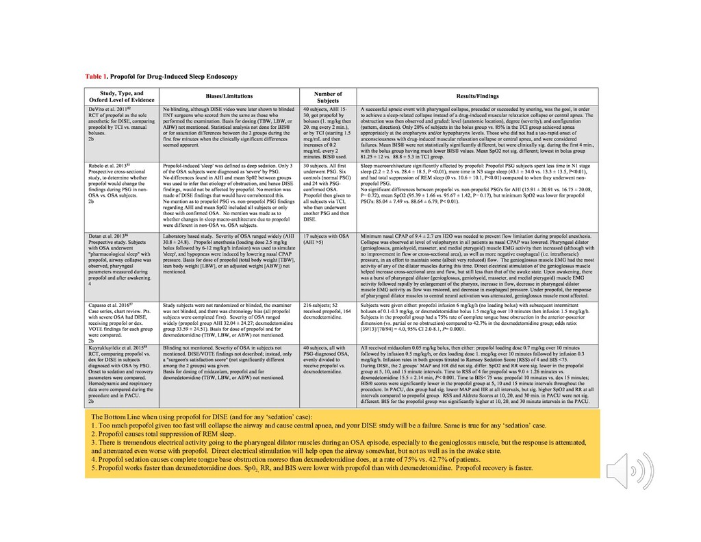
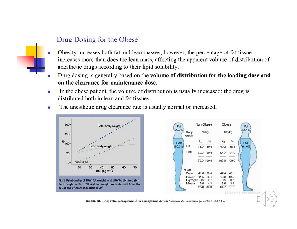
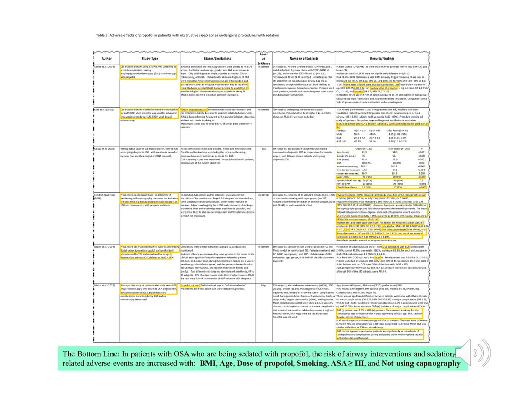
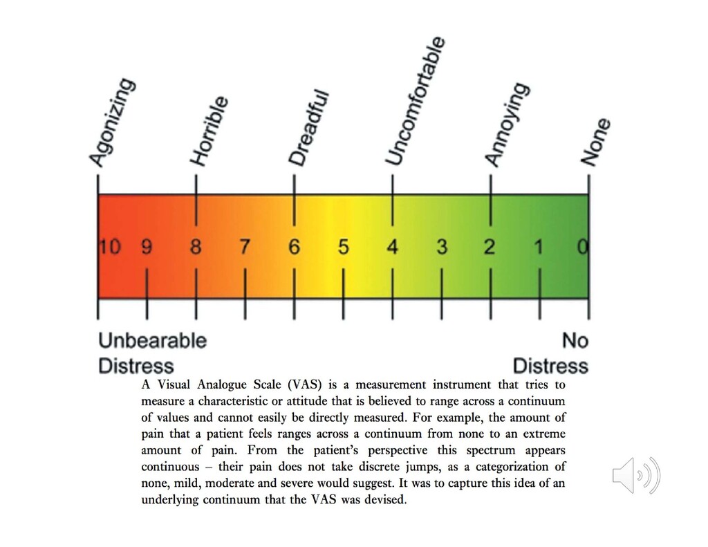
    sleep apnea undergoing procedures with sedation Author Study Type Biases/Limitations Level of Evidence Number of Subjects Results/Findings Mehta et al. (2014) Observational study, using STOP/BANG screening to predict complications during esophagogastroduodenoscopy (EGD) or colonoscopy with propofol. Both the anesthesia and endoscopy teams were blinded to the S/B scores, but factors such as age, gender, and BMI were known to them. Only brief diagnostic single procedures studied- EGD or colonoscopy, not both. Patients with a known diagnosis of OSA were excluded. Airway interventions (AI) are often routine and discretionary, and can mitigate incidents that lead to sedation- related adverse events (SRAE), but performing AI was left to the anesthesiologist's discretion without set criteria for doing AI . Most patients received opioids in addition to propofol. moderate 243 subjects. All were screened with STOP/BANG (S/B), and divided into 2 groups: those with STOP/BANG <3 (n=125), and those with STOP/BANG ≥3 (n= 118). Occurence of AI and SRAE recorded. AI defined as chin lift, placement of nasopharygeal airway, bag-mask ventilation, or unplanned intubation. SRAE defined as hypotension, hypoxia, hypopnea or apnea. Propofol used on all patients; opioids and benzodiazepines used at the anesthesiologist's discretion. Patients with STOP/BANG ≥3 were more likely to be male, >50 yrs old, BMI >25, and have HTN. Incidence rate of AI, SRAE were not significantly different for S/B ≥3. Risk of AI or SRAE did increase with BMI: for every 1 kg/m2 increase, there was an increased risk for AI (RR 1.02; 95% CI, 1.01-1.04) and for SRAE (RR 1.03; 95% CI, 1.01- 1.05). Higher rates of SRAE were also associated with: age; each 5-year increase in age (RR 1.09; 95% CI, 1.02-1.2), loading dose of propofol; 1. mg increase (RR 1.4; 95% CI, 1.1-1.8), and smoking (RR 1.9; 95% CI, 1.3-2.9). Regardless of S/B score, 47.3% of patients required an AI. One patient in each group required bag-mask ventilation, and no patient needed intubation. One patient in the S/B ≥3 group required early termination and reversal agents. Coté et al. ((2010) Observational study of sedation-related complications (AI and SRAE) when propofol was used for advanced endoscopic procedures (EUS, ERCP, small bowel enteroscopy). Airway interventions (AI) are often routine and discretionary, and can mitigate incidents that lead to sedation-related adverse events (SRAE), but performing AI was left to the anesthesiologist's discretion without set criteria for doing AI . Mallampati scores only recorded if > 4, of which there were only 4 patients. moderate 799 subjects undergoing advanced endoscopic procedures. Patients felt to be at higher risk- morbidly obese, or ASA ≥ III- were not excluded. 154 AI were performed in 115 (14.4%) patients: chin lift, modified face mask ventilation (patient needing FI02 greater than that of nasal cannulae), or nasal airway. 102 (12.8%) subjects had hypoxemia (Sa02 <90%). Procedure terminated early in 5 patients. No patient required bag-mask ventilation or intubation. BMI, male gender and ASA ≥ III were statistically significant independent predictors of AI. Subjects AI(+) = 115 AI(-) = 684 Odds Ratio (95% CI) Male 58.% 44.6% 1.75 (1.08- 2.85) BMI 29.3 ± 7.2 26.7 ± 6.3 1.05 (1.01- 1.09) ASA ≥ III 67.8% 59.2% 1.90 (1.11- 3.25) McVay et al. (2016) Retrospective study of subjects (obese vs. non-obese) undergoing diagnostic EGD, with anesthesia provided by nurse (no anesthesiologist or CRNA present). No randomization or blinding possible. Procedure time was short. Possible publication bias, concluding that non-anesthesiology personnel can safely administer propofol for EGD. OSA screening scores not mentioned. Propofol used on all patients; opioids used at the nurse's discretion. low 395 subjects: 130 consecutive patients undergoing preoperative diagnostic EGD in preparation for bariatric surgery, and 265 non-obese patients undergoing diagnostic EGD. Obese (n= 130) Non-obese (n= 265) P Age ( mean) 43.9 54.3 <0.001 Gender (% female) 76 55 <0.001 BMI (mean) 45.8 21.9 <0.001 OSA 80 (62%) 20 (8%) <0.001 Propofol dose (mean, mg) 301.1 162.8 <0.001 Procedure time (mean, min.) 12.3 9.1 <0.001 Recovery time (mean, min.) 26.9 29.2 0.038 SaO2 <90% 29 (22%) 19 (7%) <0.001 Systolic BP<90 mm Hg 16 (12%) 50 (19%) 0.1 HR< 60 BPM 17 (13%) 75 (28%) <0.001 Chin lift/jaw thrust 26 (20%) 17 (6%) <0.001 Friedrich-Rust et al. (2014) Prospective, randomized study, to determine if monitoring with capnography decreases the incidence of hypoxemia in patients undergoing colonoscopy, or EGD and colonoscopy, with propofol sedation. No blinding. Midazolam and/or ketamine also used, per the discretion of the practitioner. Propofol dosing was not standardized; some subjects received only boluses, while others received an infusion. Subjects undergoing both EGD and colonoscopy had longer procedure times and received greater total dose of propofol, and were more likely to also receive midazolam and/or ketamine. Criteria for OSA not mentioned. moderate 533 subjects, randomized to standard monitoring (n= 266) or standard monitoring with capnography (n= 267). Anesthesia performed by either an anesthesiologist, nurse (not CRNA), or endoscopist-directed. Hypoxemia (SaO2 <90%) occurred significantly less often in the capnography group: 47 (18%) [95% CI 13-23%] vs. 86 (32%) [95% CI 27-38%; P = 0.00091]. Hypoxemia incidence was reduced by 14% [95% CI 7.5-22%], with odds ratio 0.45 [95% CI 0.30-0.67; P= 0.000087]. Apnea or hypopnea was detected in 183 (69%) of the capnography group, and 23% of those patients developed hypoxemia. The mean interval between detection of apnea and onset of hypoxemia was 22 seconds. More severe hypoxemia (SaO2 < 85%) occurred in 15 (6%) of the capno group and 22 (8%) of the non-capno group (P = 0.24). Independent and statistically significant risk factors for hypoxemia were: age ≥ 55 (odds ratio [OR] 2.19 [95% CI 1.47- 3.28]) , obesity (for BMI ≥ 30, OR 3.00 [95% CI 1.78- 5.07]), OSA (OR 6.08 [95% CI 2.63- 14.08]), not using capnography (see above), total dose of propofol > 350 mg (OR 1.63 [95% CI 1.10- 2.42]) , and use of ketamine in addition to propofol (OR 3.49 [95%CI 2.34- 5.19]). Anesthesia provider was not an independent risk factor. Nagels et al. (2014) Prospective observational study of subjects undergoing dental extractions with propofol and remifentanil administered by TCI, and monitored for oxygen desaturation events (ODE, defined as SaO2 < 94%). Complexity of the dental extractions (simple vs. surgical) not mentioned. Sedation efficacy was measured by surveys taken of the nurse on the clinical team (quality of sedation questions related to patient behavior and cooperation during the procedure, ranked on a scale of excellent-good-satisfactory-poor) and the patient afterwards (asked about recall, nervousness, and recommendation of friends and family). Two different oral surgeons administered anesthesia, 67 vs. 83 subjects. 33% of subjects were male. Only 2 subjects were ASA III; the rest were ASA I-II. No mention of BIS® values or OSA diagnosis. moderate 150 subjects. Schnider model used for propofol TCI, and Minto model for remifentanil TCI. Patients monitored with BP, SaO2, capnography, and BIS®. Relationship of ODE and patient age, gender, BMI and ASA classification were analyzed. Proportion of subjects having one or more ODE correlated with BMI: underweight 20.0%, normal 47.9%, overweight 68.2%, and obese 81.8%. For each unit increase in BMI, ODE odds ratio was 1.2 (95% CI 1.1-1.3). At a fixed BMI, ODE odds ratio for a male vs. female patient was 2.6 (95% CI 1.2-5.52). Patients who had at least one ODE only spent 30% of the procedure time with SaO2 ≥ 99%. Patients with no ODE spent 70% of the time with SaO2 ≥ 99%. Age, preoperative nervousness, and ASA classification wee not associated with ODE, although 148 of the 150 subjects were ASA I-II. Mador et al. (2011) Retrospective study of patients who underwent EGD and/or colonoscopy, who also had OSA diagnosed by polysomnography (PSG). Cardiorespiratory complications occurring during EGD and/or colonoscopy were noted. Propofol not used. Sedation level kept to 'mild-to-moderate'. Procedures done with patients in lateral decubitus position. high 639 subjects, who underwent colonoscopy (68.5%), EGD (20.2%), or both (11.3%). PSG diagnosis of OSA: OSA negative, mild, moderate, or severe. Minor complications noted during procedures: hyper- or hypotension, brady- or tachycardia, oxygen desaturation (<90%), and hypopnea. Major complications noted were: chest pain, respiratory distress, cardiorespiratory arrest, or a minor complication that required intervention. Midazolam (mean, 4 mg) and fentanyl (mean, 87.5 mcg) were the sedatives used. Propofol was not used. Age (mean) 60.5 years, BMI (mean) 33.7, gender (male) 93%. PSG results: 130 negative, 509 positive (mild 135, moderate 125, severe 249). Complications: minor 19%, major 7%. There was no significant difference between patients without or with OSA in the rate of minor complications (OR 1.17, 95% CI 0.70-1.92) or major complications (OR 1.19, 95% CI 0.54- 2.63). Incidence of minor complications 17.7% in patients who were OSA (-) and 20.0% in those who were OSA (+). Incidence of major complications 6.2% in OSA (-) patients and 7.3% in OSA (+) patients. There was no tendency for the complication rate to increase with increasing severity of OSA, age, BMI, sedation dosage, or type of procedure. PSG was done prior to the endoscopy in 63.5% of patients. The mean time difference between PSG and endoscopy was 1.84 years (range 0.01- 5.3 years). Mean BMI was similar at the time of PSG and at endoscopy. OSA did not appear to predispose patients to a significantly increased rate of cardiopulmonary complications during endoscopy under mild-moderate sedation with midazolam and fentanyl. The Bottom Line: In patients with OSA who are being sedated with propofol, the risk of airway interventions and sedation- related adverse events are increased with: BMI, Age, Dose of propofol, Smoking, ASA ≥ III, and Not using capnography
  67. Figure 2. Propofol and midazolam dose-response curves for hypnosis (failure

    to open eyes on command)98, by permission of Oxford University Press.
  68. Figure 3. The propofol ED50 for hypnosis is 1.01 mg/kg

    and for general anesthesia is 1.93 mg/kg93, by permission of Oxford University Press.
  69. Table A5: Propofol, Pharmacokinetics and Obesity Study Study Design Biases/Limitations

    N Outcomes/Conclusions Oxford LOE Cortinez 2010127 PK of propofol, dosed TBW vs. ABW. Data from 3 observation studies No randomization or blinding. Some obese, some normal weight, some undergoing surgery, some not. 51 Allometric size model (clearance modeled with an allometric 3/4 power, and distribution volume was calculated with a linear [exponent of 1] model) using TBW worked best. Dosing with ABW resulted in propofol concentrations consistently lower than target concentration. 3 Servin 1993128 MO dosed by ABW and nonobese by TBW. Two observational studies of propofol PK. No randomization or blinding. Some undergoing surgery, some not. 18 Total body clearance and volume at steady state correlated linearly with body weight in obese and controls, but distribution clearance did not. Context- sensitive half time (time required for the central compartment drug concentration at the end of infusion to decrease by 50%) was much faster than elimination half-life. Propofol dosage could be based upon TBW without risk of accumulation. 4 La Colla 2009129 Propofol TCI in MO, using ABW vs. TBW. Prospective, randomized, double- blinded No mention of how many dosage adjustments were made because of cardiovascular or CNS pharmacodynamics. 24 Propofol blood levels lower than predicted in both groups, more so for the ABW group. Marsh TCI model may underestimate total body clearance and central volume of distribution at steady state, underdosing the patient, especially if ABW is used. 2 Dong 2016130 Propofol dosed by TBW or LBW in MO; non- obese by TBW. RCT Non-obese controls were undergoing a different surgery, with more potential for underlying co-morbidity or intraoperative confounders, even though all were ASA I-II. 29 MO subjects had increased systemic clearance and peripheral compartment volume, resulting in a decrease in propofol levels. Propofol maintenance infusion can be based on TBW in MO. 2 The Bottom Line: Using ABW to dose propofol may result in deficient levels of anesthesia. Using TBW may be inadvisable because of co-morbidities. Consider using other anesthetics along with propofol to balance pharmacokinetic vs. pharmacodynamic concerns.
  70. V1 Central Compartment blood, brain, heart, kidney, liver, lungs V2

    Peripheral Compartment (muscle) V3 Peripheral Compartment (fat) Effect site (brain) is within the central compartment Drug injected Ke 0  K1 2 K1 2   Fast distribution Slow distribution K3 1  K3 1  Slow distribution Elimination (clearance) phase Fast distribution phase, 1  2 Slow distribution phase, 2 1 3 Elimination phase, 3 1  0 Time Serum drug level Elimination (clearance) phase CL Q3 Q2 K1 0  Figure 1. Three-compartment pharmacokinetics. The serum drug level plotted against time is used to generate a mathematical equation. That equation- the pharmacokinetic model- derives volumes of distribution, clearances, rate constants, and allometric factors to shape the best mathematical fit for the data. The pharmacodynamics at the effect site can also be modeled, albeit using proxy methods such as the processed electroencephalogram to quantify the drug effect. Liver 60%, Kidneys 40% The 3 different parts of this curve are mathematically summarized: C(t) = Ae−αt + Be−βt + Ce−γt t is the time since the bolus C(t) is the drug concentration A, B and C are coefficients which describe the exponential functions of each phase α, β and γ are exponents which describe the shape of the curve for each phase
  71.  Abdominal (central, or android) obesity- much more strongly associated

    with Metabolic Syndrome than gynecoid (peripheral) obesity.  men with waistline >102 cm (> 40 in), women with > 88 cm (>35 in)  Atherogenic dyslipidemia-  triglycerides,  HDL cholesterol, and other lipoprotein abnormalities implicated as atherogenic.  Hypertension- not directly due to anything metabolic, but very much a part of the cluster of these obesity complications  Insulin resistance- oftens leads to glucose intolerance and Type II diabetes. Some experts recommend a glucose tolerance test for patients with Metabolic Syndrome who may not yet have diabetes.  Proinflammatory state- elevated C-reactive protein (CRP). Excess adipose tissue releases inflammatory cytokines.  Prothrombotic state- increased plasminogen activator inhibitor (PAI)-1 and fibrinogen. Fibrinogen is an acute-phase reactant like CRP, rises in response to a high-cytokine state. Thus, the proinflammatory and prothrombotic states may be metabolically interconnected. The Metabolic Syndrome …is a cluster of six metabolic complications of obesity, resulting in a multiplex risk factor for cardiovascular disease. Metabolic Syndrome https://doi.org/10.1161/CIRCULATIONAHA.106.671057 Circulation. 2007;115:e32–e35
  72. Not all fat within the body is identical.  Unlike

    peripherally deposited fat, intra-abdominal (central, or visceral) fat is highly metabolically active and is known to be a contributor to several disease states.  Patients with centrally distributed or ‘visceral’ fat are at greater peri-operative risk than those with peripherally distributed fat, and are far more likely to exhibit the Metabolic Syndrome.
  73. Adipocytes release numerous factors  hormone- leptin  complement factors-

    Factor D/adipsin  cytokines- TNF  (tumor necrosis factor)  enzymes- aromatase (estrogen synthesis)  substrates- free fatty acids, glycerol  others- plasminogen activator factor, angiotensinogen Leptin is a mediator of long-term regulation of energy balance, suppressing food intake and thereby inducing weight loss. Ghrelin, is an orexigenic hormone produced in the gastrointestinal tract, plays a role in meal initiation. In obese subjects the circulating level of the anorexigenic hormone leptin is increased, whereas surprisingly, the level of the orexigenic hormone ghrelin is decreased. It is now established that obese patients are leptin- resistant.
  74. Obesity-Related Comorbidities That Are Most Likely to Influence the Preoperative

    Cardiac Assessment and Management of Severely Obese Patients  Atherosclerotic cardiovascular disease  Heart failure  Systemic hypertension  Pulmonary hypertension related to sleep apnea and obesity hypoventilation  Cardiac arrhythmias  Deep vein thrombosis  History of pulmonary embolism  Poor exercise capacity Poirier P, Alpert M, Franklin B, et al. Cardiovascular evaluation and management of severely obese patients undergoing surgery: a science advisory from the American Heart Association. Circulation 2009;120(1):86-95. Demaria EJ, Murr M, Byrne TK, et al. Validation of the Obesity Surgery Mortality Risk score in a multicenter study proves it stratifies mortality risk in patients undergoing gastric bypass for morbid obesity. Annals of Surgery 2007; 246: 578-84. Undergoing Surgery: A Science Advisory From the American Heart Association Cardiovascular Evaluation and Management of Severely Obese Patients Obesity Surgery Mortality Risk Score for Gastric Bypass  BMI 50  Male gender  Hypertension as a comorbid condition  Pulmonary embolism (PE) risk as a comorbid condition, defined as the presence of venous thromboembolism event, previous inferior vena cava filter placement, a history of right heart failure or pulmonary hypertension, and/or a history or physical findings of venous stasis, including typical ulcerations or brawny edema  Age 45 years Six Risk Factors for Perioperative Cardiovascular Morbidity in the General Population  High risk surgery- emergency surgical procedures, major thoracic, abdominal and vascular  History of coronary artery disease  History of CHF  History of cerebrovascular disease  Patient using insulin  Preoperative serum creatinine > 2.0 mg/dL Six Risk Factors for Perioperative Cardiovascular Morbidity in the General Population  High risk surgery- emergency surgical procedures, major thoracic, abdominal and vascular  History of coronary artery disease  History of CHF  History of cerebrovascular disease  Patient using insulin  Preoperative serum creatinine > 2.0 mg/dL Surgery Mortality Risk Score for Obese Patients  BMI 50  Male gender  Hypertension as a comorbid condition  Pulmonary embolism (PE) risk as a comorbid condition, defined as the presence of venous thromboembolism event, previous inferior vena cava filter placement, a history of right heart failure or pulmonary hypertension, and/or a history or physical findings of venous stasis, including typical ulcerations or brawny edema  Age 45 years Obesity-Related Comorbidities That Are Most Likely to Influence the Preoperative Cardiac Assessment and Management of Severely Obese Patients  Atherosclerotic cardiovascular disease  Heart failure  Systemic hypertension  Pulmonary hypertension related to sleep apnea and obesity hypoventilation  Cardiac arrhythmias  Deep vein thrombosis  History of pulmonary embolism  Poor exercise capacity
  75. The cardiologist’s evaluation of a preoperative patient… Fleisher LA, Beckman

    JA, Brown KA, et al. ACC/AHA 2007 Guidelines on Perioperative Cardiovascular Evaluation and Care for Noncardiac Surgery: A Report of the American College of Cardiology/American Heart Association Task Force on Practice Guidelines (Writing Committee to Revise the 2002 Guidelines on Perioperative Cardiovascular Evaluation for Noncardiac Surgery) Circulation 2007; 116: e418 - e500. Poirer P, Alpert MA, Fleisher LA, et al. Cardiovascular Evaluation and Management of Severely Obese Patients Undergoing Surgery. Circulation 2009; 120: 86-95. History and Physical  Assess the level of functional capacity.  Look for active clinical cardiac conditions, known cardiovascular disease, and cardiac risk factors. (These 3 terms each have specific meaning to cardiologists.)  Consider the magnitude and invasiveness of the planned procedure. Testing  based upon findings of the H&P, planned procedure, age, and known or suspected comorbidities
  76. Estimating the Level of Functional Capacity • attempt to quantify

    activity level as metabolic equivalent (MET) units Fleisher LA, Beckman JA, Brown KA, et al. ACC/AHA 2007 guidelines on perioperative cardiovascular evaluation and care for noncardiac surgery. Circulation 2007; 116: e418-e500.
  77. Look for:  active clinical cardiac conditions  known cardiovascular

    disease  other cardiac risk factors Active clinical cardiac conditions (treat prior to surgery if possible)  Unstable coronary syndromes, unstable or severe angina, recent MI  Decompensated heart failure  Significant (unstable) arrhythmias  Severe valvular disease Known cardiovascular disease history (optimize, and consider noninvasive or invasive testing)  Ischemic heart disease  Compensated or prior heart failure  History of cerebrovascular disease  Diabetes mellitus  Renal insufficiency Other cardiac risk factors (if 3 or more, consider invasive testing)  Age greater than 70 years  Abnormal ECG (LVH, LBBB, ST-T abnormalities)  Rhythm other than sinus  Uncontrolled systemic hypertension
  78. Summary of the cardiologist’s preoperative evaluation of a patient having

    noncardiac surgery History and Physical  evaluate level of functional capacity  look for active clinical cardiac conditions, known cardiovascular disease, and cardiac risk factors  consider magnitude and invasiveness of the planned procedure Testing  Based upon results of H&P, planned procedure, age, and known or suspected comorbidities Address active cardiac conditions now (i.e. unstable angina, decompensated heart failure, significant arrhythmias).  ‘Low risk’ surgery can then proceed.  ‘Higher risk’ surgery needs an acceptable functional capacity of at least 4 METS.  A patient with poor or uncertain functional capacity, needing higher-risk surgery, with cardiovascular disease history and other cardiac risk factors, needs further studies to evaluate cardiac function and ischemic burden.  Further such studies should only be undertaken if they would change perioperative management.  Consider heart rate control with a beta blocker for higher risk patients.
  79. The Anesthesiologist’s Preoperative Evaluation of an Obese Patient heart lungs

    metabolic airway type of surgery type of anesthesia  Looking for evidence of pulmonary diseases: STOP-BANG to screen for OSA, symptoms and signs of OHS, right heart strain/cor pulmonale, smoking history, COPD, baseline SpO2 < 94%, elevated serum bicarbonate level.  Look for evidence of cardiovascular disease.  Poor functional capacity, newly abnormal EKG, active cardiac conditions, uncontrolled hypertension, etc.  Inquire and examine for obesity-related conditions that we know increase risk (i.e. HTN, DM, dyslipidemias, coagulation state, renal dysfunction)  Examine the airway.  Consider the type of surgery or procedure planned, and the type of anesthesia required.
  80. Preoperative Evaluation of an Obese Patient, continued  Take accurate

    vital signs, height, weight, and neck circumference, and then calculate BMI.  Room air Sp02 when supine or with head slightly elevated (do this at any point preoperatively)  Pre-admission Testing  Order labs and other studies based upon planned procedure, age, gender, known pre-existing or suspected conditions or comorbidities.  Serum bicarbonate of 28 or greater, combined with a high STOP- BANG score, increases the specificity of screening for OSA.  Request the appropriate consultations if you suspect significant untreated and uncontrolled cardiovascular, pulmonary or other diseases that need to be evaluated or treated, if it will make a difference in the perioperative management of surgery and anesthesia.
  81. Preoperative Evaluation of an Obese Patient, continued  If the

    patient was diagnosed by a pulmonologist as having OSA, and is on PAP, ask them to bring their own machine with them on the day of surgery, so it can be applied in the PACU.  If there was no pulmonology consultation, and the patient scored 5 or more on STOP-BANG, assume the patient has OSA.  If communication with the patient isn’t feasible, and you can’t get a STOP score, then a BANG score of 3 or more indicates the patient is at increased risk for OSA.
  82. In September 2011, the first guidelines were proposed by the

    Society of Bariatrics and Anaesthesia, in Chichester, UK
  83.  There is insufficient evidence in the current literature to

    support canceling or delaying surgery for a formal diagnosis (laboratory or home polysomnography) in patients with suspected OSA, unless there is evidence of an associated significant or uncontrolled systemic disease or additional problems with ventilation or gas exchange.  Additional evaluation to allow preoperative cardiopulmonary optimization should be considered in patients with diagnosed, partially treated/untreated, and suspected OSA where there is indication of an associated significant or uncontrolled systemic disease or additional problems with ventilation or gas exchange such as: (i) hypoventilation syndromes, (ii) severe pulmonary hypertension, and (iii) resting hypoxemia in the absence of other cardiopulmonary disease.  Where management of comorbid conditions has been optimized, patients with diagnosed, partially treated/ untreated OSA, or suspected OSA may proceed to surgery provided strategies for mitigation of postoperative complications are implemented.
  84. Postoperative Assessment in the PACU We realize that each patient’s

    length of stay in the PACU- and their disposition beyond the PACU- should be evaluated based upon the nature of surgery and type of anesthesia, the ease or difficulty of their intraoperative and PACU course, need for narcotic analgesics, BMI, android body habitus, patient’s age, comorbidities, home situation (for SDS patients), STOP/BANG score, use and compliance with PAP device, need for supplementary oxygen as preoperative baseline, whether they also have OHS or other Sleep Disordered Breathing, etc. How can we sort this out?
  85. Identification of Patients at Risk for Postoperative Respiratory Complications Using

    a Preoperative Obstructive Sleep Apnea Screening Tool and Postanesthesia Care Assessment. Gali, Bhargavi; Whalen, Francis; Schroeder, Darrell; Gay, Peter; Plevak, David Anesthesiology. 110(4):869-877, April 2009. DOI: 10.1097/ALN.0b013e31819b5d70 Patients with high sleep apnea clinical scores (SACS) and recurrent respiratory events in the PACU are at higher risk for postoperative respiratory complications. Fig. 1 The frequency of postoperative respiratory events is displayed according to the four patient groups defined by the combination of sleep apnea clinical score (SACS) (low/high) and recurrent postanesthesia care unit (PACU) events (no/yes). From a multiple logistic regression analysis, which included SACS group and recurrent PACU events as explanatory variables, the likelihood of postoperative respiratory events was found to be significantly associated with high SACS (odds ratio = 3.5, P = 0.001) and recurrent PACU events (odds ratio = 21.0, P Respiratory complications were defined as follows: intensive care unit admission for a new respiratory indication (e.g., respiratory failure), the need for respiratory therapy beyond standard postoperative clinical practice, the need for unanticipated noninvasive ventilatory support (e.g., continuous positive airway pressure or bilevel positive airway pressure), and the development of postoperative pneumonia (new infiltrate on chest x-ray, leukocytosis, and temperature >38°C).
  86. Abstract title: Using the RUTGERS Scoring System to Decide the

    Disposition of Patients from the Post-Anesthesia Care Unit who are at Risk for Obstructive Sleep Apnea Presenting Author: Mark H. Stein, MD, Rutgers- Robert Wood Johnson Medical School, New Brunswick, NJ Co-Authors: Geza K. Kiss, MD, Rutgers- Robert Wood Johnson Medical School, New Brunswick, NJ Stanley Z. Trooskin, MD, Rutgers- Robert Wood Johnson Medical School, New Brunswick, NJ Background: Postoperative patients of Body Mass Index (BMI) > 35 and a STOP/BANG1 score of ≥ 5 generally fall into one of three categories in our Post- Anesthesia Care Unit (PACU). 1. Patients whose preoperative status was reasonably satisfactory, intraoperative course uneventful, and PACU stay was uncomplicated: such patients can go home or to a regular floor. 2. Patients whose preoperative status was less than optimal, whose intraoperative course was very challenging, had multiple issues in the PACU, and in particular, those who had more than one2 'unexplained respiratory incident' in the PACU: such patients will go to a monitored setting postop where continuous pulse oximetry is available. 3. Patients who are in between categories 1 and 2- perhaps they had one 'unexplained respiratory incident' in the PACU, and/or their co-morbidities and other factors are cause for additional concern, and a clinical judgment is needed in order to decide their disposition beyond the PACU. A potential gray zone exists in the decision-making process. Aim: For patients at risk for Obstructive Sleep Apnea (OSA) and who fall into the third category mentioned above, we have devised the RUTGERS scoring system to aid in the decision by scoring the risk factors. Materials and Methods: Our PACU algorithm is used as the basis for deciding the disposition of patients at risk for Obstructive Sleep Apnea (OSA). We devised and added the RUTGERS scoring system (figure 1). A score of ≥ 3 warrants transfer to a monitored setting where continuous pulse oximetry is available. Results: The algorithm, combined with the scoring system, guides practitioners in their decision making by focusing the process on the ongoing abnormalities and specific risk factors. Discussion: Stratification of postoperative patients who are at increased risk for OSA-related adverse cardiopulmonary events is essential in order to most appropriately utilize the additional resources such patients require. Algorithms and guidelines3,4 currently available encourage individual institutions to formulate methods based upon the literature in the field of perioperative sleep medicine. Conclusion: Patient safety and cost-effectiveness can work hand-in-hand to provide the right patient with the right care. We believe the RUTGERS Scoring System can aid that process. We are currently gathering data on all of our perioperative patients at risk for OSA, and what strategies are most effective. References: 1. Sleep Medicine Clinics 12 (1), 123–35. 2. Anesthesiology 110 (4), 869-77. 3. Anesthesia & Analgesia 123 (2), 432-73. 4. Anaesthesia 70, 859-76.
  87.  Apnea is defined as complete cessation of breathing for

    more than 9 seconds.  Hypopnea is defined as respiratory rate less than 8 breaths per minute.  Desaturation is defined as saturation less than 90% for 30 seconds while on nasal cannula of 4 LPM. Disposition of patients who are at risk for Obstructive Sleep Apnea from the Post-Anesthesia Care Unit (cont.)
  88.  Once a patient with OSA- or deemed to be

    at high risk for OSA- is ‘recovered’ by the usual PACU Stage 1 criteria (Modified Aldrete Scoring System), they require additional observation for at least one hour in the PACU. • The additional hour of observation begins once the patient has otherwise met discharge criteria. • The additional hour of observation may be performed in any of the PACU’s, as long as continuous pulse oximetry is monitored and prompt nursing intervention is available. Continuous end-tidal CO2 monitoring, if available, may also be used to monitor ventilation. Assessing OSA patients for discharge from the PACU (to home or inpatient)
  89. Assessing OSA patients for discharge from the PACU (cont.): 

    Throughout the entire PACU stay, nurses must be on guard for:  episodes of apnea that last longer than 9 seconds  episodes of desaturation to below 90% that last at least 30 seconds  episodes of respiratory rate less than 8 breaths per minute  episodes of pain/sedation mismatch (RASS -3, -4, or -5, combined with pain VAS >5).
  90. Curtis N. Sessler, Mark S. Gosnell, Mary Jo Grap, Gretchen

    M. Brophy, Pam V. O'Neal, Kimberly A. Keane, Eljim P. Tesoro, and R. K. Elswick "The Richmond Agitation– Sedation Scale", American Journal of Respiratory and Critical Care Medicine, Vol. 166, No. 10 (2002), pp. 1338-1344. doi: 10.1164/rccm.2107138
  91. • If an episode occurs of apnea lasting longer than

    9 seconds, or desaturation below 90% lasting 30 seconds, or respiratory rate <8, or pain/sedation mismatch, then the patient needs intervention, respiratory monitoring, and re- assessment while still in the PACU.  Patient must spontaneously become aroused and breathe within 9 seconds.  SaO2 must quickly return to baseline when he/she spontaneously arouses. • An apneic/hypopneic episode lasting longer than 9 seconds with SpO2 dropping below 90% lasting 30 seconds requires an intervention and a reassessment. • An asleep hypopneic episode (i.e. heavy snoring) may be tolerated as long as the SaO2 does not drop below 90%. Assessing OSA patients for discharge from the PACU (cont.):
  92. • Reassess a patient with an apneic, hypopneic or desaturation

    episode, as there may be an explainable or correctable factor that can be addressed, such as residual anesthetics or muscle relaxant, pain, abnormal blood glucose, volume, electrolyte or acid-base disturbance, temperature abnormality, cardiac or pulmonary event, oversedation, pain/sedation mismatch, etc.  If the patient had been on CPAP, then reapply the device.  Supplementary oxygen may also be applied or increased, especially for a patient with no history of CPAP usage, but keeping in mind that that doesn’t address apnea or hypopnea. • If there is only one unexplainable apneic/hypopneic/desaturation episode throughout the PACU stay, or more than one explainable episode, do a RUTGERS score.  If the RUTGERS score is ≥ 3, there is a substantially increased risk for respiratory episodes beyond the PACU. Send the patient to a monitored setting.  If the RUTGERS score is < 3, there is relatively lower risk of respiratory episodes beyond the PACU. Assess overall perioperative course. May consider discharge to floor or home. • Admission to a monitored setting after more than one unexplainable apneic/hypopneic/desaturation episode is advisable.
  93. Patients with adjustable gastric bands in situ  Laparoscopic adjustable

    gastric banding is a recognized treatment for obesity.  However, patients with a gastric band in situ are at increased risk of pulmonary aspiration during general anesthesia owing to esophageal dysmotility and dilatation above the band.  The dilatation may persist following band deflation.  There are case reports of regurgitation of food even after prolonged fasting. A tracheal tube may be advisable in all patients who have a gastric band.  Current advice-- depending on the extent and type of surgery and anesthesia planned, a decision to deflate the band should be made on an individual basis. Our bariatric surgery group is available to deflate the band before surgery. Naef M, Mouton WG, Naef U, van der Weg B, Maddern GJ, Wagner HE. Esophageal dysmotility disorders after laparoscopic gastric banding– an underestimated complication. Annals of Surgery 2011; 253: 285–90. Koolwijk J, Schors M, el Bouazati S, Noordergraaf GJ. Airway management concerns in patient with gastric banding procedures. BMJ Case Reports 2013; doi:10.1136/bcr-2013-201009.
  94. Find out more… Society of Bariatric Anaesthetists (UK) http://www.sobauk.com/ Society

    of Anesthesia and Sleep Medicine http://anesthesiaandsleep.org/ British Obesity Surgery Patient Association www.bospauk.org British Obesity and Metabolic Surgery Society: www.bomss.org.uk American Society for Metabolic and Bariatric Surgery www.asmbs.org International Society for the Perioperative Care of the Obese Patient http://www.ispcop.org