$30 off During Our Annual Pro Sale. View Details »

CAIP 2025

CAIP 2025

Avatar for Olivier Lézoray

Olivier Lézoray

December 04, 2025
Tweet

More Decks by Olivier Lézoray

Other Decks in Research

Transcript

  1. Ensuring the Origin of Cytological Whole Slide Images Through Preparation

    and Scanner Detection Paul Barthe1,2 Romain Brixtel1 Mathieu Fontaine1 Arnaud Renouf1 S´ ebastien Bougleux2 Olivier L´ ezoray2 Datexim, Caen, France1 Universit´ e Caen Normandie, ENSICAEN, CNRS, GREYC, Caen, France2 21st International Conference in Computer Analysis of Images and Patterns, September 2025 P. Barthe et al. Ensuring the Origin of Cytological WSIs CAIP 2025 1 / 17
  2. Context: Origin Sampling Slide Whole Slide Image Cell deposition Digitization

    Coloration P. Barthe et al. Ensuring the Origin of Cytological WSIs CAIP 2025 3 / 17
  3. Context: Origin Sampling Slide Whole Slide Image Cell deposition Digitization

    Coloration Preparation Scanner Pannoramic 1000, 3DHISTECH (P1000) Pannoramic 250, 3DHISTECH (P250) NanoZoomer S360 RUO, Hamamatsu (NRUO) Aperio GT 450, Leica Biosystem (GT450) NanoZoomer S360 IVDR, Hamamatsu (NIVDR) Origin BD SurePath, Becton Dickinson (BD) ThinPrep, Hologic (ThinPrep) iLsa Diagnostic (ILSA) P. Barthe et al. Ensuring the Origin of Cytological WSIs CAIP 2025 4 / 17
  4. Context: Origin matters WSIs from different origins exhibit distinct features.

    (a) P1000 (b) P250 (c) GT450 (d) NRUO P. Barthe et al. Ensuring the Origin of Cytological WSIs CAIP 2025 5 / 17
  5. Context: Origin matters WSIs from different origins exhibit distinct features.

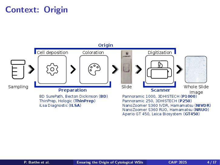
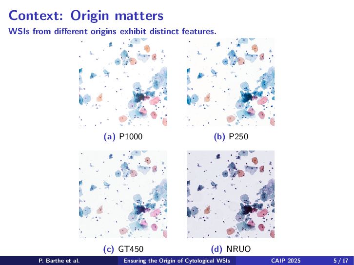
    Machine learning models are usually trained for a specific origin. Problem: At inference, how can we ensure that a WSI is suitable for a model ? Put differently: How can we ensure that a WSI comes from the correct origin ? P. Barthe et al. Ensuring the Origin of Cytological WSIs CAIP 2025 6 / 17
  6. Method: Overview CNN MIL Tile selection: Remove background tiles. Explore

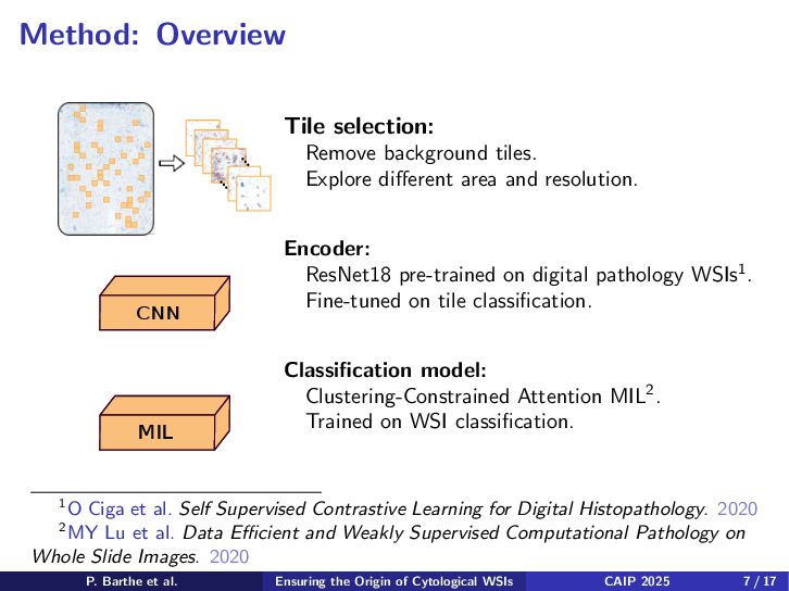
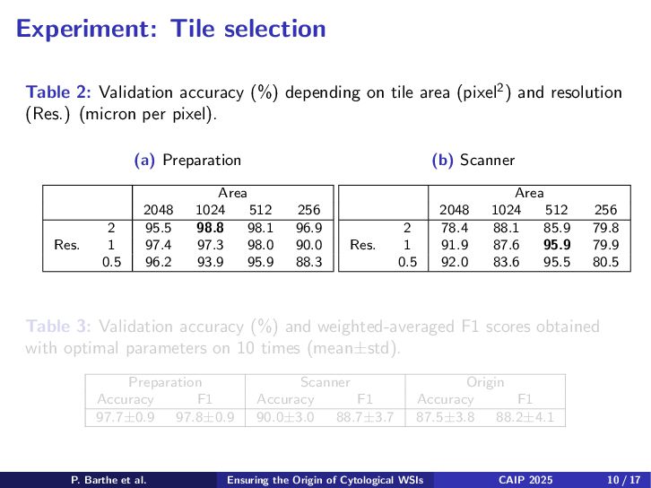
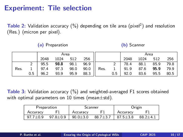
    different area and resolution. Encoder: ResNet18 pre-trained on digital pathology WSIs1. Fine-tuned on tile classification. Classification model: Clustering-Constrained Attention MIL2. Trained on WSI classification. 1O Ciga et al. Self Supervised Contrastive Learning for Digital Histopathology. 2020 2MY Lu et al. Data Efficient and Weakly Supervised Computational Pathology on Whole Slide Images. 2020 P. Barthe et al. Ensuring the Origin of Cytological WSIs CAIP 2025 7 / 17
  7. Method: Two branches - GT450 - P250 - P1000 -

    NIVDR - NRUO Scanner MIL CNN MIL CNN Preparation - BD - ThinPrep - ILSA P. Barthe et al. Ensuring the Origin of Cytological WSIs CAIP 2025 8 / 17
  8. Materials Dataset: 3473 WSIs for cervical cancer screening, ≈5 TB

    of data. Table 1: Training/validation dataset WSI distribution P1000 P250 NIVDR NRUO GT450 Total BD 395/200 0 0 0 0 395/200 ThinPrep 300/550 0 0 0 0 300/550 ILSA 232/50 300/250 300/50 300/100 246/200 1378/650 Total 927/800 300/250 300/50 300/100 246/200 2073/1400 Validation dataset: 1400 WSIs from 22 laboratories. P. Barthe et al. Ensuring the Origin of Cytological WSIs CAIP 2025 9 / 17
  9. Experiment: Tile selection Table 2: Validation accuracy (%) depending on

    tile area (pixel2) and resolution (Res.) (micron per pixel). (a) Preparation Area 2048 1024 512 256 Res. 2 95.5 98.8 98.1 96.9 1 97.4 97.3 98.0 90.0 0.5 96.2 93.9 95.9 88.3 (b) Scanner Area 2048 1024 512 256 Res. 2 78.4 88.1 85.9 79.8 1 91.9 87.6 95.9 79.9 0.5 92.0 83.6 95.5 80.5 Table 3: Validation accuracy (%) and weighted-averaged F1 scores obtained with optimal parameters on 10 times (mean±std). Preparation Scanner Origin Accuracy F1 Accuracy F1 Accuracy F1 97.7±0.9 97.8±0.9 90.0±3.0 88.7±3.7 87.5±3.8 88.2±4.1 P. Barthe et al. Ensuring the Origin of Cytological WSIs CAIP 2025 10 / 17
  10. Experiment: Tile selection Table 2: Validation accuracy (%) depending on

    tile area (pixel2) and resolution (Res.) (micron per pixel). (a) Preparation Area 2048 1024 512 256 Res. 2 95.5 98.8 98.1 96.9 1 97.4 97.3 98.0 90.0 0.5 96.2 93.9 95.9 88.3 (b) Scanner Area 2048 1024 512 256 Res. 2 78.4 88.1 85.9 79.8 1 91.9 87.6 95.9 79.9 0.5 92.0 83.6 95.5 80.5 Table 3: Validation accuracy (%) and weighted-averaged F1 scores obtained with optimal parameters on 10 times (mean±std). Preparation Scanner Origin Accuracy F1 Accuracy F1 Accuracy F1 97.7±0.9 97.8±0.9 90.0±3.0 88.7±3.7 87.5±3.8 88.2±4.1 P. Barthe et al. Ensuring the Origin of Cytological WSIs CAIP 2025 10 / 17
  11. Method: Enhancing scanner identification from residuals Scanner MIL CNN MIL

    CNN RES Preparation P. Barthe et al. Ensuring the Origin of Cytological WSIs CAIP 2025 11 / 17
  12. Method: Enhancing scanner identification from residuals In digital forensics, device

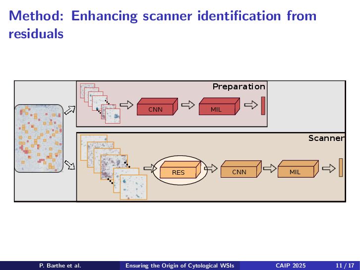
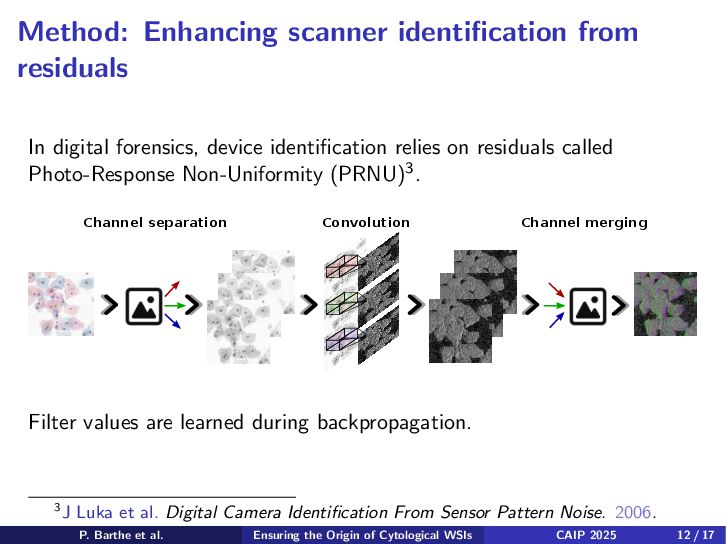
    identification relies on residuals called Photo-Response Non-Uniformity (PRNU)3. Channel separation Channel merging Convolution Filter values are learned during backpropagation. 3J Luka et al. Digital Camera Identification From Sensor Pattern Noise. 2006. P. Barthe et al. Ensuring the Origin of Cytological WSIs CAIP 2025 12 / 17
  13. Method: Enhancing scanner identification from residuals Each of the 3

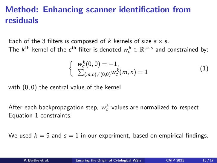
    filters is composed of k kernels of size s × s. The kth kernel of the cth filter is denoted wk c ∈ Rs×s and constrained by: wk c (0 , 0) = −1 , (m , n)̸=(0 , 0) wk c (m , n) = 1 (1) with (0 , 0) the central value of the kernel. After each backpropagation step, wk c values are normalized to respect Equation 1 constraints. We used k = 9 and s = 1 in our experiment, based on empirical findings. P. Barthe et al. Ensuring the Origin of Cytological WSIs CAIP 2025 13 / 17
  14. Illustration: Scanner identification from residuals P1000 NRUO P. Barthe et

    al. Ensuring the Origin of Cytological WSIs CAIP 2025 14 / 17
  15. Illustration: Scanner identification from residuals P1000 NRUO P. Barthe et

    al. Ensuring the Origin of Cytological WSIs CAIP 2025 15 / 17
  16. Experiment: Scanner identification from residuals Table 4: Validation accuracy (%)

    and weighted-averaged F1 scores obtained with and without residuals approach on 10 times (mean±std). Preparation Scanner Origin Accuracy F1 Accuracy F1 Accuracy F1 no residuals 97.7±0.9 97.8±0.9 90.0±3.0 88.7±3.7 87.5±3.8 88.2±4.1 residuals / / 95.6±3.0 95.8±2.8 93.8±2.6 96.9±1.3 Table 5: Validation confusion matrix. Predicted P1000 P250 NIVDR NRUO GT450 Actual P1000 0.944 0.056 0 0 0 P250 0.058 0.933 0 0.003 0.006 NIVDR 0.006 0 0.991 0.001 0.002 NRUO 0.001 0 0.001 0.988 0.001 GT450 0 0 0 0 1 P. Barthe et al. Ensuring the Origin of Cytological WSIs CAIP 2025 16 / 17
  17. Conclusion Summary: Tile selection matters. Residuals approaches for WSI scanner

    detection works. Perspectives: How to handle new origins ? Is there a correlation between machine learning performances and origin detection ? P. Barthe et al. Ensuring the Origin of Cytological WSIs CAIP 2025 17 / 17
  18. Impact of fine-tunning Table 6: Validation accuracy (%) and weighted-averaged

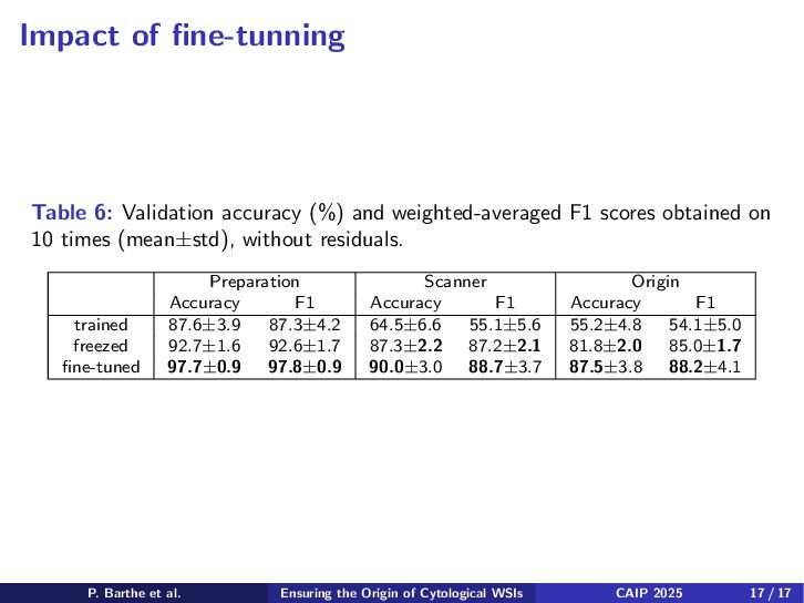
    F1 scores obtained on 10 times (mean±std), without residuals. Preparation Scanner Origin Accuracy F1 Accuracy F1 Accuracy F1 trained 87.6±3.9 87.3±4.2 64.5±6.6 55.1±5.6 55.2±4.8 54.1±5.0 freezed 92.7±1.6 92.6±1.7 87.3±2.2 87.2±2.1 81.8±2.0 85.0±1.7 fine-tuned 97.7±0.9 97.8±0.9 90.0±3.0 88.7±3.7 87.5±3.8 88.2±4.1 P. Barthe et al. Ensuring the Origin of Cytological WSIs CAIP 2025 17 / 17
  19. Impact of multiple instance learning Table 7: Accuracy (%) and

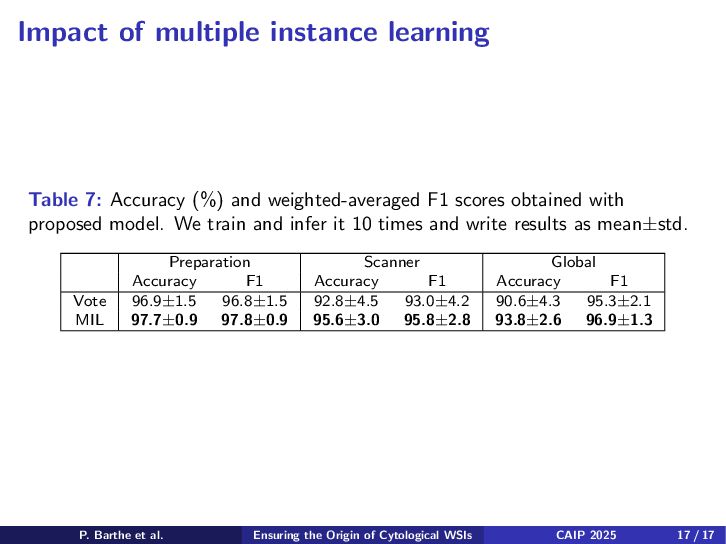
    weighted-averaged F1 scores obtained with proposed model. We train and infer it 10 times and write results as mean±std. Preparation Scanner Global Accuracy F1 Accuracy F1 Accuracy F1 Vote 96.9±1.5 96.8±1.5 92.8±4.5 93.0±4.2 90.6±4.3 95.3±2.1 MIL 97.7±0.9 97.8±0.9 95.6±3.0 95.8±2.8 93.8±2.6 96.9±1.3 P. Barthe et al. Ensuring the Origin of Cytological WSIs CAIP 2025 17 / 17