Upgrade to Pro — share decks privately, control downloads, hide ads and more …

9/16/2020 - High Yield OB Anesthesia - Dr. Malik

us414
September 16, 2020

9/16/2020 - High Yield OB Anesthesia - Dr. Malik

High Yield Board Review

us414

September 16, 2020
Tweet

More Decks by us414

Other Decks in Education

Transcript

  1. Disclaimer • Overview of HY topics, not meant to be

    in depth coverage • In depth coverage during clinical time whilst on OB rotation, OB Night float • Reflects the Exam answer, not necessarily what would be done in clinical practice • Recommend watching with the audio commentary for better understanding
  2. Anesthesia for Labor Analgesia Q. A woman in active labor

    at 5cm dilatation is having pain with each contraction. Which of the following is most likely responsible for her pain? a. Stretching of the vagina b. Active contraction of the uterine fundus c. Pressure on the perineum d. Passive stretching of the cervix
  3. Anesthesia for Labor Analgesia Labor Pain Physiology • 1st stage

    of labor – Visceral pain caused by cervical dilation and distention of lower uterine segment – Pain pathway – visceral afferents that accompany sympathetic nerves from T10-L1 – Must block T10-L1 nerve roots to achieve adequate 1st stage analgesia
  4. Anesthesia for Labor Analgesia Labor Pain Physiology • 2nd stage

    of labor – Somatic pain from stretching of perineum as fetal head descends – Pain pathway: somatic nerve fibers of pudendal nerve, which is derived from S2,3,4 nerve roots
  5. Anesthesia for Labor Analgesia Q. Which of the following anesthetic

    techniques is least likely to provide adequate analgesia for the first stage of labor? a. Combined spinal-epidural analgesia b. Lumbar sympathetic block c. Paracervical block d. Pudendal nerve block
  6. Anesthesia for Labor Analgesia Q. Which regional anesthetic technique will

    provide adequate analgesia for i. early 1st stage labor ii. Late 1st stage iii. 2nd stage?
  7. Anesthesia for Labor Analgesia Early 1st stage labor Late 1st

    stage labor 2nd stage labor Epidural Local Anesthetic yes yes yes Epidural Opioids yes ?no no CSE yes yes yes Lumbar Sympathetic Block yes yes no Pudendal Nerve Block no yes yes Paracervical Block yes no no
  8. Adverse Effects of Epidural Analgesia Q. When compared to iv

    opioids, epidural labor analgesia is most likely associated with? a. An increased incidence of C Section b. A shorter duration of labor c. An increased incidence of maternal fever d. A decreased need for oxytocin augmentation of labor
  9. Adverse Effects of Epidural Analgesia • Hypotension • Prolongation of

    2nd stage labor (~15mins) • Increase in maternal temperature • Possible increase rate of instrumental delivery
  10. Paracervical Block • Can provide labor analgesia during 1st stage

    • Generally not performed due to risk of fetal bradycardia • Highest maternal serum levels of LA among all the regional techniques used for labor
  11. Anesthesia for C Section • Regional Anesthesia – Preferred technique

    in most cases – Avoids risk of aspiration and difficult intubation – Need T4-6 level for surgical anesthesia – Contraindications • Shock/Hypotension • Coagulopathy • Sepsis • Patient refusal
  12. Anesthesia for C Section • Epidural Anesthesia is preferred technique

    when: – Anticipate difficult airway – Prolonged surgery possible – Epidural catheter already in place for labor analgesia – Select situations where raising level slowly could minimize risk of hypotension • Cardiac disease • Compromised fetal status
  13. Anesthesia for C Section Q. A patient who is receiving

    epidural labor analgesia is taken to the OR for emergent C Section due to fetal bradycardia. Her epidural should be dosed with which LA? a. 2% Lidocaine b. 0.5% ropivacaine c. 3% 2-Chloroprocaine d. 0.5% bupivacaine
  14. Anesthesia for C Section Q. Which statement about the use

    of epidural 2-chloroprocaine for cesarean delivery is most likely true? a. It enhances the efficacy of epidural morphine b. It is contraindicated when late fetal heart rate decelerations are present c. The risk of systemic toxicity is increased in a woman who is homozygous for atypical plasma cholinesterase d. It has a higher risk of systemic toxicity than ropivacaine
  15. Anesthesia for C Section • Epidural Surgical Anesthesia Dosing –

    Need T4-T6 sensory level – Lidocaine preferred for non emergent situations – Chloroprocaine preferred in emergent situations • More rapid onset • Lack of ion trapping – Additive agents • Sodium bicarbonate: speeds onset • Epi: prolongs duration of action & increases density/quality of block
  16. Anesthesia for C Section • Spinal Anesthesia for C Section

    – Preferred for elective low risk C Sections – Preferred when rapid onset required – Provides denser block than epidural – Avoid in situations where prolonged surgery is expected or difficult airway/intubation in a high risk patient who may have prolonged surgery (or use CSE technique instead)
  17. Anesthesia for C Section • Combined Spinal Epidural for C

    Section – Provides benefits of spinal anesthesia with versatility of epidural catheter – Small risk of failed epidural catheter
  18. Physiological Effects of Spinals • Cardiac – Decrease SV –

    Maintain or slightly decreased CO – Decreased SVR – Hypotension • Respiratory – Minimally affected in healthy normal weight women – 5% decrease in FVC – 10% decrease in PEFR – Larger decrease (25%) in VC in obese women
  19. Anesthesia for C Section • General Anesthesia for C Section

    – Is the anesthetic of choice for… • In cases of significant hemorrhage i.e. hypotension is a contraindication to neuraxial technique • In presence of fetal bradycardia or other instances of severe fetal distress if epidural not in place – Special concerns • Aspiration risk • Difficult airway/intubation or failed intubation
  20. Anesthesia for C Section • Management of General Anesthesia –

    Non particulate antacid – Left uterine displacement – Adequate preoxygenation – RSI
  21. Management of Difficult Airway during C Section Q. Which statement

    about the management of failed intubation after induction of general anesthesia for emergent C Section is most likely true? a. The patient should be awakened regardless of fetal status b. If mask ventilation is easily achieved, mask ventilation with cricoid pressure should be maintained throughout surgery c. If mask ventilation cannot be achieved an LMA should be inserted d. Transtracheal jet ventilation is not appropriate in a parturient
  22. Management of Difficult Airway during C Section • There is

    a difficult airway algorithm for OB • Emergency C Section – Easy mask ventilation – continue with cricoid pressure until infant delivered, then secure airway by other means – Difficult mask ventilation – insert LMA followed by securing airway after delivery of fetus • Non emergent C Section – Awaken patient and then secure airway via awake fiberoptic or reconsider regional technique
  23. Anesthesia for C Section • Maintenance of GA – Before

    delivery of baby • 50:50 Nitrous/oxygen mix • 0.5-1.0 MAC of volatile agent – After delivery • 70:30 Nitrous/oxygen mix if desired • Decrease volatile to 0.5 Mac • Iv Opioids, Tylenol, Toradol
  24. Effect of Pregnancy on Uptake of Volatile Anesthetic • Rate

    of rise of alveolar concentration is increased due to – Increased MV – Decreased FRC – More than offsets effects of increased CO (which would normally slow the rise of alveolar conc.) • Inhalational induction quicker in pregnancy
  25. Effects of GA on Infant • Nitrous oxide – Readily

    crosses placenta – Fetal levels equilibrate with maternal levels after prolonged exposure – Keep concentration at 50% to limit fetal exposure – Less depressant effect on neonate if induction to delivery time <15min • Halogenated volatile agents – Easily crosses placenta – Rapidly equilibrates with fetal tissue – Can result in neonatal depression but not with rapid delivery of fetus • FDA statement: prolonged exposure in 3rd trimester might affect neurocognitive development
  26. Anesthetic Effects on Uterine Tone Q. A patient is receiving

    GA with desflurane and N2O for emergency C Section. Which statement about the effects of GA on uterine tone is most likely true? a. N2O will decrease uterine tone b. Oxytocin induced contractions will be unaffected by desflurane c. Desflurane will decrease uterine tone to a lesser extent than sevoflurane d. The effect of desflurane on uterine tone is the same in the pregnant & non pregnant state
  27. Anesthetic Effects on Uterine Tone • Halogenated Volatile Agents –

    Dose dependent decreases in uterine tone – Desflurane – has less inhibitory effect than other agents – Maintain 0.5 Mac or less after delivery – Uterus still responsive to oxytocin at 1-2 MAC – Oxytocin responsiveness lost at >2 MAC
  28. Anesthetic Effects on Uterine Tone • Ketamine – Increases uterine

    tone (esp. if dose >1mg/kg) – Relative contraindication in uterine hypertonus (for example uterine abruption)
  29. Uterine Incision Time • Uterine incision can cause a decrease

    in uteroplacental perfusion • Prolonged uteroplacental insufficiency (>3min) increases risk of neonatal depression
  30. Anesthetics for Uterine Relaxation Q. A woman is taken to

    the OR for manual extraction of retained placenta. Her EBL is 500ml and she is hemodynamically stable. She has an epidural in situ which has been bolused to achieve a T10 Sensory level with lidocaine. As the OB attending is attempting the extraction, she requests for intense uterine relaxation. Which of the following is the best technique to provide uterine relaxation in this patient? a. Methylergonovine im b. Epidural lidocaine to increase sensory level c. Nitroglycerin iv bolus d. High concentration of desflurane after tracheal intubation
  31. Anesthetics for Uterine Relaxation • Nitroglycerin – potent smooth muscle

    relaxation – Drug of choice: rapid onset, short duration of action – Can cause hypotension, easy to treat • Volatile Anesthetic Agent – Dose dependent uterine smooth muscle relaxation – At 1 MAC causes a 50% decrease in contractile response (i.e. 50% decrease in tone)
  32. Pre-Eclampsia Labs Q. A patient with severe preeclampsia has been

    admitted to L&D at 33 Weeks Gestation in labor. She has admitting labs sent. Which of the following lab findings suggest she has compromised renal function due to pre eclampsia? a. Serum uric acid 2mg/dl b. Serum Cr of 1.1 mg/dl c. BUN 5mg/dl d. Serum ALT 100 U/L
  33. Hypertensive Disorders in Pregnancy • Gestational HTN – HTN (SBP>140mmHg

    & diastolic BP>90mmHg) – that develops after 20 weeks GA – without proteinuria or severe features – No pre-existing history of HTN • Preeclampsia
  34. Preeclampsia • Criteria for Diagnosis – HTN + Proteinuria >300mg/24hr

    (or urine P/C ratio >0.3mg/dl) – (HTN = SBP > 140mmHg & DBP >90) OR – HTN and at least 1 of the following features • PLT <100, Liver Transaminases 2x normal • Serum Cr >1.1 or 2x baseline • Pulmonary edema, visual or cerebral disturbances
  35. Preeclampsia • Mild vs severe preeclampsia are no longer terms

    that are used • Latest Terminology: Preeclampsia vs Preeclampsia with severe features • Pre-Eclampsia with Severe Features: – SBP >160 or DBP >110 (or 2 readings >4hrs apart) – PLT <100 – Impaired Liver Function (RUQ/Epigastric pain; 2x normal Transaminases) – Progressive Renal Failure (Cr >1.1 or 2x normal) – Pulmonary Edema – Cerebral or visual disturbances
  36. Preeclampsia • Eclampsia: the above plus seizures • HELLP Syndrome

    – Hemolysis – Elevated Liver enzymes – Low platelets
  37. Pathophysiology of Preeclampsia • Etiology is unclear • Likely to

    involve endothelial damage within the placenta • Release of placental vasopressor activating substances • Disease of vasoconstriction involving nearly every organ system • Thromboxane:Prostacyclin ratio increased (i.e. more thromboxane levels) – This promotes generalized arteriolar vasoconstriction – Promotes platelet aggregation • Increased endothelin levels – vasoconstrictive effects
  38. Effects of Pre-Eclampsia on Organs • Airway edema – increased

    risk of difficult intubation • Cardiovascular Effects – Increased SVR, decreasing circulating volume – Increase sensitivity to vasopressors – CO increased – Pulmonary edema • Due to increase SVR, leaky capillaries, fluid overload • LV dysfunction in small minority of patients
  39. Effects of Pre-Eclampsia on Organs • CNS – Seizures –

    Cerebral edema – Cerebral hemorrhage • Renal – Decreased GFR, Renal Perfusion – Oliguria
  40. Effects of Pre-Eclampsia on Organs • Hepatic – Increased liver

    enzymes, subcapsular hematoma, hepatic rupture • Hematology – Low PLT – Left shift of ODC • Fetus/Uteroplacental Unit – Decreased uteroplacental perfusion – Fetal hypoxia – IUGR
  41. Treatment of HTN in Preeclampsia Q. A 27 yo woman

    presents to L&D at 27 weeks GA with complaints of a headache. Her BP is 185/115 and HR 75. Urinalysis reveals 4+ Protein. Which of the following is the best medication to treat her BP? a. Magnesium b. Esmolol c. Labetalol d. Lisinopril
  42. OB Management of PreEclampsia • Delivery of fetus is considered

    definitive Treatment • Mag for seizure prophylaxis (in patients who have preeclampsia with severe features) • Treat HTN: – Labetalol or Hydralazine
  43. Side Effects of Magnesium Q. Which statement about the effects

    of administering MGSO4 to a patient with Preeclampsia is most likely true? a. Sensitivity to non depolarizing muscle relaxants is decreased b. The onset and duration of succinylcholine for intubation will not be affected c. The risk of postpartum uterine atony is decreased d. Severe hypotension is a risk in patients also receiving a calcium channel blocking agent
  44. Side Effects of Magnesium Treatment • Side effects increased with

    increasing plasma drug levels – 4-6mEq/L: normal therapeutic range – 5-10mEq/L: Prolonged PR, Wide QRS – >10mEq/L: Depressed or absent DTRs – >15mEq/L: SA node block, AV node block, Respiratory depression – >25mEq/L: cardiac arrest
  45. Side Effects of Magnesium Treatment • Increases uterine blood flow

    • Decreases uterine tone • Abnormal neuromuscular function – Decreases Ach release – Decreases motor endplate sensitivity to Ach – Increased sensitivity to NDMBs – Does not affect single dose of Sux; should use usual dose • Does not significantly increase risk of hypotension in patients also receiving CCB • Placental transfer can result in neonatal respiratory depression and poor muscle tone
  46. Anesthetic Management of Preeclampsia • Labor analgesia – Epidural is

    technique of choice – Benefits of epidural include avoiding GA, decreased maternal catecholamine levels, improvement in uteroplacental perfusion according to doppler studies – Beware of hypotension associated with sympathectomy
  47. Anesthetic Management of Preeclampsia Q. A Patient with severe preeclampsia

    requires C Section due to breech presentation. FHR tracing is normal. Her PLT count is 89,000 and had been 95,000 24 hours earlier. Her BP is 152/93. Which statement about anesthetic management of this patient is most likely true? a. GA is the anesthetic technique of choice b. Epidural anesthesia is contraindicated c. The risk of spinal induced hypotension is less in this patient than a normotensive patient d. Treatment of hypotension is not indicated until SBP <100mmHg
  48. Anesthetic Management of Preeclampsia for C Section • Epidural is

    often preferred – Avoids risk of GA – Can slowly raise level and avoid rapid hemodynamic changes • Spinal is also a good option – Concerns raised about greater risk of hypotension due to rapid onset sympathectomy – Studies have not found increased hypotension with spinal versus epidural – Hypotension is more common in normotensive patients
  49. Anesthetic Management of Preeclampsia for C Section • GA required

    in some cases – Thrombocytopenia – Severe fetal distress • Management of GA in Preeclampsia – Carefully assess the airway – Consider A line – Consider pretreatment to prevent HTN with laryngoscopy (esmolol, nitro, remi) – Nitro or SNP available for severe HTN – Use normal dose Sux but reduce subsequent doses of NDMBs
  50. Obstetric Hemorrhage Q. A 19 yo woman at 33wks GA

    presents to L&D with vaginal bleeding and abdominal pain. Uterine activity monitoring indicates tetanic contractions. The most likely diagnosis is? a. Placenta previa b. Uterine rupture c. Abruptio placenta d. Placenta accreta
  51. Obstetric Hemorrhage • Placenta Previa – Placenta lies over cervical

    os (complete, partial, marginal) – Painless vaginal bleeding – C Section required – Large bore venous access – Type & Cross match Blood – Anesthetic technique: • Induce with drugs that maintain hemodynamic stability (etomidate/ketamine) • Indications for GA: severe hemorrhage, maternal hypotension, fetal distress • Indications for RA: nonurgent, scheduled C/S; mild-mod bleeding with stable hemodynamic status
  52. Obstetric Hemorrhage • Placental Abruption – Premature separation of normally

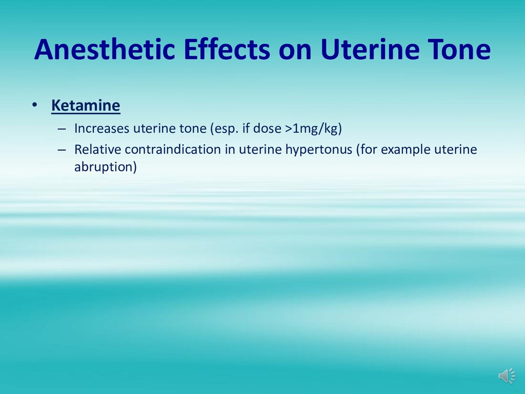
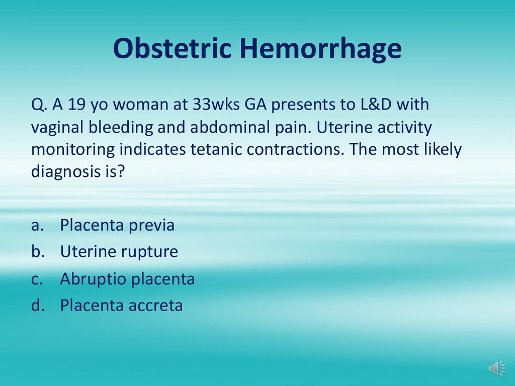
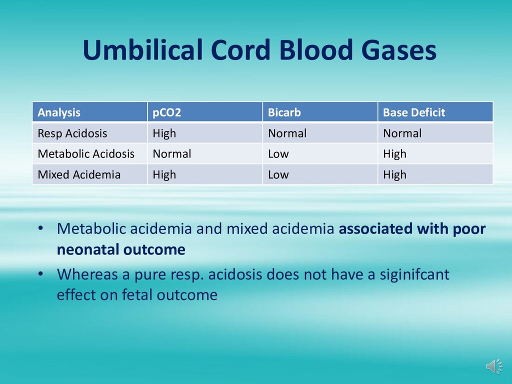
    implanted placenta – Signs & symptoms • Abdominal pain • Vaginal bleeding (concealed in 15%: retroplacental hematoma) • Increased uterine tone (tetanic contraction) • Fetal distress • IUFD – Most common cause of DIC in pregnancy – Anesthetic Management • Mild-moderate abruption (large bore access, Type & Cross, epidural if coags normal) • Severe abruption – Requires emergent C/S, large bore access, blood immediately available (MTP for OB) – fetal distress or maternal instability is often present – usually necessitating GA – If increased uterine tone, avoid ketamine for induction
  53. Uterine Rupture Q. What is the most common sign of

    uterine rupture in a patient undergoing trial of labor after cesarean (TOLAC)? a. Abdominal pain b. Vaginal bleeding c. Recession of fetal presenting part d. Fetal bradycardia
  54. Uterine Rupture • Increased risk in patients with previous C/S

    or vertical uterine scar from prior surgery • Signs & Symptoms – Fetal bradycardia – most common sign – Vaginal bleeding – Abdominal pain – Maternal hypotension
  55. Uterine Rupture • Anesthetic Management – Large bore access –

    Blood available – Catastrophic rupture generally requires GA for C section – Uterine dehiscence can usually be done with RA – Be prepared for possible hysterectomy
  56. Abnormal Placentation Q. A G4P3 woman with placenta previa presents

    for C section. She denies any recent vaginal bleeding. BP is 124/76 and HR is 84. Preop HCT is 32. She has undergone 3 previous C/S. Appropriate management of this patient will most likely include? a. Obtaining preop fibrinogen level b. Utilizing intraoperative blood salvage throughout surgery c. Preparing for massive transfusion d. Administering single shot spinal anesthesia
  57. Placenta Accreta • Placenta abnormally adherent to myometrium • Massive

    hemorrhage when attempt to separate placenta • Increased risk of accreta with prior C/S & placenta previa • Often requires aggressive fluid and blood replacement +/- hysterectomy
  58. Uterine Atony • Most common cause of PPH • Risk

    Factors – Multiple gestation – Polyhydramnios – High parity – Prolonged labor especially with augmentation – Chorioamnionitis – Tocolytic drugs
  59. Uterine Atony Q. A G5P5 with a history of asthma

    develops PPH due to uterine atony. EBL is 500ml with ongoing bleeding. Uterine massage has been unsuccessful. BP is 160/95 & HR is 95. The most appropriate management at this time is? a. Methylergonovine IM b. IV oxytocin infusion c. IM hemabate d. Uterine artery embolization in IR
  60. Uterine Atony Q. A healthy parturient inadvertently received an IV

    bolus of 20Units oxytocin after delivery of the placenta. Which of the following is the most likely adverse effect? a. Bronchospasm b. Hypertension c. Hypotension d. Pulmonary edema
  61. Treatment of Uterine Atony • Oxytocin – Causes vasodilation –

    Large bolus doses can cause hypotension – Longer duration infusions can cause hyponatremia • Methergine – Can cause HTN – Administer IM, iv dose can cause severe vasoconstriction, leading to MI, Cerebral bleed • Hemabate (prostaglandin F2 alpha; Carboprost) – Bronchospasm (relatively contraindicated in asthma) – HTN
  62. Treatment of Uterine Atony • Resuscitation (fluids/blood) • Large bore

    iv access • Bakri Balloon • Consider GA for further invasive surgical options – B-Lynch compression suture – Uterine artery ligation – Uterine artery embolization – Hysterectomy • Consider TXA in any case of PPH
  63. Amniotic Fluid Embolism • Rare complication • Often fatal (mortality

    80%) • Necessary circumstances leading to AFE – Rupture of amniotic membranes – Site of entry into maternal circulation e.g. cervical laceration
  64. Amniotic Fluid Embolism Signs & Symptoms • Shortness of breath

    • Sudden drop in O2 saturations • Hypotension • Cardiovascular collapse • Sequelae: LV failure, DIC
  65. Amniotic Fluid Embolism • Pathophysiology of AFE – Amniotic fluid

    gets into the pulmonary circulation, causing vasospasm, leading to decreased LV output, causing hypotension – Sudden Pulmonary HTN leading to cor pulmonale – Deranged Ventilation/perfusion ratio leading to hypoxemia and tissue ischemia – DIC occurs in 40% – Increase risk of uterine atony
  66. Amniotic Fluid Embolism • Management – Supportive management – Intubation

    & ventilation – Aggressive resuscitation • Fluid resuscitation • Inotropic support • Invasive lines • DIC management
  67. Cardiac Disease in Pregnancy Q. A woman with mitral stenosis

    develops hypotension following induction of epidural anesthesia for C/S. Which is the following is the best treatment for her hypotension? a. Dopamine b. Ephedrine c. Epinephrine d. Phenylephrine
  68. Cardiac Disease in Pregnancy • Mitral Stenosis Management goals: –

    Maintain Sinus rhythm, slow HR – Aggressively treat Afib – Maintain venous return – Maintain adequate SVR – Avoid increases in PVR (due to pain, hypoxemia, hypercarbia, acidosis)
  69. Cardiac Disease in Pregnancy • Mitral Regurgitation Management Goals –

    Maintain normal to slightly increased HR – Maintain sinus rhythm – Aggressively treat Afib – Prevent increases in SVR – Maintain venous return – Avoid situations that increase PVR
  70. Cardiac Disease in Pregnancy • Aortic Stenosis Management Goals –

    Maintain normal HR & sinus rhythm – Maintain adequate SVR – Maintain intravascular volume and venous return – Avoid myocardial depression during GA – Avoid spinal anesthesia
  71. Cardiac Disease in Pregnancy • Aortic Regurgitation Management Goals –

    Maintain normal to slightly increased HR – Prevent increases in SVR – Avoid myocardial depression during GA
  72. Cardiac Arrythmias in Pregnancy • Higher risk of cardiac arrythmias

    during pregnancy; the most common arrythmia is SVT • Most antiarrhythmic agents can be used safely in pregnancy e.g. adenosine • Amiodarone associated with adverse fetal effects, so use only for refractory cases of dysrhythmias • Cardioversion is safe – Use same energy levels as in non pregnant cases – FHR monitoring recommended; transient FHR abnormalities may occur – Actual current reaching the fetus is minimal
  73. Congenital Heart Disease • CHD is the most common form

    of heart disease in pregnancy (2nd is valvular disorders) • Physiologic CVS changes in pregnancy can lead to return of symptoms or worsening symptoms in a patient who is stable and doing well post surgical repair • Need to have a good understanding of patients pathophysiology & anatomy to develop an anesthetic plan • MDT approach to such patients is essential
  74. Cardiac Disease in Pregnancy Q. Which statement about pulmonary HTN

    & pregnancy is most likely true? a. Maternal mortality does not differ between primary & secondary pulmonary HTN b. Epidural analgesia is contraindicated c. C/S is the recommended mode of delivery d. Inhaled nitric oxide can safely be used during L&D
  75. Pulmonary HTN & Pregnancy • High maternal mortality (Primary 30%;

    Secondary 60%) • Vaginal delivery preferred unless OB indication for C Section • Labor analgesia & Invasive monitors needed • Pulmonary vasodilators used in pregnancy, including nitric oxide (potential fetal methemoglobinemia at high concentrations) • Maternal deaths usually post partum – RV failure, dysrhythmias, PE
  76. Cardiac Arrest in Pregnancy • Follow ACLS guidelines • Maintain

    manual left uterine displacement during CPR • Proceed to immediate C Section if unable to obtain ROSC after 4 mins (2 cycles), even in the L&D room, do not transport patient
  77. Intrapartum Fever Management • Treatment to lower maternal temperature •

    Identify cause of fever • Treatment based on underlying cause – Chorioamnionitis: Antibiotics – Epidural: no need for Abx
  78. Antiphospholipid Syndrome • Prothrombotic disorder with presence of anticardiolipin antibody

    or lupus anticoagulant • At risk of venous and arterial thrombosis • Nearly 25% of VTE events occurring during pregnancy • Increased risk of fetal loss (esp. 2nd and 3rd trimester) • Lupus anticoagulant has no anticoagulant activity but results in prolonged PTT due to lab artifact • Does not preclude neuraxial anesthesia • Most patients will be on prophylactic or therapeutic LMWH
  79. Non Obstetric Surgery during Pregnancy Q. Which of the following

    recommendations BEST reflects the ASA statement on non obstetric surgery during pregnancy? a. If the fetus is previable, no assessment of FHR is recommended b. If the fetus is viable, continuous intraoperative FHR monitoring must be performed c. If possible, surgery is best performed in the 2nd trimester d. Midazolam should be avoided due to teratogenic effects
  80. Non Obstetric Surgery during Pregnancy Considerations • Don’t forget about

    the physiological changes in pregnancy and adjust anesthetic technique accordingly • Be cognizant of teratogenic drugs • Ensure maintenance of uteroplacental perfusion and fetal oxygenation • Prevention of premature labor
  81. Non Obstetric Surgery during Pregnancy Teratogenicity of Anesthetic Agents •

    All benzodiazepines are Class D drugs in pregnancy – Previous FDA system classed Drugs A-X for teratogenicity – Class D = evidence of harm in pregnancy – Chronic use of Valium is associated with fetal anomalies • Nitrous oxide is controversial – Evidence is mixed, but no good data to suggest it is teratogenic or increases fetal loss/miscarriage
  82. Non Obstetric Surgery during Pregnancy Anesthetic Technique • No specific

    drug or technique shown to improve fetal outcome • Regional anesthesia is often preferred if possible – Advantages: minimizes drug exposure to fetus, avoids increased maternal risk associated with GA • Consider left uterine displacement after GA20wks • Avoid maternal hypercapnia (fetal acidosis) • Avoid maternal hyperventilation (maternal alkalosis can cause uterine artery vasoconstriction)
  83. Non Obstetric Surgery during Pregnancy Effect of anesthetics on FHR

    tracing Anesthetic drugs that decrease or eliminate fetal heart rate variability: • Induction agents • Opioids • Inhalational agents Can only assess: Baseline FHR & Absence of Presence of decelerations
  84. Non Obstetric Surgery during Pregnancy Preterm Labor • Incidence of

    preterm labor increased in general • Greatest risk during 3rd trimester and surgery in abdomen/pelvic
  85. Anesthesia for Cerclage • Neuraxial technique most commonly performed (spinal)

    • T10-S4 sensory level preferred • Generally avoid sedative/anxiolytic agents
  86. Meconium Stained Amniotic Fluid • Routine suctioning/intubation no longer recommended,

    even in non vigorous infants • Recommendation in a vigorous infant with meconium stained amniotic fluid is to follow routine neonatal care • Non-vigorous infant – Follow same resuscitation guidelines as an infant without meconium – Intubate and suction only if airway is obstructed – Don’t intubate/suction all infants before trying bag-mask ventilation
  87. Neonatal Myasthenia Gravis • Maternal antibodies to Ach Receptor crosses

    the placenta • And so approx. 16% of infants born to mothers with MG will have neonatal MG • Develop symptoms within 4 days of life (feeding issues, hypotonia, respiratory distress) • Resolves in 2-4 weeks with metabolism of antibodies • May require treatment with anticholinesterase
  88. Fetal Heart Rate Monitoring Q. Which statement about FHR patterns

    is most likely true? a. Early decelerations indicate fetal hypoxia b. Maternal drug administration does not influence fetal heart rate c. Beat to beat variability is an insensitive indicator of fetal well being d. Sustained FHR >180 may be indicative of fetal hypoxia
  89. Fetal Heart Rate Monitoring • Have a systematic approach to

    interpreting FHR tracing • Parameters monitored – Baseline HR – Variability – Periodic patterns (acceleration versus deceleration) – Uterine activity
  90. Fetal Heart Rate Monitoring • Baseline Fetal Heart Rate –

    Normal: 120-160 beats/min – Fetal tachycardia >160 bpm – Fetal bradycardia <120bpm
  91. Fetal Heart Rate Monitoring • Causes of Fetal tachycardia –

    Chronic fetal asphyxia due to inadequate uteroplacental perfusion – Maternal fever/sepsis – Maternal thyrotoxicosis – Maternal drug administration • Atropine • Ephedrine • Beta mimetic tocolytic agents e.g. terbutaline
  92. Fetal Heart Rate Monitoring • Causes of Fetal Bradycardia –

    Acute fetal asphyxia – Fetal acidosis – Local anesthetic toxicity from paracervical block – Maternal Beta Blockers – Congenital heart block
  93. Fetal Heart Rate Monitoring • HR variability – Beat to

    beat variability = short term variability • Most sensitive indicator of fetal well being – Long term variability • Another indicator of fetal well being but not as sensitive
  94. Fetal Heart Rate Monitoring • Causes of decreased Beat to

    Beat Variability – Fetal hypoxia & acidosis – Normal fetal sleep cycles (scalp stimulation) – Maternal Drugs (opioids, barbiturates, volatile, Benzos, Anticholinergics)
  95. Fetal Heart Rate Monitoring • Periodic Patterns – Accelerations •

    FHR >15bpm with fetal movement or uterine contraction – Decelerations • Early: onset and peaks with contraction, HR >100bpm • Variable: shape, peak & onset times vary, HR may be <100bpm • Late: onset after peak of contraction, HR usually >100bpm
  96. Fetal Heart Rate Monitoring • Early Decelerations – Etiology: vagal

    reflex due to head compression during contraction – Not concerning – Does not require treatment
  97. Fetal Heart Rate Monitoring • Variable decelerations – Etiology is

    always a vagal response – Vast majority of which is caused by umbilical cord compression during contraction – Variable decels occurring in late labor may be due to dural stimulation resulting from fetal head compression as it traverses vaginal canal – Recurrent variable decels that are deep and long duration are concerning – Occasional decels that are of short duration and not deep are not concerning
  98. Fetal Heart Rate Monitoring • Late Decelerations – Etiology is

    usually inadequate uteroplacental perfusion – Always concerning regardless of depth of decel – Initial management: • Maternal supplemental oxygen • Left uterine displacement • Fluid bolus • Vasopressors to treat hypotension
  99. Fetal Heart Rate Monitoring • Fetal Heart Tracing Categories –

    Category 1: Reassuring • Normal tracing, strongly predictive of normal fetal acid- base status at the time of tracing – Category 2 • Indeterminate, not predictive of abnormal fetal acid- base status, inadequate evidence to classify as normal or abnormal – Category 3 : Non-reassuring • Abnormal, predictive of abnormal fetal acid-base status at the time of tracing, requires prompt evaluation
  100. Physiologic Changes in Pregnancy Q. When is the cardiac output

    of a parturient at its highest level? a. 1st stage of labor b. Immediately postpartum c. 2nd stage of labor d. 2nd trimester
  101. Physiologic Changes in Pregnancy Q. Which of the following is

    most likely to represent an abnormal finding in a woman at 38 weeks gestation? a. Left axis deviation on an EKG b. Pulmonary artery occlusion pressure of 22mmHg c. Mild tricuspid regurgitation d. Presence of a 3rd heart sound on auscultation
  102. CVS Changes in Pregnancy • See Lecture on Maternal Physiology

    for details • CO increases throughout pregnancy • Increases approx. 50% by 3rd trimester – Due to increase in SV (30%) & HR (15%) • Further increased during Labor & Delivery – Active labor (30%) – 2nd stage (45%) – Immediate postpartum (80%)
  103. CVS Changes in Pregnancy • Blood volume increase 35% –

    Due primarily to increase plasma volume • SVR decreases 20% – Leads to mild decrease in BP • Central pressures (CVP/PCWP) remain unchanged due to dilation of vessels
  104. Physiologic Changes in Pregnancy Q. Which of the following is

    most likely to be decreased in a healthy parturient at 36 weeks gestation? a. Total RBC volume b. Tidal Volume c. Serum Creatinine level d. Factor VII level
  105. Respiratory Changes in Pregnancy • Minute ventilation – Increases up

    to 50% at term – Increases primarily by marked increase in TV – Small increase in respiratory rate • Lung Volumes & Capacities – ERV & RV decreases – Inspiratory reserve volume increases slightly – In normal sitting position FRC decreases 20% due to decrease in ERV & RV (this decreases even further in supine position) • TLC and VC unchanged
  106. Respiratory Changes in Pregnancy • Oxygen consumption – Increases 20%

    during pregnancy – Increases 100% during labor • Oxygen delivery – Right shift of maternal ODC – Facilitates oxygen delivery to fetus
  107. Physiologic Changes in Pregnancy Q. A normal ABG in a

    non laboring parturient in the upright position at 37 weeks gestation is most likely? a. 7.40/40/100/24 b. 7.35/40/90/22 c. 7.44/35/103/24 d. 7.44/30/103/20
  108. ABGs in Pregnancy • paCO2 decreases to 26-32mmHg due to

    increase in MV • Resulting in a mild respiratory alkalosis with pH 7.44 • Metabolic compensation results in decrease in serum HCO3 to 18-22mEq • Small increase in paO2 (103-107)
  109. GI Changes in Pregnancy • Hormonal changes – Increase in

    progesterone leads to reduced LES tone • Mechanical changes from cephalad displacement of stomach – Increased intragastric pressure – Distorted GE junction leads to gastric reflux • Clinical implications of GI changes – Increase risk of aspiration
  110. Hematologic Changes in Pregnancy • 20% increase in red cell

    mass • 45% increase in plasma volume • Leads to dilutional anemia (Normal HCT 35%) • Hypercoagulable state – PE is a leading cause of maternal mortality
  111. Coagulation Factor Changes • Increased: Factors I, VII, VIII, IX,

    X & XII • Unchanged: Factor II & V • Decreased: Factor XI & XIII • TEG: Hypercoagulable • PLT count: unchanged or decreased (gestational thrombocytopenia) • Plasminogen: increased • Fibrin degradation products: increased
  112. CNS Changes of Pregnancy • MAC of volatiles decreases 30-40%

    • LA requirements decrease 30% • Increased endorphins & enkephalin levels (elevated pain threshold)
  113. Uterine Blood Flow • Uterus is not capable of autoregulation

    • Uterine perfusion is dependent on: – Adequate driving pressure i.e. maternal BP – Low uterine venous pressure – Low uterine vascular resistance
  114. Uteroplacental Perfusion What decreases uteroplacental perfusion? a. Low uterine artery

    pressure b. Increased uterine venous pressure - Vena caval compression, uterine contractions - Uterine hypertonus, Valsalva c. Increased uterine vascular resistance - Endogenous and exogenous vasoconstrictor (not the epidural test dose) - High concentration LA - Preeclampsia, DM
  115. Choice of Vasopressor • Ephedrine – Alpha and beta effects

    – Less increase in uterine vascular resistance compared to phenylephrine – Previously was vasopressor of choice in OB • Phenylephrine – Pure alpha effect; was thought to worsen resistance – Cord pH higher in infants whose mothers received phenylephrine compared to ephedrine
  116. Maternal Fetal Drug Transfer Factors affecting placental drug transfer •

    High maternal concentration of drug • Drug diffusion constant of the drug • DDC is increased by: – Low molecular weight – Low protein binding (only unbound drug crosses) – High lipid solubility – Low degree of ionization (only nonionized drugs crosses)
  117. Maternal Fetal Drug Transfer Factors affecting placental drug transfer •

    Placental binding • Placental metabolism • Factors that alter placental perfusion – Aortocaval compression – Hypovolemia – Vasoconstrictors
  118. Factors Affecting Fetal Drug Concentrations • Drug Metabolism – immature

    enzyme pathways in the fetus, contribute to elevated drug levels • Fetal protein binding – lower protein binding capacity compared to mother can lead to high free drug levels • Fetal pH
  119. Fetal pH & Local Anesthetics Q. A patient who is

    receiving epidural labor analgesia is taken to the OR for emergent C Section due to fetal bradycardia. Her epidural should be dosed with which LA? a. 2% Lidocaine b. 0.5% ropivacaine c. 3% 2-Chloroprocaine d. 0.5% bupivacaine
  120. Fetal pH & Local Anesthetics • Ion Trapping – In

    acidotic fetus, LA that crosses the placenta to the fetus becomes ionized and therefore cant cross back over to the maternal side – i. e. the LA gets “ionized and trapped” – Fetal accumulation of LA can lead to high drug levels and floppy baby • Chloroprocaine drug of choice in fetal distress – Ion trapping is not an issue because short maternal half life leads to minimal drug transfer to the fetus
  121. Maternal Fetal Drug Transfer Q. The least amount of placental

    transfer will occur with which of the following anesthetic drugs? a. Isoflurane b. Sufentanil c. Succinylcholine d. Ketamine
  122. Maternal Fetal Drug Transfer Drugs that don’t cross placenta in

    significant amounts – Heparin (large molecule, highly water soluble) – Insulin (large molecule with no placental transfer) – Neuromuscular blocking drugs • Non depolarizing drugs do cross but in clinical insignificant amounts • Minimal drug transfer of muscle relaxants due to their ionization and high molecular weight • Rarely affects neonatal muscle tone • Succinylcholine essentially does not cross (v. large molecule)
  123. Maternal Fetal Drug Transfer Maternal Fetal Distribution of Bupivacaine •

    High degree of maternal protein binding and high pKa limits transfer to fetus • Lesser degree of fetal protein binding leads to fetal concentration of free drug equilibrating with maternal concentration • Total fetal concentration is less than maternal concentration – BUT concentration of active drug is equal
  124. Maternal Fetal Drug Transfer • Placental transfer of anticholinergics –

    Directly correlates with ability to cross BBB – Atropine easily crosses and can affect FHR – Scopolamine easily crosses – Glycopyrrolate minimally crosses placenta (has little to no effect on FHR)
  125. Maternal Fetal Oxygen Transport • Placental O2 transfer occurs via

    diffusion – Transport determined by difference in pO2 between maternal and fetal circulation • Bohr Effect – Concomitant fetal to maternal transfer of CO2 makes maternal blood more acidic and fetal blood more alkalotic – Leads to right shift of maternal ODC curve and left shift of fetal ODC curve – Favors unloading of oxygen to fetus
  126. Fetal Oxygenation • Fetal pO2 is rarely > 50-60mmHg –

    Placenta has high rate of O2 consumption – Fetal arterial blood is a mixture of oxygenated umbilical venous blood and deoxygenated IVC blood
  127. Fetal Hb & Oxygen Transport • Fetal oxygenation depends on

    maternal/fetal O2 gradient and differences in type of Hb • Hemoglobin F – Greater affinity for oxygen – Lower affinity for 2,3-DPG – Leftward shift of fetal ODC – P50 lower in fetal blood than adult blood • Hb F and higher Hb concentrations leads to fetal arterial blood oxygen concentration just minimally lower than adult (even though there is a lower oxygen tension)
  128. Maternal paO2 & Fetal Oxygenation • At normal fetal paO2,

    increases in maternal FiO2 has only slight effect on increasing fetal paO2 • At decreased fetal paO2, increases in maternal FiO2 could be beneficial because fetal ODC is steep in that range • Hence on L&D when fetus is in distress, one of the first resuscitation maneuvers is to place high flow oxygen to the mother to increase maternal FiO2
  129. Umbilical Cord Blood Gases Umbilical Vein Umbilical Artery pH 7.34

    7.28 pO2 30 15 pCO2 35 45 Bicarb 22 20 Base Deficit 5 7 • Umbilical cord blood gases – Umbilical artery cord gas is reflection of what is happening on the fetal side – Umbilical vein cord gas is more a reflection of what is happening on the maternal side and placenta
  130. Umbilical Cord Blood Gases Analysis pCO2 Bicarb Base Deficit Resp

    Acidosis High Normal Normal Metabolic Acidosis Normal Low High Mixed Acidemia High Low High • Metabolic acidemia and mixed acidemia associated with poor neonatal outcome • Whereas a pure resp. acidosis does not have a siginifcant effect on fetal outcome